

 PDF
PDF Citation
 Print
Print


INTRODUCTION
Coelomycete is an emerging group of pathogens capable of causing soft tissue infections in immunocompromised patients. These infections are classified as phaeohyphomycosis, and they are caused by dematiaceous fungi (pigmented hyphae)123. Microsphaeropsis arundinis, a member of the class Coelomycetes, was first detected from the “giant reed” Arundo donax in Pakistan, and it usually inhabits terrestrial plant hosts and is ubiquitous in soil and fresh water environments3. Herein, we experienced a rare case of subcutaneous and intranasal phaeohyphomycosis caused by M. arundinis; we identified the fungal organism based on internal transcribed spacer (ITS) sequence analysis. To the best of our knowledge, this is the first such case in South Korea.
CASE REPORT
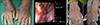
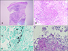
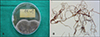
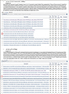
A 76-year-old Korean man presented with cutaneous lesions on both his forearms and dorsal hands. He was a farmer and was in close contact with soil for several decades. When he visited our clinic with enlarged skin lesions that had first appeared 6 months ago, he also complained of asymptomatic diffuse erythematous plaques and swelling with scales and pustules on both his forearms and dorsal hands (Fig. 1A). Subsequently, he reported having experienced rhinalgia a month ago. There was no fever or lymphadenopathy, and both respiratory and neurological examinations were unremarkable. The patient was under medication for hypertension, angina pectoris, and diabetes mellitus. He was suspected as having bacterial, atypical mycobacterial, or deep fungal infections, and a skin biopsy was subsequently performed from the lesion. Bacterial and fungal cultures were taken, and the cutaneous tuberculosis (TBC) polymerase chain reaction (PCR) test (BAG Health Care GmbH, Lich, Germany) was performed. In addition, the patient was referred to the otorhinolaryngology department for rhinalgia. Otorhinolaryngology examinations revealed necrotic changes accompanied by hemorrhage in the nasal septum (Fig. 1B); subsequently, a biopsy was performed at that site. The findings raised a suspicion of mucormycosis. Hence, other fungal infections, including mucormycosis, were considered as differential diagnoses, and further examinations were performed. The patient was tested for aspergillus antigen and (1-3)-β-D-glucan. Invasive involvement of both arms as well as nasal invasion was observed; therefore, chest x-ray and bone scan were performed to check for more invasive lesions. Bone scan revealed cellulitis; however, chest x-ray showed no remarkable findings. Histopathologic examination of the skin showed dense infiltrates of inflammatory cells such as neutrophils, monocytes, histiocytes, and giant cells extending from the upper dermis through the subcutaneous fatty layer (Fig. 2A, B). Staining for acid-fast bacilli showed negative results; similarly, PCR for mycobacteria and bacterial culture showed negative results. Gomori's methenamine silver staining and periodic acid-Schiff staining showed scattered fungal spores and hyphae (Fig. 2C, D). The histopathologic findings of the nasal septal lesions were similar to the results of skin biopsy. Through consultation with the Department of Infectious Diseases, the blood tests revealed negative results for aspergillus antigen and positive results for (1-3)-β-D-glucan (100.0, positive >80). The cultures of the biopsy specimen and nasal septum tissue grew a dematiaceous (pigmented) fungus; microscopic examination showed short and thick irregularly shaped septate hyphae (Fig. 3). Owing to the absence of conidia, the organism could not be morphologically identified. Hence, gene sequencing analysis was performed for molecular identification of the organism. The sequence of the D1/D2 domain of ribosomal RNA gene regions and ITS1-5.8S-ITS2 regions, using primers and standard sequencing methods, showed identity with M. arundinis (100% sequence identity to M. arundinis cystathionine beta-synthase (CBS) 100243 [GenBank accession number JX496123.1] and 100% sequence identity to M. arundinis 0012 [GenBank accession number KY992587.1])4. Based on these findings, the patient was diagnosed with subcutaneous and intranasal phaeohyphomycosis caused by M. arundinis (Fig. 4).
The patient was initially administered fluconazole 200 mg and cefbuperazone 2 g daily. This was changed to itraconazole 200 mg/d and isoconazole topical cream after the presence of pathogenic organism was confirmed; subsequently, itraconazole was administered for more than 5 months. Thereafter, the lesion completely disappeared and the patient no longer complained of rhinalgia; there was no subsequent relapse of infection in cutaneous lesions (Fig. 1C).
DISCUSSION
In 2004, Pendle et al.1 described infections caused by M. arundinis in patients with diabetes mellitus and chronic kidney disease. Since then, cutaneous infections caused by the same pathogen have been reported in the United States, Japan, and Australia, although it is still a rare opportunistic infection that occurs in immunocompromised patients235. Especially in South Korea, phaeohyphomycosis due to Exophialia spp. has been reported in several cases678; however, to the best of our knowledge, this is the first case of phaeohyphomycosis caused by M. arundinis. In addition, it is a very rare case owing to the involvement of the nasal septum as well as the skin.
Phaeohyphomycosis, first named in 1974, can occur in various clinical forms from subcutaneous mycosis to deep organ infections9. Especially, the subcutaneous form tends to occur at the distal limbs, which might be easily exposed to trauma or infections6. M. arundinis infection can occur in the form of subacute onset on the limbs and can spread around by direct inoculation of mold-contaminated materials5. In this case, as there were no signs indicative of systemic involvement, the occurrence of phaeohyphomycosis in the nasal septum could be attributed to the direct contact of M. arundinis through the hands of the patient.
The histopathologic findings of phaeohyphomycosis are very typical, as observed in the present case, although identifying the causative pathogen is always challenging. The coelomycete fungi produce a blister-like fruiting structure called conidiomata, which differ from other fungi, and the size and shape of the conidia of M. arundinis differ from those of other coelomycete genera. However, the morphological diagnosis of M. arundinis is always challenging owing to poor sporulation1011. Therefore, species identification is often dependent on DNA sequencing using ITS, as in this case.
Phaeohyphomycosis is primarily treated by surgical excision alone, in cases with localized and small well-defined lesions. However, in case of wide or ambiguous cutaneous lesions, surgical treatment is difficult and systemic medications should be administered78. In all the eight cases of M. arundinis infections reported so far, diffuse infection was observed and no surgical excision was performed; treatment involved use of antifungal agents and thermotherapy. This suggests that M. arundinis infection may demonstrate a diffuse form rather than localized or solitary nodular and cystic subcutaneous appearance. Triazole antifungal agents have been considered as the first-line therapy for phaeohyphomycosis. Besides, clinical data related to non-azole antifungal therapy for M. arundinis infection are limited, and the minimum inhibitory concentration of itraconazole for M. arundinis has been reported as ≤0.25 mg/L2512. Therefore, oral itraconazole was used for treatment of phaeohyphomycosis in this case, although the lack of susceptibility test was considered as a limitation.
To the best of our knowledge, this is a rare case of subcutaneous and intranasal phaeohyphomycosis caused by M. arundinis, and our patient was also immunocompromised, undergoing treatment for diabetes mellitus, similar to the patients in previous reports. He might have been misdiagnosed with mucormycosis based on the findings of nasal septal lesion biopsy and ENT examinations, although the pathogen was finally confirmed using gene sequencing analysis. Positive (1-3)-β-D-glucan in blood is indicative of invasive or disseminated fungal infections. This test can be helpful for the detection of Aspergillus, Candida, and Pneumocystis jiroveci, which do not produce (1,3)-β-D-glucan13. This could help to rule out mucormycosis. Clinically, subcutaneous phaeohyphomycosis manifests mainly in nodular or cystic form in the limbs of immunosuppressed patients, whereas primary cutaneous mucormycosis is relatively rare, and malodorous brown or black necrotic lesions with an erythematous border are mainly observed14. Clinical differential diagnosis and gene sequencing analysis were used to diagnose phaeohyphomycosis in our case. Efforts should be made to identify the pathogenic strains that can influence the prognosis and treatment of this infectious disease.
 XML Download