

 PDF
PDF Citation
 Print
Print


INTRODUCTION
Gastric cancer is a major global health issue, particularly in Asian countries including Korea, Japan, and China.1 Despite advances in endoscopic and surgical resection equipment and techniques, gastric cancer is the third leading cause of cancer mortality globally. Detection of gastric cancer in the early stages and prompt treatment are associated with favorable prognosis.23 Patients diagnosed with early gastric cancer (EGC) during screening endoscopy show low risk of lymph node metastasis and can undergo endoscopic submucosal dissection (ESD) as curative local treatment for stomach preservation. However, most patients with EGC present with non-specific symptoms. Previous studies have shown that the prevalence of advanced gastric cancer was higher in patients undergoing diagnostic examination without previous screening endoscopy to investigate the causes of gastrointestinal symptoms.4 Thus, a mass screening program for gastric cancer has been adopted in Korea and Japan. A recent study evaluated the effect of gastric cancer screening via biennial upper endoscopy or gastrography in adults aged ≥ 40 years, which is the Korean National Cancer Screening Program. This study reported the effectiveness of such assessment in reducing the gastric cancer-related mortality risk.4 However, there is a report about the incidence of interval gastric cancer (IGC) in patients who received the periodic endoscopic examination in Korea.5 Therefore, meticulous endoscopic examination is mandatory to reduce false-negative rates.
Several factors should be considered to optimize the diagnostic yield of endoscopy. First, the administration of antispasmodics and mucolytic agents such as pronase facilitates a careful examination as the pretreatment.6 Second, adequate air insufflation and removal of mucus for optimal visualization of the mucosal surface are essential for accurate detection of EGC as technical aspect of examination. Third, a systematic screening protocol to map the entire stomach without blind spots reduces the risk of missed lesions as the observation procedure.7 In addition to a technically accurate endoscopic procedure, knowledge of the features of suspicious lesions is important to detect EGC. Key signs of EGC are lesions with an irregular surface (elevated or depressed lesions) and color (reddish or discolored) changes, and spontaneous bleeding (Fig. 1). If EGC or dysplasia is suspected, the chromoendoscopy using indigo carmine can be helpful to define the surface morphology of suspicious lesions. Finally, advanced imaging techniques such as magnifying endoscopy with narrow band image provide detailed information regarding small gastric lesions. This review discusses EGC locations detected from screening and surveillance endoscopy, especially in Korea. This article will contribute significantly to the literature by providing a better understanding of EGC and will guide endoscopists in performing careful endoscopic examination and surveillance.
 | Fig. 1Images of upper endoscopy for early gastric cancers. (A) Elevated and reddish lesion sized 1.5 cm with spontaneous bleeding (arrow) is found on the antrum greater curvature. (B) Depressed and discolored lesion sized 1.0 cm is found on the lower body lesser curvature (arrow). (C) Flat lesion sized 0.8 cm with inhomogeneous erythema is found on the cardia (arrow).
|
INTRAGASTRIC LOCATION OF EGC INDICATIVE ESD
Mucosal cancers that on histopathological examination measure < 2 cm in size and show well-differentiated features without ulceration can be effectively treated by ESD owing to their low risk of lymph node metastasis.8 A recent multicenter single-arm confirmatory trial (JCOG0607) performed by the Japan Clinical Oncology Group suggested that ESD should be the standard treatment (preferred over gastrectomy) for intestinal-type EGC that fulfills the expanded indications.9 For undifferentiated EGC sized less than 2 cm without ulceration, a meta-analysis including 972 undifferentiated type EGC from 14 retrospective studies reported that the ESD showed favorable short-term outcome.10 Although long-term data after ESD are further needed to confirm the safety and efficacy, the endoscopic resection has been performed for the curative intention in clinical practice.11 However, endoscopists should consider that the curative resection rate is lower than that of ESD for differentiated type EGC.12
Early detection of EGC is important to achieve curative resection through ESD.
Several retrospective studies conducted in Korea demonstrated that the lower part is the most common site to perform ESD for the treatment of EGC (Table 1). One study in which 647 consecutive EGC lesions were resected by ESD also showed that the most frequent site of occurrence of EGC was the lower part of the stomach (89.6%). This study has reported that the lesser curvature was the most common site of involvement (43.6%).13
Table 1
Locations of early gastric cancer that received endoscopic resection
| Author | Year | No. | Study design | Tumor location (%) |
|---|---|---|---|---|
| Min et al.36 | 2015 | 1,497 | Single center | Body (21.8) |
| Fundus (21.8) | ||||
| Cardia (21.8) | ||||
| Antrum (78.2) | ||||
| Angle (78.2) | ||||
| Kim et al.3 | 2015 | 165 | Single center | Upper (6.1) |
| Middle (14.5) | ||||
| Lower (79.4) | ||||
| Choi et al.37 | 2015 | 961 | Single center | Upper (6.0) |
| Middle (21.2) | ||||
| Lower (72.8) | ||||
| Ahn et al.38 | 2011 | 1,370 | Single center | Upper (7.1) |
| Middle (32.3) | ||||
| Lower (60.6) | ||||
| Shin et al.39 | 2015 | 1,105 | Multicenter | Upper (4.3) |
| Middle (28.5) | ||||
| Lower (67.2) |
![]()
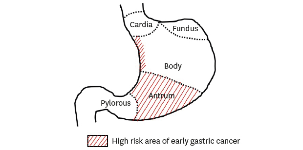
The lower part of the stomach includes the antrum-pylorus and the lower third of the stomach corpus including the angle. The mid-to-upper part refers to the middle and upper thirds of the stomach corpus including the cardia and the fundus. H. pylori infection causes gastric mucosal inflammation. Intestinal-type gastric cancer is preceded by an inflammation-induced cascade of precancerous lesions induced by inflammation (the atrophy-metaplasia-dysplasia-adenocarcinoma sequence).14 Atrophic mucosal changes advance from the antrum to the corpus along the lesser curvature.15 The distribution pattern of atrophy explains the higher incidence of EGC in the lower parts and the lesser curvature of the stomach. Although the inflammation-induced carcinogenesis cascade by H. pylori infection is not the primary cause of undifferentiated type gastric cancer, it is speculated that inflammation itself promotes development of these gastric cancers. Therefore, the undifferentiated-type EGC commonly occurs in the lower portions of the stomach.1316
CHARACTERISTICS OF SURGICALLY RESECTED GASTRIC CANCER BASED ON ITS LOCATION
Various studies performed in Korea showed that the lower part is the most frequent site to detect EGC in patients who received the surgical resection (Table 2). One study evaluated 313 consecutive surgically resected small gastric cancers measuring < 2 cm in size, to obtain information regarding the common sites of origin of this type of gastric cancer.17 EGCs were primarily detected in the antrum-angle (42.2%) and the cardia (26.2%). Most (68%) small gastric cancers originated along the lesser curvature. This result concurs with a study showing EGC resected by ESD. Undifferentiated EGCs occur more commonly in the distal stomach than the proximal site.1819
Table 2
Locations of early gastric cancer that received surgical resection
| Author | Year | No. | Study design | Tumor location (%) |
|---|---|---|---|---|
| Jeon et al.40 | 2018 | 275 | Single center | Upper (4.4) |
| Middle (33.1) | ||||
| Lower (62.5) | ||||
| Kim et al.3 | 2015 | 292 | Single center | Upper (7.2) |
| Middle (14.4) | ||||
| Lower (78.4) | ||||
| Ryu et al.41 | 2016 | 144 | Multicenter | Upper (4.9) |
| Middle (32.9) | ||||
| Lower (62.2) | ||||
| Hahn et al.42 | 2018 | 1,206 | Multicenter | Upper (8.3) |
| Middle (32.8) | ||||
| Lower (58.9) |
![]()
Clinicopathological studies comparing early adenocarcinoma of the gastric cardia and early adenocarcinoma of the distal stomach have revealed that proximal gastric cancer shows the following characteristics: 1) elderly male predominance, 2) greater tendency toward submucosal invasion and, 2) better histopathological differentiation.2021 In these studies, proximal gastric cancer was defined as a tumor with its epicenter located within 3 cm below the gastroesophageal junction (to avoid misclassification of esophageal cancer as gastric cancer). These reports would help endoscopists to investigate the antrum-angularis and the lesser curvature more intensively, as well as to evaluate the cardia more closely in elderly men in geographical areas with a high prevalence of H. pylori infection. Notably, endoscopic examination in young women with a family history of gastric cancer needs to focus on the gastric corpus, particularly the area near the greater curvature.22
CHARACTERISTICS OF MISSED SYNCHRONOUS GASTRIC LESIONS
Several reports have described missed synchronous gastric cancer after ESD or gastrectomy.2324 The delayed detection of synchronous lesions may lead to an additional treatment including ESD or surgery that can result in patient's discomfort, and increased medical cost.
Early detection of synchronous gastric lesions before these progress to invasive cancers is important to improve the prognosis of patients. A meta-analysis including 22 studies (six studies conducted in Korea) reported that the rate of missing synchronous lesions after ESD was 9.0%.25 An excessive focus on primary gastric neoplasms during endoscopy can result in that other synchronous lesions might be missed.26 One study reported that more than half of cases with missed synchronous lesion had images of missed lesion captured during the previous endoscopy examination.27 The lower parts of the stomach is the most frequent location to find missed synchronous lesions (Table 3). Factors associated with missed synchronous lesions were origin in the small size, and non-elevated morphology.2328 Non-elevated and small synchronous lesions are likely to be missed because these are difficult to detect on endoscopy. Therefore, endoscopists should consider the possibility of synchronous gastric neoplasms and carefully examine the entire stomach to evaluate not only primary lesions, but also check for synchronous lesions.
Table 3
Locations of missed synchronous gastric lesions after endoscopic resection in Korea
| Author | Year | No. | Study design | Tumor location (%) |
|---|---|---|---|---|
| Nam et al.26 | 2018 | 77 | Single center | Upper (5.9) |
| Middle (21.3) | ||||
| Lower (72.7) | ||||
| Yoo et al.28 | 2013 | 29 | Single center | Upper (6.9) |
| Middle (31.0) | ||||
| Lower (62.1) | ||||
| Lee et al.24 | 2010 | 12 | Single center | Upper (6.3) |
| Middle (43.8) | ||||
| Lower (50.0) | ||||
| Kim et al.43 | 2017 | 141 | Multicenter | Upper (7.1) |
| Middle (32.6) | ||||
| Lower (60.3) |
![]()
CHARACTERISTICS OF METACHRONOUS GASTRIC CANCER (MGC)
MGC is generally defined as a gastric cancer that developed at a location distant from the primary resection site during follow-up endoscopy after index ESD.29 Retrospective studies have reported that incidence of metachronous lesions ranged from 3.6% to 6.1%.3031 Therefore, endoscopic surveillance should be continued after gastric ESD. In a retrospective study including 2,779 patients who received ESD for EGC and endoscopic surveillance in Korea, the 5 and 10 year cumulative incidence of MGC or high grade dysplasia were 4.7% and 11.3%.30 Lower third of the stomach was the most common location for metachronous lesions (Table 4). Other features of MGC are small, differentiated intramucosal cancers sized less than 2 cm.29 Therefore, the regular endoscopic surveillance can allow repeat endoscopic resection for MGC.

CHARACTERISTICS OF IGC
The high detection rate of EGC in Korea and Japan is attributable to regular screening endoscopy performed in these countries4; however, several studies have reported cases of IGC.2532 The concept of IGC is derived from colon cancer and refers to cancer that is diagnosed between the time of screening and the next scheduled screening endoscopy. Therefore, this term is applicable only in countries such as Korea and Japan where a national screening program for gastric cancer is operational. A Korean study that evaluated the clinicopathological characteristics of IGC reported that the incidence of IGC diagnosed between the time of screening and post-screening endoscopy was 2.0% (16/81,762 subjects).5 Characteristics of IGC were cancer detected in the lower corpus of the stomach (70.6%) and histopathological evidence of undifferentiated lesions (70.6%). The doubling time of gastric cancer is approximately 2–3 years.33 Therefore, most IGC lesions in this study could be lesions that progressed from missed gastric mucosal abnormalities. A meta-analysis investigating the rate of missed gastric cancer during upper endoscopy showed a 9.4% incidence of missed gastric cancer. Identification of cancer within an interval of 6 months to 3.5 years after a negative endoscopy was defined as missed cancer. Thus, the incidence of missed lesions may be higher.25 Most missed gastric cancers were poorly differentiated lesions located in the gastric antrum and lower body.34 This result concurs with a previous study describing IGC. The anatomical variations in different parts of the stomach contribute to missed gastric cancer. Small-sized gastric cancer lesions occurring in the corpus might be obscured within the gastric folds. Frequent belching is associated with a difficult examination of the stomach corpus.35
CONCLUSION
Early detection improves the prognosis and reduces the economic burden of gastric cancer. Additionally, ESD is curative treatment for stomach preservation in such patients when EGC has not progressed to a stage where ESD is not feasible. Sound knowledge of standard endoscopic techniques and endoscopic features of EGC are important to improve the detection rate of EGC. Therefore, endoscopists need appropriate training to improve their technical skills and knowledge of various gastric lesions. This article focuses on the importance of understanding the common sites of occurrence of EGC as a clue for the accurate detection of gastric cancer in the early stages. The lesser curvature and the lower part of the stomach were observed to be the predominant locations affected by EGCs that are resectable by ESD or gastrectomy. The incidence of gastric cancer affecting the cardia was higher in elderly men. Most IGCs were poorly differentiated on histopathological examination and were located in the gastric antrum and lower body. Endoscopists should be mindful of the possibility of non-elevated synchronous lesions. Finally, newly detected EGC and MGC are frequently found in the lower third, and the majority of MGCs found during endoscopic surveillance are small, differentiated intramucosal cancer. In conclusion, knowledge regarding the location of EGC is useful for accurate endoscopic examination for screening and surveillance.
 XML Download