

 PDF
PDF Citation
 Print
Print


INTRODUCTION
Over the past 20 years, improvements in neonatal intensive care units have resulted in better survival outcomes for extremely-low-birth-weight (ELBW) neonates.1234 However, the incidence of acute abdominal problems has increased, causing gastrointestinal complications to be one of the significant risk factors in ELBW neonates' survival.5
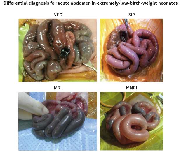
Acute abdomen in ELBW neonates can be differentially diagnosed into four disease entities, which are necrotizing enterocolitis (NEC), spontaneous intestinal perforation (SIP), meconium related ileus (MRI), and meconium non-related ileus (MNRI).67891011 Emergent operation is performed in patients with suspicion of bowel perforation or with clinical deterioration despite medical treatment. Laparotomy and peritoneal drainage are two methods of treatment for intestinal perforation in ELBW neonates, and the treatment of choice is still controversial.1213 Moreover, in laparotomy, it is still debatable whether to perform primary anastomosis or to perform diversion.14
The aim of this study was to review our experience in abdominal surgery for ELBW neonates, and to evaluate characteristics and clinical outcome according to the differential diagnosis.
METHODS
Data collection
Eight hundred five ELBW neonates who were admitted to the neonatal intensive care unit of Seoul National University Children's Hospital between January 2003 and December 2015 were retrospectively reviewed. From the medical records and electronic databases, we identified 65 ELBW neonates who underwent abdominal surgery for acute abdomen, including NEC, SIP, MRI, and MNRI.
NEC was defined as necrosis of the mucosal and submucosal layers of the bowel, and Bell's stage III cases indicated for surgery was enrolled in this study. SIP was defined as focal punched-out intestinal perforation without mechanical obstruction or necrotic change.78910 MRI was defined as functional ileus characterized by impaired meconium excretion, small-sized colon extending to the distal ileum, and dilated proximal ileum filled with sticky meconium.1115 MNRI was defined as functional ileus and small-sized colon without meconium impaction, and it was not associated with Hirschsprung's disease or intestinal atresia.
Definitions
The Clavien-Dindo classification Grade III is defined as a postoperative condition that requires surgical, endoscopic, or radiological intervention.16 Grade IV is defined as a life-threatening complication that requires intensive care unit management. Grade V is defined as death. Parenteral nutrition-associated liver dysfunction (PNALD) was defined as a total bilirubin level > 5 mg/dL with total parenteral nutrition (TPN) use. Z-score is defined as the standard deviations from the mean of the general population. The World Health Organization Anthro Plus program was used to calculate z-score of bodyweight and height.
Statistical analysis
Statistical analyses were performed using SPSS 23.0 statistical software program for Windows (IBM Corporation, Armonk, NY, USA). Descriptive data are reported using parameters such as frequency, mean, and standard deviation. The difference between disease groups was analyzed with one-way analysis of variance. Tukey's multiple comparison test was used to compare the difference between each pair of means with appropriate adjustment for the multiple testing. The cumulative survival rates and curves were calculated using the Kaplan-Meier method and compared using a log-rank test. The hazard ratios (HRs) were calculated and multivariate analysis was performed with Cox's proportional hazards model. The level of significance was set at 0.05.
RESULTS
Data
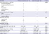
Of 805 ELBW patients, 65 (8.1%) received abdominal surgery. Patient characteristics are described in Table 1. There were 29 (44.6%) cases of NEC, 18 (27.7%) of SIP, 13 (20%) of MRI, and 5 (7.7%) of MNRI. Mean gestational age was 26+1 weeks (22+3–33+0), and mean birth weight was 695.9 g (355–990 g). Mean maternal age was 32.2 years (24–57 years). There were 5 (7.7%) cases of maternal preeclampsia, 2 (3.1%) cases of chorioamnionitis, 3 (4.6%) cases of hypothyroidism, 2 (3.1%) cases of thyrotoxicosis, and 1 (2.3%) case of long-term steroid usage. Infantogram and abdominal ultrasonogram were performed in all patients preoperatively. Fifty-one (78.5%) patients showed free air on the preoperative infantogram. Eight (12.3%) patients had portal air, 17 (26.2%) showed intramural gas, and 13 (20.0%) showed ascites on ultrasonograms. The only significant difference between the diagnoses was mean gestational age.
Table 1
Patient characteristics
P value with bold highlights: P < 0.05.
NEC = necrotizing enterocolitis, SIP = spontaneous intestinal perforation, MRI = meconium-related ileus, MNRI = meconium non-related ileus, SD = standard deviation, RDS = respiratory distress syndrome, PDA = patent ductus arteriosus.
aThe same letters indicate a non-significant difference between groups based on Tukey's multiple comparison test.
![]()
Operative characteristics are depicted in Table 2. Mean age at operation was 12.4 days. Mean body weight at operation was 710.7 g. Peritoneal drainage was performed in 3 cases, but because of worsening prognosis, all these patients underwent laparotomy. There were three pan intestinal involvement cases, two colonic involvement cases for NEC, and three colonic perforation cases for SIP. Rest of the cases were small bowel involved cases. Ostoma formation of any fashion was performed in 61 (93.8%). Four patients underwent primary anastomosis without ostoma formation (3 patients with SIP underwent primary anastomosis without proximal ostoma formation, and 1 patient with MRI underwent enterotomy, removal of the packed meconium, and primary closure of the enterotomy site). All patients without ostoma formation encountered leakage of the anastomosis site. Segmental resection and double barrel ileostomy were the most common procedure for NEC. Five patients with segmental resection with end ileostomy underwent second-look operations. Mean postoperative duration of TPN was 39.7 days, and 26 patients had PNALD. Mean hospital stay was 107.8 days. Comparing two group of primary anastomosis and ostoma formation, early complication for anastomosis leakage, and mean hospital stay was significantly higher in primary anastomosis group.
Table 2
Operative characteristics
P value with bold highlights: P < 0.05.
NEC = necrotizing enterocolitis, SIP = spontaneous intestinal perforation, MRI = meconium-related ileus, MNRI = meconium non-related ileus, SD = standard deviation, TPN = total parenteral nutrition, PNALD = parenteral nutrition-associated liver dysfunction.
aThe same letters indicate a non-significant difference between groups based on Tukey's multiple comparison test.
![]()
Outcomes of surgery
Details of postoperative complications, mortalities, and follow up growths are presented in Table 3. Twenty-three (35.4%) postoperative complications occurred before 30 days after the operation. Fourteen were complications more than grade III according to the Clavien-Dindo classification. There were 8 (12.3%) 30-day mortalities, and 7 of them were NEC. The most common cause of death was septic shock. Mean follow up period for growth was 62 months. Mean z-score for bodyweight was −1.52, and height was −1.16. There was no significant difference between the differential diagnoses.
Table 3
Outcome of abdominal surgery according to differential diagnosis
NEC = necrotizing enterocolitis, SIP = spontaneous intestinal perforation, MRI = meconium-related ileus, MNRI = meconium non-related ileus.
aWound complication and incisional hernia occurred after the ostoma repair; bZ score measured on the last follow up visit to clinic.
![]()
Outcomes according to surgical procedure is depicted in Table 4. There were four primary anastomosis cases and all four cases encountered anastomosis leakage. Late complication and mortality was not different between the two groups. Mean hospital stay was higher in primary anastomosis group (P = 0.041).
Table 4
Outcome of abdominal surgery according to surgical procedure
P value with bold highlights: P < 0.05.
TPN = total parenteral nutrition, SD = standard deviation, PNALD = parenteral nutrition-associated liver dysfunction.
aZ score measured on the last follow up visit to clinic.
![]()
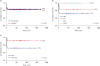
The long-term survival rate is shown in Fig. 1. The 5-year overall survival (OS) rate of the non-surgical and surgical groups were 84% and 83%, respectively (P = 0.967) (Fig. 1A). The 5-year OS rates for patients with NEC, SIP, MRI, and MNRI were 72%, 83%, 100%, and 100%, respectively. Survival outcomes between the four disease entities were not significantly different (P = 0.109) (Fig. 1B). However, compared with the non-NEC group, the NEC group showed a significantly poor survival outcome (P = 0.033) (Fig. 1C).
Multivariate analysis of survival
The logistic regression test for survival was performed with covariates of patient characteristics and follow-up variables. Even after adjusting for potentially confounding factors, NEC was still a statistically significant factor for survival (HR, 3.52; 95% confidence interval, 1.05–13.29; P = 0.033) (Table 5).
Table 5
Multivariate proportional hazards analysis for survival

P value with bold highlights: P < 0.05.
CI = confidence interval, Op. = operation, NEC = necrotizing enterocolitis.
![]()
DISCUSSION
ELBW neonates are known to have high morbidity and mortality; however, recent improvements in neonatal ventilation and monitoring techniques have made it possible for ELBW neonates to live. The incidence of ELBW neonates in South Korea has grown over the years. The treatment guidelines for ELBW neonates have evolved over the years, but many portions of them are controversial because of the rarity of ELBW neonates. There have been few reports about the differential diagnosis of acute abdomen in ELBW neonates, and some reports have discussed treatment modalities for acute abdomen, for example, peritoneal drainage or laparotomy. However, clinical characteristics and prognosis of acute abdomen according to the differential diagnosis among ELBW neonates have not been established well and have been reported only for each disease category. Our single institutional study provides data in support of the safety of laparotomy for ELBW neonates with acute abdomen with respect to complications and OS according to the differential diagnosis.
Four different disease entities lead to acute abdomen in ELBW neonates, but they are seldom emphasized. Decision for surgical intervention is made when a bowel perforation is suspected with free intra-abdominal air or infants with clinical deterioration despite maximal medical treatment with radiographic findings of portal venous gas or intramural gas. When bowel perforation is suspected, it is important to make a precise diagnosis, however, it is difficult to make a diagnosis before the exploration of the intestine. Suspecting diagnosis with pre-operative radiographic findings or clinical symptoms is often misdiagnosed. Making the correct diagnosis may aid physicians for different post-operative treatment strategy. SIP is defined as isolated single intestinal perforation that is typically found at the terminal ileum.789 Absences of traditional clinical signs of Bell's criteria and histologic signs of typical NEC characterize SIP as a distinct entity. Some authors reported that radiographic findings associated with NEC such as pneumatosis intestinalis and portal venous air are indicative factors for distinguishing between SIP, but in the current series, it was statistically difficult to make the differential diagnosis preoperatively. A report by the Pediatrix Medical Group17 mentioned that the clinical manifestation of neonates with SIP occurs at an earlier age than those with NEC (median age 7 vs. 15 days), but this was not adjusted in the current study. Most of the studies reported that the mortality of SIP is generally lower compared to NEC, however, Koivusalo et al.18 and his colleagues described that Morbidity after surgical treatment of SIP and NEC were similar for preterm infants weighing less than 1,500 g. Kubota and his colleagues have demonstrated histopathological characteristics of SIP in abrupt discontinuation of intestinal musculature, however, there were only three cases of muscular discontinuation stated in the pathology report in this study.15 Kubota et al.11 first used the term MRI in their report defining bowel obstruction with meconium in neonates without cystic fibrosis. They suggested that inspissated meconium is the result of excessive water absorption in the hypoperistaltic bowel before birth, and they reported 13 ELBW neonates with intestinal perforation secondary to MRI. MNRI is defined as ileus without a definite sticky meconium, and it is not associated with Hirschsprung's disease or intestinal atresia.192021 It is often hypothesized that zonal hypoganglionosis may be the pathophysiology of the disease. Although the macroscopic findings of NEC, SIP, MRI, and MNRI are clearly different from each other, it is often difficult to make a definitive diagnosis without laparotomy.
Abdominal surgery for ELBW neonates was performed for infants on suspicion of bowel perforation diagnosed by pneumoperitoneum on infantogram or for infants with clinical deterioration despite medical treatment. In this series of cases, all patients underwent serial examinations with infantogram and abdominal ultrasonogram preoperatively. In most cases, loop type or double barrel-type ostoma formation was performed. However, primary closure was performed in 3 patients with SIP and 1 patient with MRI, and all underwent additional surgery for proximal diversion due to anastomosis leakage. Although positive results of primary peritoneal drainage have been reported,131422 the current study adjusted data for primary peritoneal drainage in 3 patients with SIP who eventually received laparotomies after worsening of the disease. Because of the small percentage of adjustments, it is difficult to conclude the effectiveness of peritoneal drainage through this study.
The 5-year OS for NEC in this study was 72%, which is similar or higher than that in previous reports.623 The 5-year OS for SIP was 83%, which is also higher than that in previous studies.78924 There is still no consensus on which surgical technique may be most beneficial to the patient. Numerous centers have stated that primary anastomosis for NEC or SIP showed a decent outcome.25 Herein, we performed intestinal resection and primary anastomosis in 3 selected cases, but all resulted in complication of leakage at the anastomosis site. Although SIP, MRI, and MNRI have focal intestinal perforation and limited inflammation around the perforation site, we recommend performing ostoma formation in ELBW neonates.
The survival rate of ELBW neonates is known to increase because of the development of perinatal health care in various studies.123 In the study by the Japanese Society of Pediatric Surgeons, mortality of intestinal perforation for ELBW neonates in the last 15 years was 31.6%.5 There is no Korean study on the OS rate of ELBW neonates. However, the Korean Neonatal Network database system was launched recently and reported a survival rate of 69.6% for very-low-birth-weight neonates.326 In this study, the 5-year OS rates for ELBW neonates who underwent abdominal surgery and those who did not receive abdominal surgery were 84% and 83%, respectively. As far as we are concerned, this is the first study to report survival outcome for ELBW neonates with abdominal surgery using Korean data. The OS rate of the surgery group did not show a significant difference compared to the non-surgical group, which was different from the finding of other previous studies. Regarding risk factor analysis, NEC was a single significant prognostic factor.
The limitation of this study was that it was retrospective review, and the relatively small patients might have influenced the results. And the classification of the four disease entities were not yet established in international consensus. Previous articles describing acute abdomen in ELBW neonates have been using various terms with mixed definitions. However, we have suggested four disease entities to explain the abdominal surgical indications. As the incidence of ELBW neonates is increasing worldwide, a long-term, detailed single-center study may aid clinicians and surgeons in their medical decisions.
In conclusion, abdominal surgery for ELBW neonates is feasible with a good survival outcome. Ostoma formation can lead to reduced complications compared to primary anastomosis. The proper surgical technique are indispensable especially for patients with NEC, in whom most of the postoperative morbidity and mortality were encountered.
 XML Download