

 PDF
PDF ePub
ePub Citation
 Print
Print


Abstract
Purpose
Methods
Results
References
Fig. 1.
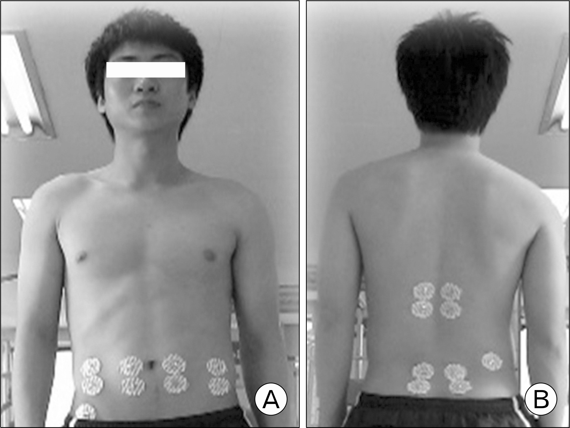
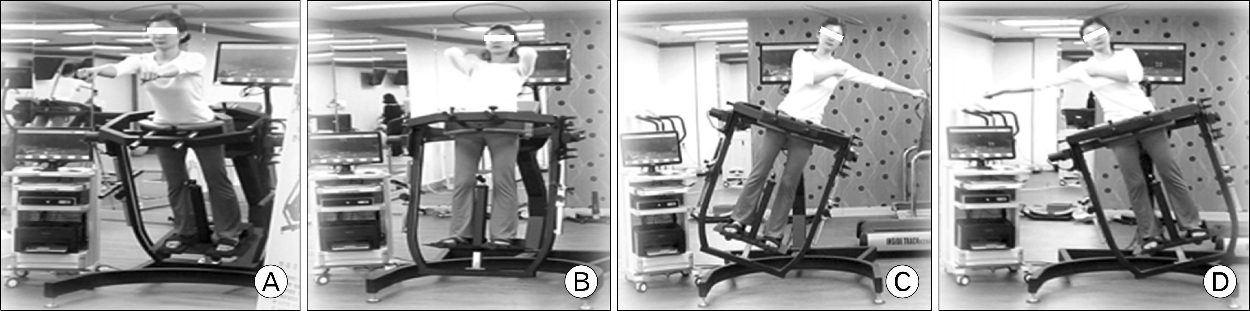
Table 1.
Table 2.
Values are presented as mean±standard deviation.
EMG: electromyography, RA: rectus abdominis, EO: external oblique, LT: longissimus thoracis, MF: multifidus.
Significant difference with pre at the same motion degree (∗p<0.05); significant difference with 0° at the same time point († p<0.05, ‡p<0.01, §p<0.001); significant difference with 10o at the same time point (||p<0.05, ∗∗p<0.01, ¶p<0.001); significant difference with 20o at the same time point (‡‡p<0.01, ††p<0.001).
Table 3.
Values are presented as mean±standard deviation.
EMG: electromyography, RA: rectus abdominis, EO: external oblique, LT: longissimus thoracis, MF: multifidus.
Significant difference with pre at the same motion degree (∗p<0.05); significant difference with 0° at the same time point (†p<0.05, ‡p<0.01, §p<0.001); significant difference with 10° at the same time point (||p<0.05, ∗∗p<0.01, ¶p<0.001); significant difference with 20° at the same time point (††p<0.05).
Table 4.
Values are presented as mean±standard deviation.
EMG: electromyography, RA: rectus abdominis, EO: external oblique, LT: longissimus thoracis, MF: multifidus.
Significant difference with pre at the same motion degree (∗p<0.05); significant difference with 0° at the same time point (†p<0.05, ‡p<0.01, §p<0.001); significant difference with 10° at the same time point (∗∗p<0.01, ¶p<0.001); significant difference with 20 degree at the same time point (||p<0.01).
Table 5.
Values are presented as mean±standard deviation.
EMG: electromyography, RA: rectus abdominis, EO: external oblique, LT: longissimus thoracis, MF: multifidus.
Significant difference with pre at the same motion degree (∗p<0.05); significant difference with 0° at the same time point (†p<0.05, §p<0.01, ‡p<0.001); significant difference with 10° at the same time point (∗∗p<0.05, ¶p<0.01, ||p<0.001); significant difference with 20° at the same time point (††p<0.05).
 XML Download