

 PDF
PDF ePub
ePub Citation
 Print
Print


Abstract
Purpose
To investigate the alteration of lower extremity movement during maintaining balance test with their eyes closed in chronic ankle instability (CAI) patients compared to healthy group with and without plantar cutaneous sensation.
Methods
Ten healthy volunteers (age, 23.40±2.22 years; height, 165.42±6.67 cm; weight, 60.93±13.42 kg) and 10 CAI patients (age, 23.90±2.56 years; height, 166.89±10.50 cm; weight, 67.43±12.96 kg), were recruited. Subjects immersed both feet in an ice water for 10 minutes and performed three trials of a single-leg stance balance test with their eyes closed while standing on a force plate for 10 seconds.
Results
CAI group showed increased knee flexion, reduced knee external rotation, and hip internal rotation compared to the healthy group from single-limb stance with eyes closed after diminished plantar cutaneous sensation. However, there was no significant interaction between group and time
Conclusion
These findings indicate that the postural kinematic analyses revealed that individuals with CAI used different strategy of controlling their lower extremities, which alters transverse plane motion of hip and knee compared to the healthy group in order to compensate for their ankle deficits after freezing the plantar cutaneous.
References
1. Massion J. Postural control system. Curr Opin Neurobiol. 1994; 4:877–87.

2. McGuine TA, Greene JJ, Best T, Leverson G. Balance as a predictor of ankle injuries in high school basketball players. Clin J Sport Med. 2000; 10:239–44.
3. Hertel J. Functional anatomy, pathomechanics, and pathophysiology of lateral ankle instability. J Athl Train. 2002; 37:364–75.
4. Holmer P, Sondergaard L, Konradsen L, Nielsen PT, Jorgensen LN. Epidemiology of sprains in the lateral ankle and foot. Foot Ankle Int. 1994; 15:72–4.
5. Drewes LK, McKeon PO, Kerrigan DC, Hertel J. Dorsiflexion deficit during jogging with chronic ankle instability. J Sci Med Sport. 2009; 12:685–7.
6. Freeman MA. Instability of the foot after injuries to the lateral ligament of the ankle. J Bone Joint Surg Br. 1965; 47:669–77.
7. McKeon PO, Hertel J. Spatiotemporal postural control deficits are present in those with chronic ankle instability. BMC Musculoskelet Disord. 2008; 9:76.
8. McKeon PO, Hertel J, Bramble D, Davis I. The foot core system: a new paradigm for understanding intrinsic foot muscle function. Br J Sports Med. 2015; 49:290.
9. Horak FB, Nashner LM. Central programming of postural movements: adaptation to altered support-surface configurations. J Neurophysiol. 1986; 55:1369–81.
10. Chua MC, Hyngstrom AS, Ng AV, Schmit BD. Movement strategies for maintaining standing balance during arm tracking in people with multiple sclerosis. J Neurophysiol. 2014; 112:1656–66.
11. Meyer PF, Oddsson LI, De Luca CJ. The role of plantar cutaneous sensation in unperturbed stance. Exp Brain Res. 2004; 156:505–12.
12. Nurse MA, Nigg BM. The effect of changes in foot sensation on plantar pressure and muscle activity. Clin Biomech (Bristol, Avon). 2001; 16:719–27.
13. Powell MR, Powden CJ, Houston MN, Hoch MC. Plantar cutaneous sensitivity and balance in individuals with and without chronic ankle instability. Clin J Sport Med. 2014; 24:490–6.
14. McKeon PO, Hertel J. Plantar hypoesthesia alters time-to-boundary measures of postural control. Somatosens Mot Res. 2007; 24:171–7.
15. Magnusson M, Enbom H, Johansson R, Pyykko I. Significance of pressor input from the human feet in anterior-posterior postural control: the effect of hypothermia on vibration-induced body-sway. Acta Otolaryngol. 1990; 110:182–8.
16. Roll R, Kavounoudias A, Roll JP. Cutaneous afferents from human plantar sole contribute to body posture awareness. Neuroreport. 2002; 13:1957–61.
17. Horak FB. Clinical measurement of postural control in adults. Phys Ther. 1987; 67:1881–5.
18. Tropp H, Odenrick P. Postural control in single-limb stance. J Orthop Res. 1988; 6:833–9.
19. Diener HC, Dichgans J, Bootz F, Bacher M. Early stabilization of human posture after a sudden disturbance: influence of rate and amplitude of displacement. Exp Brain Res. 1984; 56:126–34.
20. Gribble PA, Delahunt E, Bleakley C, et al. Selection criteria for patients with chronic ankle instability in controlled research: a position statement of the International Ankle Consortium. Br J Sports Med. 2014; 48:1014–8.
21. Hale SA, Hertel J. Reliability and sensitivity of the foot and ankle disability index in subjects with chronic ankle instability. J Athl Train. 2005; 40:35–40.
22. Hertel J, Buckley WE, Denegar CR. Serial testing of postural control after acute lateral ankle sprain. J Athl Train. 2001; 36:363–8.
23. Eils E, Nolte S, Tewes M, Thorwesten L, Volker K, Rosenbaum D. Modified pressure distribution patterns in walking following reduction of plantar sensation. J Biomech. 2002; 35:1307–13.
24. Eils E, Behrens S, Mers O, Thorwesten L, Volker K, Rosenbaum D. Reduced plantar sensation causes a cautious walking pattern. Gait Posture. 2004; 20:54–60.
25. Dyck PJ, O'Brien PC, Kosanke JL, Gillen DA, Karnes JL. A 4, 2, and 1 stepping algorithm for quick and accurate estimation of cutaneous sensation threshold. Neurology. 1993; 43:1508–12.
26. Doherty C, Delahunt E, Caulfield B, Hertel J, Ryan J, Bleakley C. The incidence and prevalence of ankle sprain injury: a systematic review and metaanalysis of prospective epidemiological studies. Sports Med. 2014; 44:123–40.
27. Hallen LG, Lindahl O. The “screw-home” movement in the knee-joint. Acta Orthop Scand. 1966; 37:97–106.
28. Kozanek M, Hosseini A, Liu F, et al. Tibiofemoral kinematics and condylar motion during the stance phase of gait. J Biomech. 2009; 42:1877–84.
29. Hill PF, Vedi V, Williams A, Iwaki H, Pinskerova V, Freeman MA. Tibiofemoral movement 2: the loaded and unloaded living knee studied by MRI. J Bone Joint Surg Br. 2000; 82:1196–8.
30. Horak FB, Nashner LM, Diener HC. Postural strategies associated with somatosensory and vestibular loss. Exp Brain Res. 1990; 82:167–77.
Fig. 1.
Lower-body plug-in-gait marker set. RASI: right anterior superior iliac spine, LASI: left anterior superior iliac spine, RTHI: right thigh, LTHI: left thigh, RKNE: right knee, LKNE: left knee, RTIB: right tibia, LTIB: left tibia, RANK: right ankle, LANK: left ankle, RTOE: right toe, LTOE: left toe, LPSI: left posterior superior iliac spine, RPSI: right posterior superior iliac spine, LHEE: left heel, RHEE: right heel.
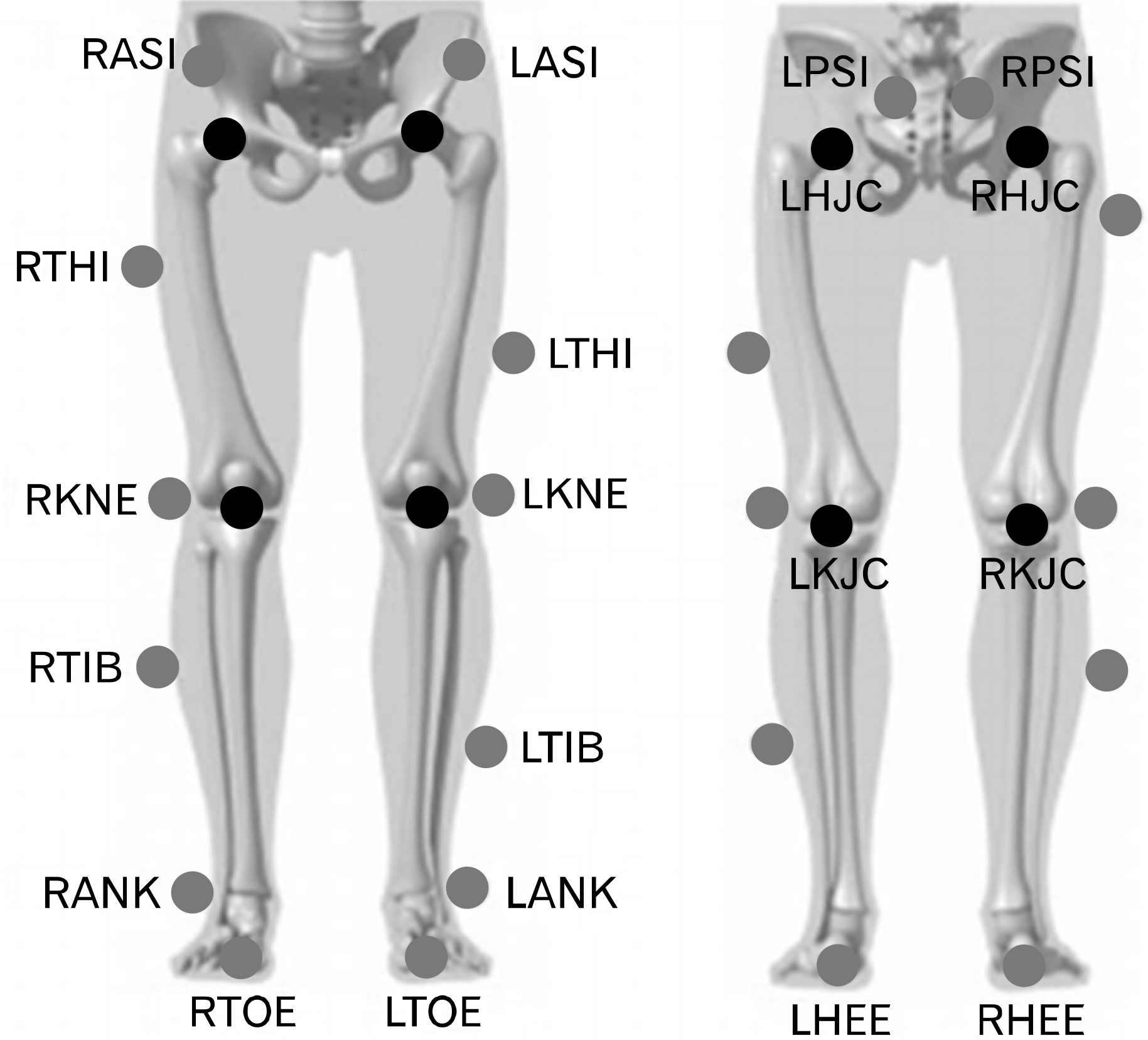
Fig. 2.
Semmes-Weinstein monofilaments results. ∗p <0.001: significant difference between pre-ice and ice 10 minutes; ∗∗p<0.001: significant difference between pre-ice and after ice 10+1 minutes.
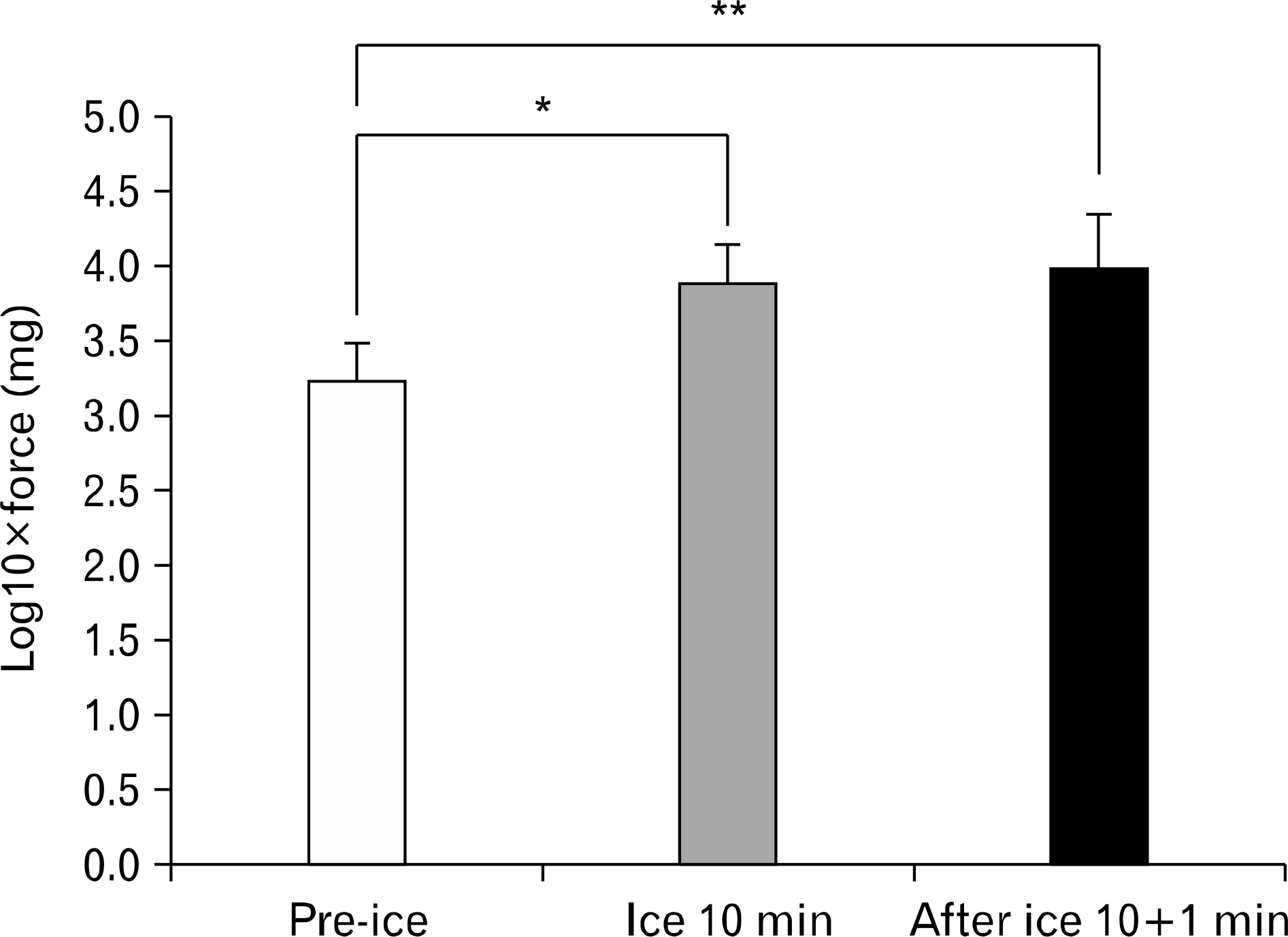
Fig. 3.
Ensemble curves of the lower extremity joint during the single-leg stance test. (A) Hip flexion (Flex)/extension (Ext), hip adduction (ADD)/abduction (ABD), hip internal rotation (IR)/external rotation (ER). (B) Knee Flex/Ext, knee varus (Var)/valgus (Val), knee IR/ER. (C) Ankle dorsi flexion (DF)/plantar flexion (PF), ankle inversion (IV)/eversion (EV), ankle IR/ER. CAI: chronic ankle instability, CI: confidence interval, Diff: difference.
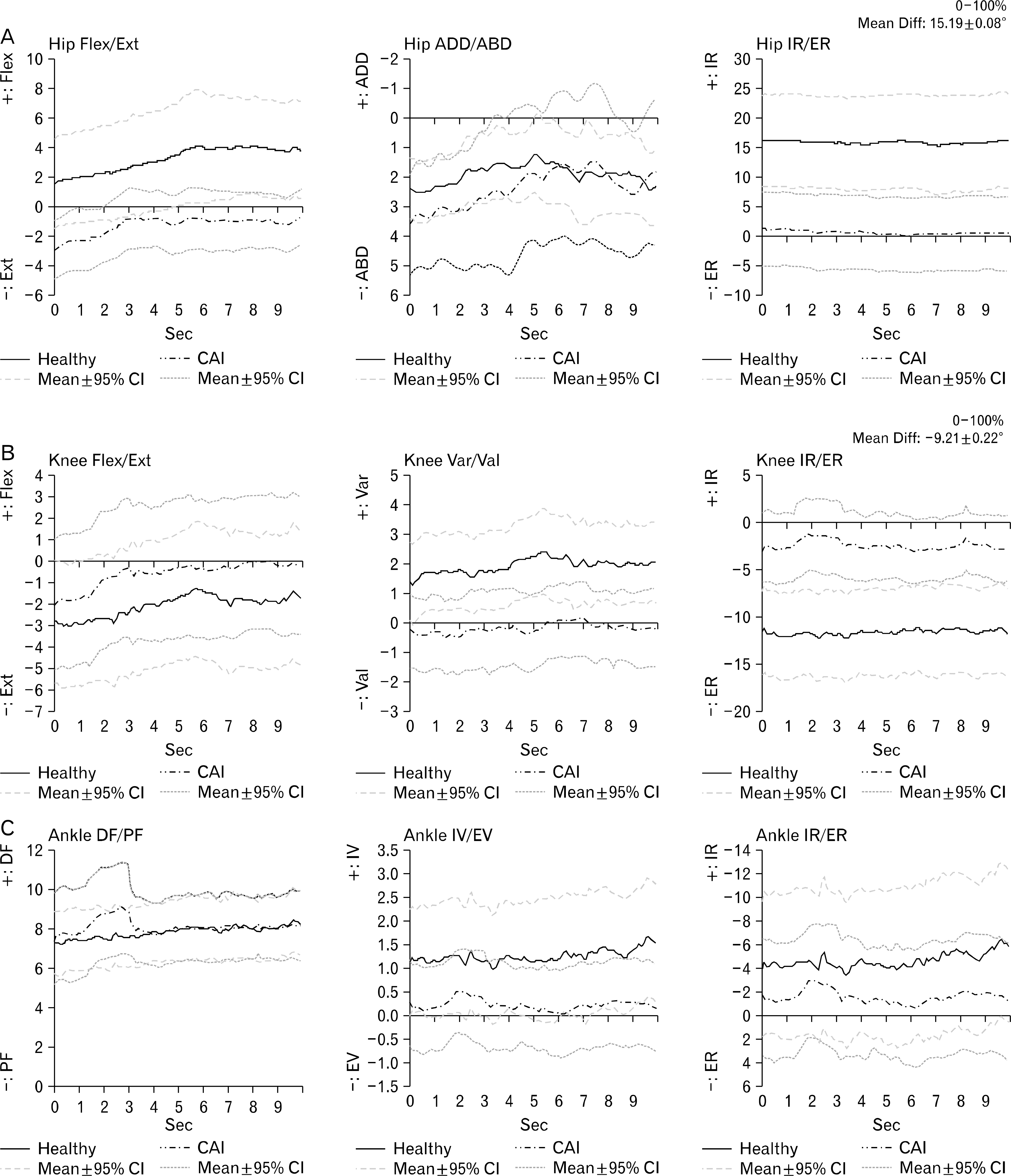
Table 1.
Physical characteristics of subjects
| Variable | CAI (N=10) | Healthy (N=10) |
|---|---|---|
| Male:female | 5:5 | 5:5 |
| Age (yr) | 23.90±2.56 | 23.40±2.22 |
| Weight (kg) | 67.43±12.96 | 60.93±13.42 |
| Height (cm) | 166.89±10.50 | 165.42±6.67 |
Table 2.
Single limb balance test with eyes closed
Values are presented as mean±standard deviation.
CAI: chronic ankle instability, Flex: flexion, Ext: extension, DF: dorsal flexion, PF: plantar flexion, ADD: adduction, ABD: abduction, Var: varus, Val: valgus, IV: inversion, EV: eversion, IR: internal rotation, ER: external rotation.
∗Group significant, at p<0.05.
 XML Download