

 PDF
PDF Citation
 Print
Print


INTRODUCTION
Inflammatory bowel disease (IBD) is characterized by chronic inflammation of the gastrointestinal tract. Various immunological factors play an important role in this group of chronic inflammatory disorders. IL-18 is a primary mediator of IBD (12). A balance of Th17 cell differentiation and Treg function is crucial for chronic intestinal inflammation, which is reported to be regulated by IL-18.
Metformin is a first-line medication used for the treatment of type 2 diabetes (T2D) and is widely prescribed in patients with hyperglycemia and obesity-related metabolic syndrome. Metformin treatment has been reported to have various beneficial effects, such as anti-inflammatory and anti-cancer effects (34). Recent studies have demonstrated that metformin ameliorates the symptoms of IBD (5).
Gut microbiota is an important environmental factor that affects energy metabolism and immune homeostasis (678). In recent studies, modulation of the gut microbiota by metformin therapy led to a consistent metabolic improvement with a significant increase in the abundance of Akkermansia muciniphila (9101112). Moreover, a significant increase in butyrate-producing bacteria, such as Bacteroides and Butyricimonas species, was observed in an aged mouse model (13).
Fecal microbiota transplantation (FMT) is widely used to confirm the causative effects of modulated gut microbiota by certain interventions. In this study, we aimed to evaluate the effects of allogeneic FMT on metabolic parameters by a transplant of fecal material from metformin-treated mice to antibiotic-treated mice, and investigate immunological changes following FMT to clarify the anti-inflammatory effects of metformin via gut microbiota modulation.
MATERIALS AND METHODS
FMT
Fecal material was collected from a metformin-treated mice model as described in a previous study (13). Briefly, 6-week-old male C57BL/6N mice were fed a high-fat diet (HFD) (45% kcal fat; FeedLab Inc., Hanam, Korea) for 39 weeks to induce metabolic disorders. Metformin (250 mg/kg) was administered daily for the final 16 weeks of HFD feeding (HFD-Met mice group). Prior to FMT, the mice were provided drinking water containing antibiotics (penicillin G procaine [2,000 IU/L] and streptomycin [2.5 mg/L]) for 5 days. Fecal material (0.1 g) from the metformin-treated mice was pooled in 1 mL of PBS. After centrifugation at 2,000×g for 2 min, 500 µL supernatant was administered using an oral gavage to the antibiotic-treated mice that were fed with an HFD for 48 wk (HFD-fMet, n=6). All experimental protocols were approved by the Institutional Animal Care and Use Committee (IACUC) of Sahmyook University and were conducted in accordance with the Guide for the Care and Use of Laboratory Animal (SYUIACUC 2015001).
Metabolic parameters
Body weight, serum glucose level, and food intake were recorded once every second week. Serum glucose level was estimated using an Accu-Chek Performa system (Roche Diagnostics, Mannheim, Germany) following fasting for 12 h. Intraperitoneal glucose tolerance testing (IPGTT) was performed at 16 wk after metformin administration. The mice were intraperitoneally injected with glucose solution (2 g/kg in PBS), and the glucose level was estimated 30, 60, 90, and 120 min after injection. The total cholesterol was estimated using a biochemical analyzer (AU480; Beckman Coulter, Brea, CA, USA).
Gut microbiota analysis
Total DNA was extracted using a PowerSoil DNA Isolation Kit (MO BIO Laboratories Inc., Carlsbad, CA, USA) from cecum samples including fecal material according to the manufacturer's instructions. Based on a previous study, the relative abundance of 3 bacterial genera (Akkermansia, Bacteroides, and Butyricimonas) in the cecum was confirmed using SYBR Green PCR Master Mix (Applied Biosystems, Foster City, CA, USA) and a StepOnePlus real-time PCR system (Applied Biosystems). The genus-specific primer sets for target bacterial 16S rDNA were described in previous study (13).
Transcriptome analysis
The expression of IL-1β (forward primer: 5′-CAGGATGAGGACATGACACC-3′, reverse primer: 5′-CTCTGCAGACTCAAACTCCAC-3′), IL-6 (forward primer: 5′-GTACTCCAGAAGACCAGAGC-3′, reverse primer: 5′-TGC TGG TGA CAA CCA CGG CC-3′), TGF-β1 (forward primer: 5′-GCGGACTACTATGCTAAAGAGG-3′, reverse primer: 5′- GTAGAGTTCCACATGTTGCTCC-3′), IL-4 (forward primer: 5′- GAGCCATATCCACGGATGCGACAA-3′, reverse primer: 5′- CATGGTGGCTCAGTACTACGAGTA-3′), TNF-α (forward primer: 5′-GCCTCTTCTCATTCCTGCTTG-3′, reverse primer: 5′-CTGATGAGAGGGAGGCCATT-3′), NF-κB (forward primer: 5′-GAAATTCCTGATCCAGACAAAAAC-3′, reverse primer: 5′-ATCACTTCAATGGCCTCTGTGTAG-3′), and IL-18 (forward primer: 5′- CAGGCCTGACATCTTCTGCAA-3′, reverse primer: 5′-TCTGACATGGCAGCCATTGT-3′), TLR1 (forward primer: 5′-CAATGTGGAAACAACGTGGA-3′, reverse primer: 5′-TGTAACTTTGGGGGAAGCTG-3′), TLR2 (forward primer: 5′-AAGAGGAAGCCCAAGAAAGC-3′, reverse primer: 5′-CGATGGAATCGATGATGTTG-3′), TLR4 (forward primer: 5′-ACCTGGCTGGTTTACACGTC-3′, reverse primer: 5′-CTGCCAGAGACATTGCAGAA-3′), TLR5 (forward primer: 5′-AAGTTCCGGGGAATCTGTTT-3′, reverse primer: 5′-GCATAGCCTGAGCCTGTTTC-3′), TLR6 (forward primer: 5′-TTCCCAATACCACCGTTCTC-3′, reverse primer: 5′-CTATGTGCTGGAGGGTCACA-3′), GLP-1 (forward primer: 5′-GGGTCTCTGGCTACATAAGGACAAC-3′, reverse primer: 5′-AAGGATGGCTGAAGCGATGAC-3′), DPP4 (forward primer: 5′-AAATGGGATTTGTGGACAGCAAG-3′, reverse primer: 5′-CCGATCCCAGGACCATTGAG-3′), and GAPDH (forward primer: 5′-AACTTTGGCATTGTGGAAGG-3′, reverse primer: 5′-ACACATTGGGGGTAGGAACA-3′) were determined in the ileum including the Peyer's patches. Total RNA was extracted using a RiboEx (GeneAll, Seoul, Korea) and cDNA synthesis was performed using a HyperScript RT premix (GeneAll) according to the manufacturer's instructions. SYBR Green PCR Master Mix (Applied Biosystems) and a StepOnePlus real-time PCR system (Applied Biosystems) was used to quantify the mRNA levels. GAPDH was used as an internal control. The primer sets for transcriptome were described in previous study (13).
Western blot analysis
Ileum tissue were homogenized in RIPA cell lysis buffer (GenDEPOT, Katy, TX, USA) added with protease inhibitor cocktail solution (GenDEPOT). To perform the western blot analysis of IL-18, protein lysates (80 µg) were separated by electrophoresis. The blots were blocked for 1–2 h with 5% BSA in TBST solution (Tris buffered saline containing 0.05% Tween-20), and incubated overnight at 4°C with primary antibodies: anti-GAPDH and anti-IL-18 polyclonal antibodies (Abcam, Cambridge, UK). After repeated washings, anti-rabbit IgG HRP-conjugated antibody (GenDEPOT) were added for 1 h, immunoreactive bands were visualized using a chemiluminescent peroxidase substrate (ECL Plus; GenDEPOT) and image system (Chemidoc XRS system; Bio-Rad, Hercules, CA, USA).
Statistical analysis
All data are presented as the means±SEM. The 2−ΔΔCt relative quantification method [ΔΔCt=(Ct.Target−Ct.β-actin)Group 1−(Ct.Target−Ct.β-actin)Group 2] was used to quantify the in vivo mRNA levels relative to the internal control (GAPDH). The statistical significance was assessed by one-way ANOVA followed by Duncan's post hoc test. All statistical analyses were performed using R Studio software (RStudio, Inc., Boston, MA, USA). The statistical significance was determined at p<0.05.
RESULTS
Effects of FMT on metabolic parameters and the gut microbiota
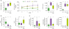
Serum glucose level (154.2±8.4 mg/dL) and IPGTT (991.5±184.8) increased more after FMT involving fecal materials from the HFD-Met mice group than from the HFD group (192.0±27.6 mg/dL and 1,243.5±137.5, respectively). This effect lasted for 4 weeks, although the increase in IPGTT was not significant (Fig. 1A). Body weight and total cholesterol did not differ significantly between the HFD and HFD-fMet groups. GLP-1 expression was significantly higher in the HFD-fMet group than in the HFD group (Fig. 1B). The relative abundances of Akkermansia, Bacteroides, and Butyricimonas were not statistically significant between the HFD and HFD-fMet groups (Fig. 1C).
Figure 1
Changes in the metabolic profiles and gut microbiota after FMT (A) metabolic profiles are shown. Six-week-old mice were fed on HFD for 43 wk after which pooled fecal material from metformin-treated mice (HFD-fMet, n=6) was orally transferred once to the antibiotic-treated mice. Body weight, IPGTT, and total cholesterol were measured 4 wk after FMT [RD (n=5), HFD (n=4)]. The serum glucose level was measured after fasting for 12 h. (B) Relative mRNA level of DPP4 and GLP-1 as determined by quantitative PCR in the ileum. (C) The relative abundance of bacterial genera determined by quantitative PCR in the cecum. Different superscript letters indicate significant differences (p<0.05) according to Duncan's post hoc test.
*Statistical significance between HFD and HFD-fMet groups (p<0.05).
Effects of FMT on immune responses in the ileum
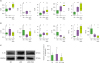
TLR1 and TLR4 expression levels were significantly higher in the HFD-fMet group than in the HFD group (Fig. 2A). TLR2, TLR5, and TLR6 did not differ significantly between the HFD and FMT groups. The expression of TLRs were not significantly different between RD and HFD groups. Among the pro-inflammatory cytokines, IL-18 expression was significantly lower in the HFD-fMet group than in the HFD group; this was the only significant difference (Fig. 2B). In addition, relative protein expression level of IL-18/GAPDH was 1.24±0.11, 1.37±0.72, and 0.95±0.28 in RD, HFD, and HFD-fMet group, respectively (Fig. 2C).
Figure 2
Immunological changes in the ileum after FMT. (A) Relative mRNA levels of TLRs. (B) Relative mRNA levels of the inflammatory cytokines. (C) Western blot analysis of IL-18 in the ileum. Different superscript letters indicate significant differences (p<0.05) according to Duncan's post hoc test.
DISCUSSION
Recently, modulation of the gut microbiota by metformin treatment has been shown to be a causative factor for metabolic improvement, such that FMT from metformin-treated donors improved the glucose tolerance of germ-free mice (1013). As anti-inflammatory effect of metformin was reported as well, which is expected to have a relation with the modulation of the pre-existing gut microbiota by metformin treatment.
Interestingly, gut microbiota modulation by metformin treatment was related to an increase in the short-chain fatty acid (SCFA)-producing bacteria (13), Akkermansia, Butyricimonas, and Bacteroides. Akkermansia (A. muciniphila), considered to be a next-generation probiotic, is a mucin-degrading species that produces propionate, the abundance of which is associated with anti-inflammatory effects, antioxidant effects, and metabolic improvements (141516). Moreover, the abundance of Akkermansia has been reported to be significantly higher in patients with T2D treated with metformin and was found to be increased by metformin treatment in an animal model regardless of the age (91113). Bacteroides is an SCFA-producing bacterium that is abundant in non-obese individuals (17). Several strains of Bacteroides produce propionate and succinate, which protect against various obesity-related metabolic disorders, such as insulin resistance (18). Recently, the dipeptidyl peptidase-4 inhibitor vildagliptin has been shown to decrease the abundance of butyrate-producing bacteria including Bacteroides in the intestines of diabetic rats (19). However, after FMT using fecal material from the HFD-Met mouse group, which contained abundant Akkermansia and Bacteroides populations, only the glucose parameters improved but not body weight or lipid parameters. FMT also increased the abundance of Butyricimonas, suggesting that Butyricimonas plays an important role in the regulation of hyperglycemia after FMT. The increased abundance of Butyricimonas was associated with improved metabolic parameters in the aged and adult mice treated with metformin (9). Butyrate, which is produced by Butyricimonas spp., protects against diet-induced obesity (20). Moreover, butyrate increased the mucosal layer thickness in the intestine. This parameter is also regulated by Akkermansia, which has been associated with metabolic improvements, including glucose tolerance (2122). On the other hands, pro-inflammatory cytokines were induced by SCFAs in a time- and dose-dependent manner (23). Pro-inflammatory roles of activated free fatty acid receptors, involved in inflammation and immune responses by SCFAs, were also reported (24). Therefore, the adequate amount of SCFAs should be considered for beneficial effects on anti-inflammation and metabolic improvements.
TLR expression patterns provide insights into the mechanism of FMT in glucose regulation. During the treatment of Clostridium difficile infection, FMT modulates the tight junctions and mucus structure and stimulates the mucosal immune system in the intestine (25). However, the mechanism of FMT in metabolic improvements has remained unclear. To clarify this mechanism, we investigated the regulation of TLRs that recognizes different molecular patterns of the microorganism. TLR1 recognizes bacterial lipoproteins and peptidoglycans and is known to mediate cytokine production along with TLR2. Recently, TLR1 has been reported to play an important role in the regulation of chronic inflammation by sensing the gut microbiota (26). In another study, TLR1 signaling was shown to regulate inflammation during intestinal infection, and TLR1 deficiency led to vulnerability to intestinal inflammation and tissue injury by dysbiosis (27). Moreover, drug efficacy has been shown to be affected by gut microbiota, such that TLR1 was upregulated in healthy individuals with good glycemic control without any complication (28). TLR4 recognizes lipopolysaccharides derived from bacteria, insulin resistance, and inflammation by TLR4 signaling mediated by NF-κB (29). Dysbiosis contributes to the development of colonic inflammation and metabolic disorders including T2D via TLR4 signaling (30). However, in the current study, TLR4 expression was increased by FMT, and NF-κB remained unchanged, although the regulatory effects of TLR4 on hyperglycemia not mediated by NF-κB was considered. Recently, TLR4 was reported to play an important role as a recognition receptor controlling the sensitivity of GLP-1, which is a therapeutic target for T2D (31). Therefore, mediation of gut microbiota modulation by TLR1 and TLR4 signaling may be associated with hyperglycemia regulation, although the specific roles of TLR1 and TLR4 remain unknown.
GLP-1 upregulation may play a key role in glucose regulation after FMT therapy. GLP-1 is an insulinotropic hormone, whereas dysbiosis is responsible for GLP-1 resistance (31). Therefore, glucose regulation after FMT was caused by the therapeutic modulation of intestinal dysbiosis by metformin. Although activation of AMP-activated protein kinase is a well-known mechanism of action of metformin, it also induces GLP-1 secretion, which may be associated with modulation of gut microbiota. Interestingly, GLP-1 was increased in chronic inflammatory states and related to immune responses in the gut (32). In addition to the anti-hyperglycemic effects, GLP-1-based therapies have shown anti-inflammatory effects in chronic inflammatory disease, such as T2D and atherosclerosis (3334).
Metformin has beneficial effects on glucose regulation and causes improvement in the symptoms of IBD (5), which may be associated with modulation of the gut microbiota. IL-18 is an IL-1 family inflammatory cytokine that contributes to IBD pathology and acts as an indicator of IBD progression (35). IL-18/IL-18R signaling in the intestinal epithelial cells plays a pivotal role in the progression of IBD. The regulation of CD4+ T cells and Foxp3+ Treg cells are modulated during intestinal inflammation (136). Therefore, neutralization of IL-18 activity was suggested to ameliorate intestinal inflammation (37). Metformin therapy was reported to inhibit IL-18 secretion in patients with T2D, and interestingly, consistent results were observed following FMT treatment in the current study (38). Additionally, recently reported anti-cancer and anti-aging effects of metformin might have been mediated by the modulation of the gut microbiota (3).
In summary, based on recent results that metformin improves metabolic disorders including obesity, hyperglycemia, and hyperlipidemia via gut microbiota modulation, we determined that FMT using fecal material from metformin-treated mice modulated the anti-inflammatory immune responses as well as glucose regulation. The mechanism underlying the effects of FMT on metabolic improvements is unclear; however, the use of FMT by upregulation of GLP-1 may play a key role in glucose regulation mediated by TLR1 and TLR4 after FMT. Above all, the anti-inflammatory effects of metformin may be associated with inhibition of IL-18 via gut microbiota modulation.
 XML Download