

 PDF
PDF ePub
ePub Citation
 Print
Print


INTRODUCTION
A number of studies have shown that aurora kinase (AK) overexpression and amplification are closely related to various human cancers [1234]. AKs participate in mitotic events and influence essential processes in human carcinogenesis, telomerase activity, collagen I-induced cell migration and anchorage-independent growth [3]. Therefore, there has been a great deal of research interest in the development of potential AK inhibitors [5]. Three members of the AK family, Aurora-A, Aurora-B, and Aurora-C, are associated with multiple aspects of mitosis. Aurora-A localizes to centrosomes/spindle poles and is required for spindle assembly, whereas Aurora B is a chromosome passenger protein required for phosphorylation of histone H3, chromosome segregation, and cytokinesis. Aurora-A and Aurora-B are frequently overexpressed in a variety of tumors in humans and promote cell cycle progression. Although Aurora-A has attracted the most attention with regard to the link with human cancer, Aurora-B may be a more suitable anti-cancer drug target because inhibition of Aurora-B rapidly results in mitotic arrest and cell death. Aurora-C may rescue the Aurora B-silenced multinucleation phenotype in human cells because it shows functional overlap with Aurora-B [67].
ZM447439 (ZM) is a potent and selective inhibitor of Aurora A and B kinases, with similar 50% inhibitory concentrations (IC50) seen for both kinases [8]. Aurora kinase inhibitors (AKIs), including daurinol, alisertib, and TC-A2317, were shown to induce apoptosis in vitro and in vivo [91011]. In 2010, Li et al. [12] reported that ZM-induced apoptosis was associated with the upregulation of p53, breakdown of the mitochondrial membranes and activation of caspase-3.
The development of newer targeted anti-cancer agents, including some kinase inhibitors, has increased the life expectancy of patients with several types of cancer. However, a number of drugs used in oncology can affect the heart in various ways [1314151617]. Cardiotoxicity covers a wide range of side effects, including arrhythmias, especially torsades de pointes induced by QT-prolonging drugs, and dysfunction of myocardial contraction and/or relaxation [18]. Therefore, it is necessary to verify the cardiac safety of novel anti-cancer drugs. Several recent studies have confirmed the anti-cancer effects of ZM in various cancer cell lines. However, there have been no studies regarding the cardiac safety of ZM using electrophysiological tools and image-based contractility assays. In this study, we demonstrated the potential effects of ZM on cardiac repolarisation using a whole-cell patch-clamp technique with human induced pluripotent stem cell-derived cardiomyocytes (hiPSC-CMs). The hiPSC-CMs are a potentially ideal source of human cardiomyocytes (CMs) for pharmacological screening, with no associated ethical issues and no differences among species. We also analyzed the mechanism of action of ZM using cells with heterogeneous cardiac ion channel expression. Furthermore, we performed image-based contractility assays with enzymatically isolated rat ventricular myocytes to determine the effects of ZM on myocardial contraction/relaxation. This approach improved our understanding of the potential mechanism underlying ZM-induced cardiotoxicity and provided insight into the usefulness of hiPSC-CMs as a new human model for assessing the safety of AKI-related drugs.
METHODS
Animals
Contractility assay using the left ventricular myocytes was performed using Sprague-Dawley rats (female, 250–350 g). The animals were kept in a storage room under standard conditions of constant temperature (23 ± 1℃), relative humidity (50 ± 10%) and illumination (12 h light/dark cycles) until initiation of the experiment. These studies were conducted in facilities approved by the Association for Assessment and Accreditation of Laboratory Animal Care International. All procedures were approved by the Institutional Animal Care and Use Committee (IACUC) at the Korea Institute of Toxicology (IACUC approval No. RS13006).
Drug and solutions
ZM was purchased from Toronto Research Chemicals (North York, ON, Canada) and it was formulated into a stock solution with dimethyl sulfoxide (DMSO). The stock solution was further diluted in the bath solution to yield final perfusion solutions with 0.1% DMSO and appropriate drug concentrations. All chemicals for solution preparation were purchased from Sigma-Aldrich Co. (Sigma-Aldrich, St. Louis, MO, USA).
Cell culture and transfection for ion channel studies
The hiPSC-CMs (Cardiosight-S; NEXEL, Co., Ltd., Seoul, Korea) were used for single-cell action potential assay. The cells were cultured according to the manufacturer's instructions. To recording of cardiac APs using a patch-clamp system, the cells were cultured to four-well plates containing Matrigel (Corning, Cat. # 354234)-coated glass coverslips at a low density to yield single uncoupled cells. The cells maintained in a culture incubator at 37℃ in an atmosphere of 95% air and 5% CO2 and used within 4 weeks after thawing.
For various aspects of cardiac ion channel study, human embryonic kidney (HEK293; ATCC, Manassas, VA, USA) cells were transiently transfected using lipofectAmin2000 (Gibco BRL, New York, NY, USA) according to the manufacturer's instructions. The hERG (human ether-ago-go-related gene corresponding to IKr), KCNQ1/KCNE1 (the gene corresponding to IKs), KCNJ2 (the gene corresponding to IK1) or SCN5A (the gene corresponding to INa) cDNA was co-transfected with green fluorescence protein, the surface marker protein, to allow assessment of the transfection efficiency. For the calcium current, a Cav1.2-expressing cell line (human Cav1.2/β2/α2δ1 calcium channel cell line, Cat. #CT6004) was purchased from Charles River (Cleveland, OH, USA).
Whole-cell voltage-clamp recordings
The external solution for recording the IhERG, IKs and INa channel currents was normal Tyrode's solution. The internal solution for IhERG contained the following (in mM): 130 KCl, 5 ethylene glycol bis(2-aminoethylether)-N,N,N′,N′-tetraacetic acid (EGTA), 10 N-(2-hydroxyethyl)piperazine-N′-2-ethansulfonic acid (HEPES), 1 MgCl2, 5 Mg-ATP (pH 7.25 with KOH), and for IKs in the KCNQ1/KCNE1-cotransfected HEK293 cells, 150 KCl, 5 EGTA, 10 HEPES, 2 MgCl2, 1 CaCl2 and 5 Na2-ATP (pH adjusted 7.25 with KOH). The internal solution for IK1 in KCNJ2-transfected HEK293 cells contained (in mM): 130 K-Asp, 15 KCl, 10 HEPES, 1 MgCl2, 5 Na2-ATP, 5 EGTA (pH 7.25 with KOH), and for the sodium current in SCN5A-transfected HEK293 cells, 105 CsF, 35 NaCl, 10 EGTA, 10 HEPES (pH 7.25 with NaOH). The calcium current was measured in a Cav1.2-expressing CHO cell line, the cells were superfused with an external solution that consisted of (in mM): 145 NaCl, 5.4 KCl, 10 HEPES, 1 MgCl2, 5 glucose, 1.8 CaCl2 (pH 7.4 with NaOH), whereas the intracellular solution used to fill the pipette had the following ionic solution (in mM): 20 CsCl, 120 Cs-aspartate, 5 NaCl, 10 EGTA, 10 HEPES, 20 TEA-Cl, 5 Mg-ATP (pH 7.25 with CsOH).
Cell viability assay (MTS assay)
A549 (human lung carcinoma cell line; ATCC CCL-185), HepG2 (human liver cancer cell line; ATCC HB-8065), MCF7 (human breast cancer cell line; ATCC HTB-22), NCI-H1299 (human non-small cell lung carcinoma cell line, ATCC CRL-5803) and human lung fibroblast (HLF; ATCC PCS-201-013) cells were plated (1 × 10³ cells) in 96-well plates in basal medium (10% foetal bovine serum [FBS] in Dulbecco's modified Eagle's medium [DMEM]). After 24 h, the medium was replaced with serum-free DMEM. At the same time, cells were treated with ZM at various concentrations (0.01, 0.03, 0.1, 1, 3, and 10 µM). After 24, 48, and 72 h, each well was treated with 20 µl of the tetrazolium compound 3-(4,5-dimethylthiazol-2-yl)-5-(3-carboxymethoxyphenyl)-2-(4-sulfophenyl)-2H-tetrazolium (MTS; Promega, Madison, WI, USA) for 2 h in a 37℃ incubator. Optical density was then read directly at 492 nm using a SpectraMax M3 plate reader (Molecular Devices LLC, San Jose, CA, USA).
Invasion assay
A549 cells were plated (1 × 10⁶ cells) on 10-cm cell culture dishes in basal medium (10% FBS in DMEM). After 24 h, the medium was replaced with serum-free DMEM. At the same time, cells were treated with ZM at a concentration of 100 nM, 1 µM or 10 µM in individual plates. After 24, 48, and 72 h, A549 cells were reseeded (6 × 10⁴ cells) in Matrigel and fibronectin (BD Biosciences, San Jose, CA, USA)-coated Falcon cell culture inserts with a basal medium. The invasion assay was carried out for 24 h in a tissue culture incubator. After 24 h, the cells were fixed with 4% formaldehyde dissolved in phosphate-buffered saline (PBS; Santa Cruz Biotechnology, Inc., Dallas, TX, USA). After fixing for 1 min, the chambers were rinsed once in PBS and then stained with hematoxylin and eosin (Merck, Mendota Heights, MN, USA) for 3–5 min. These cells were counted under a microscope with a 4× objective.
Migration assay (wound healing assay)
A549 cells were plated on 10-cm cell culture dishes in basal medium (10% FBS in DMEM). The next day, the media were replaced with serum-free DMEM and cells were treated with 3 µM ZM for 72 h. A549 cells were then reseeded at 105 cells per well in 48-well plates. After 24 h, wounds were made in each well and the medium was replaced with fresh medium. The wounds were observed under a microscope with a 4× objective.
Myocyte contractility assay
To study the effect of ZM on myocytes contractility, we isolated single rat ventricular myocytes (rVMs) using the Langendorff system. Rats were anesthetized with pentobarbital sodium (30 mg/kg, i.p.), and the hearts were extracted and rapidly mounted onto the Langendorff perfusion system which were then perfused with a Ca2+-free solution for 10 min (in mM; NaCl 135, KCl 5.4, MgCl2 3.5, glucose 5, HEPES 5, Na2HPO4 0.4, taurine 20 at pH of 7.4, NaOH), followed by a further 8-min perfusion with the same solution containing collagenase (1 mg/ml, Worthington Biochemical Co.; protease, 0.133 mg/ml, BSA 1.65 mg/ml; Ca2+ 0.05 mM). Afterward, the LV free wall was dissected and incubated in a fresh collagenase-only solution. The rVMs were harvested following a further 10-min digestion period, washed and resuspended in storage solution (in mM; NaCl 120, KCl 5.4, MgSO4 5, CaCl2 0.2, Na-pyruvate 5, glucose 5.5, taurine 20, HEPES 10, mannitol 29, pH 7.4, NaOH). The myocyte suspension was stored at room temperature and cells were used within 8 h of isolation. Changes in sarcomere length were measured in LV myocytes by using a video-sarcomere detection system (IonOptix Corp., Milton, MA, USA). All experiments were carried out at 36 ± 1℃ and paced at a frequency of 2 Hz. Measurements from at least 10 steady-state contractions were averaged for each myocyte and for each stage of the experimental protocols. Data analysis was performed offline using IonWizard software (IonOptix Corp.). Sarcomere waveforms were collected and analyzed at baseline and at each testing concentration.
Statistical analysis
pCLAMP (Axon Instruments, Foster City, CA, USA), Origin 8 (OriginLab Corp, Northampton, MA, USA), Excel (Microsoft, Redmond, WA, USA), and GraphPad Prism (GraphPad Software, San Diego, CA, USA) were used for data acquisition and analysis. The concentration-response relationships for drug-induced blockage were calculated using SigmaPlot (Systat Software, San Jose, CA, USA). The IC50 values, the drug concentration that reduced the ionic currents by 50%, were obtained using the sigmoidal Hill equation: f = xH / (IC50H + xH), where x is the concentration, H is the Hill coefficient, and f is the inhibition ratio. Data are presented as the means ± standard error of the mean (SEM) or standard deviation, and n represents the number of experimental replicates. Statistical significance was determined using the Student's t-test and one-way ANOVA with post hoc testing using Dunnett's method; p < 0.05 was considered to indicate statistical significance.
RESULTS
Inhibitory effects of ZM on cancer cell viability
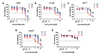
The MTS in vitro cell proliferation assay is one of the most widely used assays for evaluating preliminary anticancer activity. To assess the in vitro anticancer activity of ZM, MTS assays were performed with various cancer cell lines—A549 (non-small cell lung cancer), MCF-7 (breast cancer), NCI-H1299 (non-small cell lung cancer), HepG2 (human hepatocellular carcinoma) and HLF cells—after treatment with ZM at several concentrations (0.01, 0.03, 0.1, 1, 3, and 10 µM). After 24, 48, or 72 h of incubation, cell viability was examined by MTS assay. ZM showed concentration- and time-dependent cytotoxicity in all cancer cell lines but the inhibitory potential was slightly different among cell lines. In A549 cells, the IC50 values were 3.2 µM with treatment for 48 h and 3.3 µM with treatment for 72 h. In H1299 cells, the IC50 values were 1.1 and 0.7 µM with treatment for 48 and 72 h, respectively. In the MCF-7 cell line, the IC50 values were 3.1 µM with treatment for 48 h and 0.8 µM with treatment for 72 h. The IC50 values were 3.3 and 0.6 µM for the HepG2 cell line with treatment for 48 and 72 h, respectively. However, the HLF cell lines did not show significant growth inhibition with ZM up to 10 µM for 72 h. The results of cell viability assays with IC50 doses are presented in Fig. 1. These data suggest that ZM has greater cytotoxicity against cancer cells than normal cells.
Effects of ZM on invasion of A549 cells
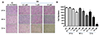
The invasion assay is widely used to study cell behavior and assess the metastatic potential of tumor cells. The Transwell invasion assay with Matrigel is one of the most widely used and convenient techniques to study cell invasion. To evaluate its effects on invasive activity, A549 cells were incubated for 24, 48, or 72 h with ZM at a concentration of 0.1, 1, or 10 µM. The invasive activity determined by counting the cells crossing the membrane pores was decreased by ZM in a dose- and time-dependent manner (Fig. 2). Cell invasion was significantly inhibited by ZM in a dose- and time-dependent manner. At the highest dose tested (10 µM) for 24, 48, and 72 h, ZM reduced invasion of A549 cells by 23.9 ± 1%, 42 ± 1.7% and 90.6 ± 2.7%, respectively (Fig. 2B, each n = 3).
Effects of ZM on migration of A549 cells
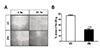
A549 cells were grown to confluent monolayers, which were wounded by scratching with a pipette tip and incubated with ZM at a concentration of 3 µM. The cells in the DMSO-treated control group continuously migrated into, and almost completely closed, the scratch within 24 hours. The mean percentage of wound healing was 94.3 ± 2.9% in three repeated experiments (Fig. 3). On the other hand, the migration ability of cells in the ZM-treated group was significantly inhibited and they showed only 43.2 ± 4.0% wound healing (Fig. 3B, each n = 3).
Effects of ZM on cardiac action potential of hiPSC-CMs
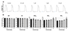
To confirm the effects of ZM on the action potential (AP) of hiPSC-CMs, we measured the spontaneous APs in contracting cells using the whole-cell patch-clamp technique. Most cells showed ventricular-type APs with relatively more negative maximal diastolic potential (MDP) and rapid AP upstroke, with a long plateau phase and AP duration at 90% (APD90) longer than 200 ms. The MDP, maximum upstroke velocity (dV/dtmax), AP amplitude (APA), AP duration at 50% (APD50) and APD90 values were analyzed; only ventricular-type cells were included in the analysis of the effects of ZM. Under control conditions, the values of the AP parameters were as follows: −66.6 ± 0.8 mV for MDP, 99.9 ± 6.8 V/s for dV/dtmax, 258.4 ± 17.2 ms for APD90, 215.1 ± 17.4 ms for APD50 and 113.3 ± 1.9 mV for APA (n = 3, mean ± SEM). The effects of ZM on the AP parameters were normalized relative to the control value in each cell and summarised in bar graphs (Fig. 4). Both APD50 and APD90 were significantly prolonged at 10 µM ZM versus the other concentrations (n = 3). At concentrations up to 10 µM, ZM did not affect any other AP parameters (MDP, dV/dtmax or APA). The APD50 and APD90 prolongation induced by ZM showed partial recovery after drug washout.
Effects of ZM on cardiac ion channels
The effects of ZM on cardiac ion channels were evaluated using the whole-cell patch-clamp technique. We measured the peak current amplitude and inhibitory percentage for various concentrations of ZM. In the studies of cardiac ion channels, ZM did not significantly affect IKs, IK1, INa or ICa, but reduced IhERG in a dose-dependent manner. As shown in Fig. 5A, ZM dose-dependently decreased hERG tail current at 0.3, 1, 3, and 10 µM by 11.1%, 28.3%, 38.9%, and 55.4%, respectively, with an IC50 for IhERG of 6.53 ± 0.03 µM (n = 4). The IC50 values for IKs, IK1, INa, and ICa were around 100 µM (Fig. 5C–F).
Effects of ZM on cardiac contraction
Before testing ZM, we have tested a positive control drug as representative negative inotropes, nifedipine, extensively to confirm the assay reliability. As shown to Fig. 6A, nifedipine produced a concentration-dependent decrease in sarcomere shortening, contraction velocity and relaxation velocity. However, ZM did not significantly affect these parameters of contractility (Fig. 6B). Mean and SEM values for parameters of contraction/relaxation were summarized in Table 1.
DISCUSSION
The AKs are only expressed and active during mitosis [1920]. Therefore, non-proliferating healthy cells would be less adversely affected by AK inhibitors than proliferating cancer cells. Consistent with previous studies, we confirmed that proliferating A549, H1299, MCF-7 and HepG2 tumor cells were killed by treatment with ZM, with IC50 values in the submicromolar range, while this agent showed little effect on normal HLF cells (Fig. 1). Evidence has shown that ZM has different effects in tumorous and normal cells, such as proliferating epithelial cells of the oral and gut mucosa, according to the presence or absence of the p53-mediated post-mitotic checkpoint [2021]. These tumor cells underwent additional cycles with aberrant mitosis and failure of cytokinesis, which would be predicted to result in cell death. In addition to the cytotoxicity of ZM, this agent also inhibited the invasion and migration of A549 cells (Figs. 2 and 3). This in vitro efficacy suggested that ZM could be a potent anti-cancer chemotherapeutic drug.
One of the major concerns in drug development is drug-induced cardiotoxicity. Indeed, many drugs, including anti-cancer drugs, have been shown to induce fetal arrhythmia and death. In this study, we conducted several in vitro assays to examine ZM-induced cardiotoxicity with hiPSC-CMs, which are considered a suitable screening model for drugs that could be used in humans. In addition, to evaluate the potential effects of ZM on cardiac repolarisation, whole-cell patch-clamp experiments were performed using cells with heterogeneous cardiac ion channel expression for depolarizing INa, ICa and repolarizing IhERG, IKs, and IK1. The results indicated that at the highest concentration tested in this study, i.e., 10 µM, ZM induced prolongation of APD due to hERG channel inhibition. However, ZM at any of the other test concentrations, i.e., from 0.1 µM to 3 µM, did not significantly affect AP parameters or cardiac ion channel currents (Figs. 4 and 5).
The use of stem cell technology is a promising approach for drug screening due to hiPSCs enable to overcome the species differences and to have the potential for high-throughput screening of drugs in vitro [2223]. The results for the effects of ZM on cardiac AP in hiPSC-CMs and ion channel assay show that hiPSC-CMs recapitulate the electrophysiological effects of ZM on cardiac ion channels. These results improve the insight into the usefulness of hiPSC-CMs as a new human-based model for AKIs-related drug safety assessment.
We also conducted contractility assays with rat ventricular myocytes, and the results showed that ZM did not significantly affect myocardial contraction/relaxation at concentrations up to 10 µM (Fig. 6), consistent with the results of ion channel assay using Cav1.2-expressing CHO cells (Fig. 5E). Intracellular calcium plays a key role in excitation-contraction coupling of heart and has important functional interactions with other cations including sodium, potassium, and magnesium, etc. Calcium transients associated with contraction-relaxation cycles of the heart [24]. Intracellular calcium is regulated by transport proteins and ion channels. Among them, ICa is one of the major sources of calcium influx [25]. Because the balance between ICa and IK (IKr and IKs) determines the amplitude and duration of the plateau phase of cardiac AP, pharmacological inhibition of the associated ion channels has been a critical issue of cardiac toxicity [26]. Fortunately, these studies for the effects of ZM on ICa and contractility of rVMs suggest that ZM did not have adverse effects on the cardiac calcium currents and contractility up to 10 µM.
In this study, ZM dose-dependently decreased hERG tail current with an IC50 for IhERG of 6.53 ± 0.03 µM which is higher compared to effective doses to kill the tumorous cells ranging from 0.6 µM to 3.3 µM in this study. In addition, ZM did not significantly affect myocardial contraction/relaxation and any other ion channel currents, IKs, IK1, INa or ICa, at concentrations up to 10 µM. The most common mechanism of drug-induced QT prolongation and associated ventricular arrhythmias involves blockade of the IKr [27]. As the hERG gene encodes the pore-forming subunit of the IKr channel, blockade of hERG channels could result in APD and QT prolongation [28]. However, as with QT prolongation, hERG inhibition is also a known risk factor, and it is widely accepted that hERG inhibition is neither absolutely necessary nor sufficient for a drug to increase the risk of torsadogenesis [29]. Short QT syndrome (SQTS) is a rare disease associated with short QT intervals and increased risk for atrial and ventricular fibrillation [30]. A gain-of-function mutation in KCNH2 (hERG) is linked to SQTS type 1 (SQT1). A gain-of-function mutation on KCNH2 causes a significant increase of IKr during the action potential plateau. For cancer patients with SQTS, ZM could be a primary chemotherapeutic agent.
In summary, ZM showed potent broad-spectrum anti-tumor activity, but relatively low levels of cardiac side effects compared to the effective doses to tumor. Therefore, ZM has a potential to be a candidate as an anticancer with low cardiac toxicity.
 XML Download