

 PDF
PDF ePub
ePub Citation
 Print
Print


INTRODUCTION
Intracranial aneurysms (IAs) leading to subarachnoid hemorrhage (SAH) are significantly linked with patient mortality and morbidity. Despite advancements in managing such events, the overall case fatality rate remains high (1). For this reason, many patients whose IAs remain unruptured are candidates for preventive surgical or endovascular intervention. Currently, IAs are found in approximately 3% of the general population throughout their lifetimes (2).
De novo intracranial aneurysms (DNIAs) are newly identified aneurysms arising from vessels previously documented as normal in imaging studies (3). Although rare, the potential development of DNIAs after endovascular embolization is nevertheless a matter of concern. Such lesions are typically detected during long-term follow-up imaging in patients with IAs. There have been some prior studies regarding DNIAs in this setting (456789), offering a broad range of incidence (0.3–4.4% per patient-year) due to differences in study population volumes, observation periods, and follow-up protocols (3456789101112). Reported risk factors for DNIA formation, such as age, female sex, hypertension (HTN), and smoking history, have also been inconsistent (3567891012131415161718), and there is little guidance regarding the frequency and interval of scheduled imaging after endovascular treatment. We therefore generated estimates of DNIA development during long-term observation of coiled cerebral aneurysms, determining DNIA incidence rates and related risk factors.
Go to : 
MATERIALS AND METHODS
Study Population
A total of 1035 consecutive coil embolization procedures performed at a single institution between January 2008 and December 2010 were assessed for this retrospective review. Non-saccular aneurysms (n = 70) were excluded, leaving 965 saccular aneurysms eligible for the study. The endpoint was DNIA development during extended follow-up of coiled aneurysms. Sixty-six lesions with < 6-month follow-up were further excluded. Ultimately, 773 patients with 899 aneurysms were investigated. During the same period, 133 different patients with 148 aneurysms were treated with surgical clipping. However, they could not be included in this study because regular imaging follow-up was not recommended according to our institutional protocol for patients who underwent clipping. This study was approved by our Institutional Review Board, which waived the requirement for written informed consent.
Information regarding multiple patient variables, including sex, age, comorbidities (HTN, hyperlipidemia, diabetes mellitus), smoking, alcohol intake, and clinical presentation (unruptured intracranial aneurysm vs. subarachnoid hemorrhage [SAH]), were retrieved from medical records. Angiographic data on coiled aneurysms were also collected, namely, size of the aneurysm (overall and neck dimensions), location (anterior vs. posterior circulation), nature of the aneurysm (side-wall vs. bifurcation type), aneurysm status (initial vs. second embolization), and multiplicity of lesions. In patients with multiple coiled aneurysms, data of the largest aneurysm were used for analysis. Procedural variables for stent usage were also reviewed.
Study Evaluation
Follow-up radiologic examinations of patients with coiled aneurysms were performed at 6, 12, 24, and 36 months post-procedure with time-of-flight magnetic resonance angiography (TOF-MRA) using three-dimensional reconstruction and source images (19). If magnetic resonance angiography (MRA) was not feasible or if recanalization was suspected by MRA, conventional angiography was performed to determine the need for further treatment. Thereafter, examinations at intervals of 2–3 years were suggested, particularly in younger patients and in lesions involving SAH presentation, other untreated aneurysms, retreatment, or recanalization during follow-up. Anatomic outcomes of coiled aneurysms were gauged as complete occlusion or recanalization (minor or major) by using the Raymond-Roy criteria for occlusion in coiled aneurysms (20). Repeat embolization was recommended for patients showing major recanalization.
DNIA was defined as an aneurysm > 1.5 mm in size arising from a parent vessel previously normal in imaging studies, either MRA or conventional angiography (3). The full array of follow-up diagnostic examinations was randomly ordered, allowing two experienced neurointerventionists (10 years and 15 years) to independently review follow-up MRAs (maximum intensity projection images and source images) and conventional angiographic studies. Each reviewer was blinded to pertinent clinical and radiologic information. In the event of disagreement, a consensus was established by a third interventional neuroradiologist (> 25 years of experience).
Statistical Analysis
Interobserver agreement for the evaluation of TOF-MRA (and/or conventional angiography) findings was analyzed by means of weighted kappa statistics. Continuous data were expressed as mean ± standard deviation. Chi-squared and Fisher's exact tests or unpaired t test were used to assess categorical or continuous variables, respectively. Univariate analysis of parameters impacting DNIA formation after coiling relied on Cox proportional hazards regression by using designated outcomes (p < 0.20) in a multivariable model to determine risk factors. The average annual risk of DNIA in patients with coiled aneurysms was calculated as the total DNIAs divided by the patient-year total for follow-up periods. Kaplan-Meier product-limit estimator and generalized Wilcoxon tests were applied to compute the cumulative survival rate by risk factors in the absence of DNIA. Statistical significance was set at p < 0.05, employing standard software (SPSS v19; SPSS Inc., Chicago, IL, USA) for all purposes.
Go to :
RESULTS
Clinical and Radiologic Characteristics in the Study Population
We monitored the coiled aneurysms of 773 patients for extended periods (mean, 52.7 ± 29.7 months; range, 6–126 months). Patients were predominantly female (525/773, 67.9%) and the mean age of the subjects was 57.9 ± 10.7 years. A majority of patients (491/773, 63.5%) had HTN, but only 141 (18.2%) were smokers. Anterior (713/773, 92.2%) rather than posterior (20/773, 7.8%) circulation was primarily involved, and side-wall aneurysms (420/773, 54.3%) were slightly more common than bifurcation aneurysms (353/773, 45.7%). In 140 patients, the aneurysms were accompanied with SAH. Moreover, 225 patients had multiple aneurysms, and the aneurysms of 53 patients (6.9%) were retreated lesions (second treatments). Mean estimated aneurysm size was 5.6 ± 3.0 mm; 406 aneurysms (52.5%) were ≤ 5 mm and 51 (6.6%) were > 10 mm in size. Mean neck size was 3.7 ± 1.7 mm, with 223 aneurysms (28.8%) qualifying as wide-necked lesions (> 4 mm). During the procedure, stents were deployed in 210 patients. During follow-up evaluations, 198 patients showed recanalization of coiled aneurysms (minor, 89; major, 109). Patient characteristics are detailed in Table 1.
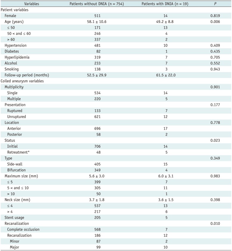
Table 1
Demographic and Angiographic Characteristics of Patients with Prior Coiled Aneurysms (n = 773)
![]()
Characteristics for DNIA Formation
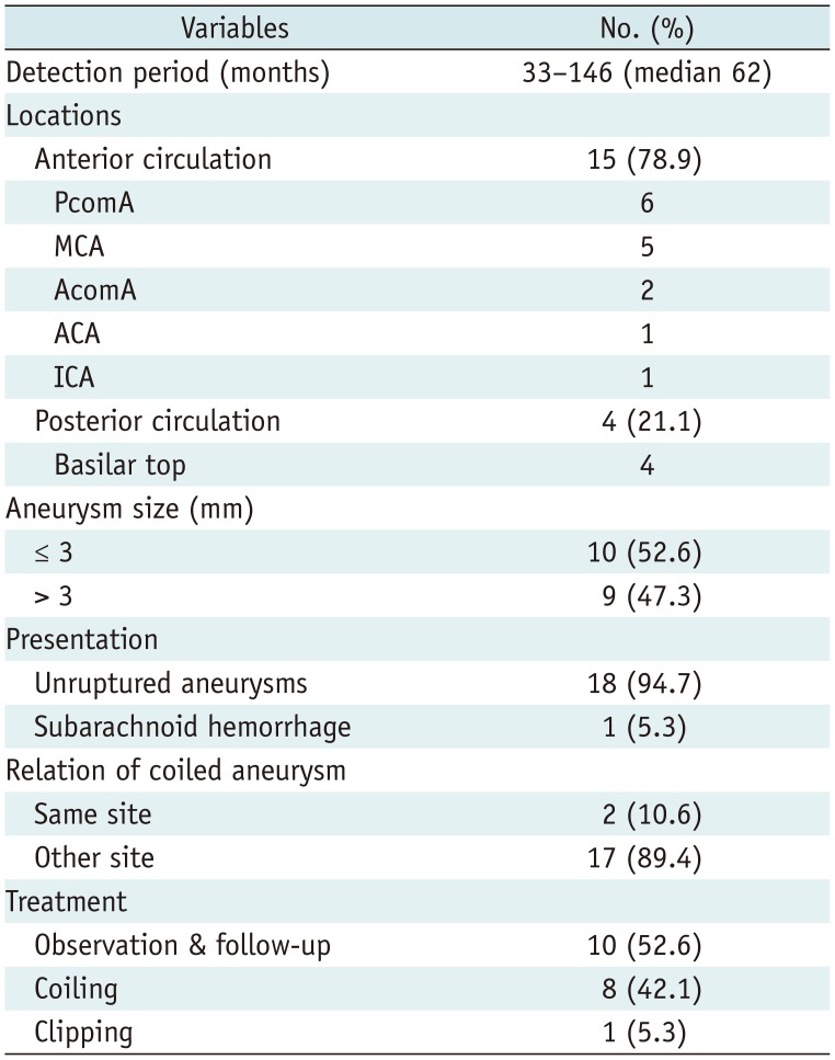
Of the 773 patients with coiled aneurysms under observation for > 6 months, DNIAs were confirmed in 19 (2.5%) during continued monitoring (3395.3 patient-years); 9 (47.4%) DNIAs surfaced within 60 months and 10 (52.6%) appeared thereafter. Within this time frame, inter-observer agreement in assessing DNIA was excellent (κ = 0.875; range, 0.820–0.930). The characteristics of DNIAs detected are summarized in Table 2. Mean size of DNIAs was 3.3 ± 1.6 mm (range, 1.5–8.7 mm; median, 3.0 mm). In one patient, the DNIA was identified in the midst of SAH presentation, whereas the remaining 18 aneurysms were documented during regular follow-up and were unruptured. The posterior communicating artery (PcomA; n = 6) was the most common site of DNIAs, followed by the middle cerebral artery (n = 5), basilar top (n = 4), anterior communicating artery (n = 2), and distal segments of the anterior cerebral or internal carotid artery (n = 1, each). In two patients, DNIAs developed adjacent to coiled aneurysms (Fig. 1), and one of the DNIAs emerged from the infundibulum of PcomA origin. Of the 19 DNIAs identified, 9 aneurysms (including one accompanied with SAH) ≥ 3 mm in size were treated (endovascular coiling, 8; surgical clipping, 1). Further follow-up was recommended for the remaining 10 aneurysms, which were smaller (< 3 mm).
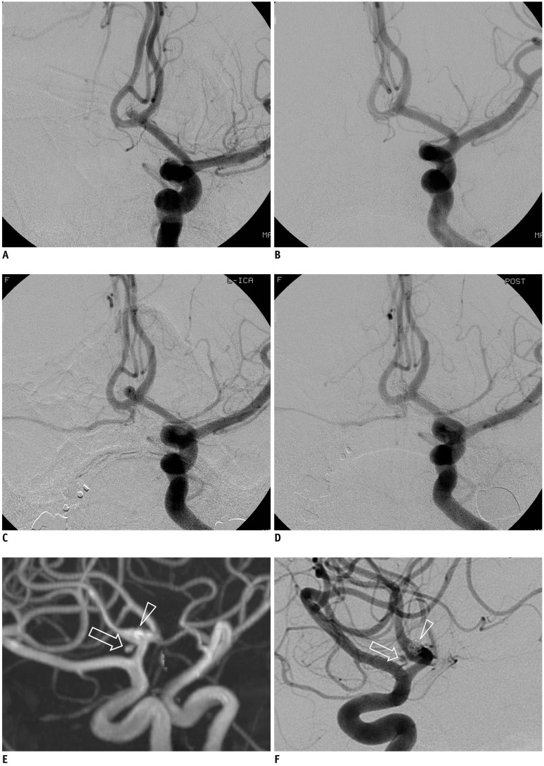 | Fig. 1Representative case of de novo aneurysm.
(A) Pre- and (B) post-embolization angiographic images of ruptured anterior communicating artery aneurysm. C. Conventional angiography with 6-month follow-up shows major recanalization of coiled aneurysm. D. Post-procedural angiography after additional coiling confirms successful occlusion of aneurysm. (E) Magnetic resonance and (F) conventional angiography in 60-month follow-up show de novo aneurysm (arrows) adjacent to coiled aneurysm (arrowheads).
|
Table 2
Characteristics of DNIAs (n = 19)
![]()
Risk Factors for DNIA Formation
In univariate analysis, patient age, retreatment, and recanalization during follow-up differed significantly between patients without (n = 754) and with (n = 19) DNIAs (Table 1). Binary logistic regression analysis indicated that younger age (hazard ratio [HR] = 1.045, 95% confidence interval [CI]: 1.010–1.081; p = 0.010) and recanalization of coiled aneurysms (HR = 2.560, 95% CI: 1.012–6.481; p = 0.047) were independently correlated with the development of DNIAs in patients with coiled aneurysm (Table 3).
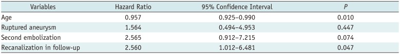
Table 3
Logistic Regression Model Assessing Risk of DNIAs in Patients with Prior Coiled Aneurysms
![]()
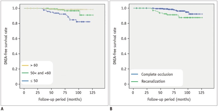
During follow-up, DNIAs were detected in a mean of 61.5 ± 22.0 months (median, 62 months; range, 33–102 months). The overall DNIA development rate was 0.56% per patient-year. Annual rates of DNIA formation for specific risk factors were estimated as follows: 1) age (≤ 50 years, 1.51% per patient-year; > 50 and ≤ 60 years, 0.35% per patient-year; > 60 years, 0.14% per patient-year) and 2) recanalization (complete occlusion, 0.31% per patient-year; recanalization, 1.05% per patient-year). Kaplan-Meier rate estimates of cumulative survival without DNIA are presented in Figure 2. The overall survival rate was 98.1%, but 84-month estimates differed by age (≤ 50 years, 93.5%; > 50 and ≤ 60 years, 98.4%; > 60 years, 99.4%) and recanalization (complete occlusion, 99.0%; recanalization, 94.4%). Cumulative survival rates without DNIA reached significance for age (p < 0.001) and recanalization (p = 0.006) via generalized Wilcoxon analysis.
Go to :
DISCUSSION
In the present study, we found that DNIAs developed in 19 (2.5%) of 773 patients with coiled aneurysms in follow-up imaging, yielding a cumulative estimated incidence of 0.56% per patient-year. This result was slightly higher than the outcomes of a prior systematic review (incidence, 2%; incidence density, 0.3% per patient-year) (3). The incidence of DNIA formation in the surgical clipping group was about 2.6–3.3% (0.3–0.6% per patient-year), which was not significantly different from the results of our study (1617).
DNIA may be a phenotype of metachronous multiple IAs, the incidence of which is variably reported to be 15–30% (21). According to a systematic review and meta-analysis, the risk factors for multiple IAs are as follows: female sex, smoking, HTN, older patient age, and DNIA formation (22). The precise mechanisms of DNIA formation are as yet unclear, but it is likely that multiple factors are involved. According to several relevant reports, the risk factors associated with DNIA formation are female sex, younger age, HTN, past history of SAH, and smoking (571315182324252627282930), similar to the factors associated with multiple IAs. These findings suggest that DNIA formation may be influenced by the systemic profiles of patients rather than being affected solely by local forces. However, other researchers partially refute this, claiming that female sex, HTN, and smoking are not culpable, as we also have concluded (1216283132). Therefore, the factors contributing to DNIA formation are still controversial.
In the present study, we identified two significant risk factors for DNIA formation among the various clinical and anatomic variables analyzed. Younger age at initial IA diagnosis proved to be an independent risk factor for DNIA formation in binary logistic regression analysis (HR = 1.045, 95% CI, 1.010–1.081; p = 0.010), particularly in patients aged ≤ 50 years. In younger patients, the annual DNIA formation rate was 10-fold greater, relative to patients > 60 years (1.51% vs. 0.14% per patient-year); and compared with elder patients, the younger subset displayed a significantly lower cumulative survival rate without DNIA (p < 0.001). These findings are perhaps related to the propensity of IA development in the general population. The overall prevalence of IA in a meta-analysis was estimated to be 3.2% in a population without comorbidity (2), at a mean age of 50 years. In addition, the prevalence of IA was higher in adults aged > 60 years, in comparison with younger adults. Therefore, it seems that the majority of the aneurysms develop in patients aged < 60 years. In our series, DNIAs were detected in only two (0.6%) of 339 patients > 60 years old, which is remarkably lower than the rate of DNIA detection in patients aged < 50 years (7.1%). Hence, an extended monitoring beyond routine follow-up for coiled aneurysms may be more appropriate in younger patients, given their greater risk. Lindgren et al. (5) and Lecler et al. (33) have likewise reported correlations between younger age and the likelihood of DNIA formation.
The second risk factor for DNIA formation that we identified was recanalization of coiled aneurysms during follow-up (HR = 2.560, 95% CI: 1.012–6.481; p = 0.047). In reviewing the current literature, no publications have yet addressed this proclivity. Consequently, we cannot readily offer any corroborating scientific evidence. However, it is plausible that arterial wall deformation and remodeling are common morphologic adaptations of both phenomena. Recanalization after coiling is related to aneurysmal growth to greater or lesser degrees (3435). Several experimental studies using computational fluid dynamics in a patient-specific vascular model have further indicated that the elasticity of the vascular wall may play a fundamental role in deformation of the arterial wall (363738). A rigid (vs. elastic) wall serves to increase wall shear stress acting on the vascular wall in a pulsatile flow, potentially accentuating the hemodynamic impact on the endothelial cell, intima, and smooth muscle cell. Thus, vascular wall elasticity may be closely associated with arterial wall deformation, promoting both DNIA formation and growth of coiled aneurysms.
DNIAs rarely occur (about 2% incidence and < 1% per patient-year), and only one case involved SAH among the 19 DNIAs studied herein. Data from the International Subarachnoid Aneurysm Trial indicate an annual rupture rate of 0.04–0.06% due to DNIAs in patients with coiled aneurysms (39). Although the major concern for patients first diagnosed with IA is the risk of rupture, DNIA rupture seldom occurs. Therefore, serial imaging studies beyond a given routine protocol may not be cost-effective in most patients with coiled IAs. However, based on our study results, we suggest that careful follow-up with periodic scheduled imaging (e.g., intervals of 2–5 years after 3-year postembolization follow-up) may be reasonable as a long-term approach to exclude DNIAs in selected patients, particularly those aged < 50 years at initial IA diagnosis or with recanalization of coiled aneurysms.
There were several limitations in this study, the first being the potential for selection bias in a retrospective review. The patients with aneurysms treated by surgical clipping were not included, inevitably due to a lack of the follow-up imaging data. Furthermore, the follow-up protocol was not well standardized within the designated population, with differing imaging modalities and variable exam intervals among individual subjects. Finally, due to the low probabilities of DNIA development, a small number of DNIAs observed in this study may have skewed our discovery of independent risk factors for DNIA formation. Further prospective studies examining a broader array of lesions are need for more conclusive support.
In conclusion, we have demonstrated that DNIAs developed in 2.5% of patients with coiled aneurysms and that the cumulative incidence was 0.56% per patient-year. Although the incidence of DNIA formation is rare, particular attention should be paid to patients aged < 50 years at initial diagnosis of IA and those with recanalized coiled aneurysm after endovascular treatment.
Go to :
 XML Download