

 PDF
PDF ePub
ePub Citation
 Print
Print


INTRODUCTION
Long-standing tachycardia is a well-established cause of left ventricular (LV) systolic dysfunction.1)2)3) Tachycardia-induced cardiomyopathy (T-CMP) can be defined as a heart failure (HF) syndrome originating from increased atrial or ventricular rates.4) Since the first description of T-CMP in the early 20th century, multiple studies have established a cause-relationship between tachycardia and cardiomyopathy in both animal models and humans.5) T-CMP is one of the major causes of congestive HF, which is a major health concern with high burden of morbidity.6)7) T-CMP can be dramatically improved by appropriate management of tachycardia and HF. Thus, early recognition of T-CMP is important.1)2)3)4) In this review, we will discuss the proposed pathophysiologic mechanism, diagnostic strategy, and treatment of T-CMP.
CAUSES OF TACHYCARDIA-INDUCED CARDIOMYOPATHY
T-CMP can be induced by various tachyarrhythmias, including atrial fibrillation (AF), incessant supraventricular tachycardias, and ventricular arrhythmias.5) Although the incidence of T-CMP has not been investigated thoroughly in previous literature, T-CMP can develop as early as a week after the onset of tachycardia or it can take several years following the presentation of symptoms of tachycardia.3)8) AF with rapid ventricular response is the most common cause of T-CMP.3) As AF facilitates development of HF regardless of rapid ventricular response and HF itself is predisposed to AF,9)10) it is difficult to determine the exact proportion of T-CMP in these patients. However, earlier studies regarding rhythm control or rate control in patients with AF and systolic dysfunction suggest that the tachycardia-induced cardiomyopathy component is common in those patients.10)11)12)13)14) Benefits of heart rate control were independent of atrioventricular synchrony and atrial contraction in those patients who received rate control with atrioventricular (AV) junction ablation and permanent pacemaker implantation.12)13) Studies investigating the time course of hemodynamic improvement following cardioversion in patients with AF and HF show that there is a time lag between restoration of sinus rhythm and LV systolic function, suggesting the presence of underlying myocardial failure.14)15)16) Focal atrial tachycardia (AT) is a well-known cause of T-CMP.15)17) Although focal AT is usually benign,18) it can occasionally show an incessant nature and may cause T-CMP. In a series of focal AT patients who underwent catheter ablation, 10% of those patients presented with T-CMP had improved LV function following catheter ablation.17) It has been reported that T-CMP can be induced by atrioventricular nodal reentrant tachycardia (AVNRT), atrioventricular reciprocating tachycardia (AVRT), or permanent junctional reciprocating tachycardia (PJRT).19)20)21)22)23) Given that reentrant supraventricular tachycardias (SVT), including AVNRT and AVRT, are mostly paroxysmal and rarely incessant, reentrant SVT induced T-CMP is uncommon.15) Dual AV node physiology can facilitate T-CMP through dual atrioventricular nodal non-reentrant tachycardia, which is uncommon.15)24) Dual AV nodal non-reentrant tachycardia is the double firing of a single sinus beat which are conducted into both fast and slow pathways, simultaneously. If this phenomenon repeats rapidly, tachycardias can occur, and catheter ablation of the slow AV nodal pathway is curative in these patients.24) Ventricular tachycardia (VT) is another cause of T-CMP. VTs causing T-CMP commonly originate from outflowing tracts or coronary cusps,15) and idiopathic left ventricular tachycardia (ILVT) may also induce T-CMP.25) There is a consensus that tachycardias of higher heart rate and longer duration could lead to cardiomyopathy (CMP).3) However, the exact heart rate level that may induce T-CMP is not well defined.1)3) Deterioration of left ventricular function could be induced at pacing rate of over 240 beats/min and 450 beats/min in the dog and rabbit model, respectively.5) Those pacing rates are approximately two times faster than normal heart rates of dogs (120 bpm) and rabbits (240 bpm). In our cohort of T-CMP, most of patients presented with heart rate of greater than 110-120 beats/min.26) Another clinical trials also have reported that patients with T-CMP had similar range of heart rate.17)23)27) In this context, the heart rate of 110-120 beats/min which is approximately two times faster than normal heart rate of human can be considered as a threshold of T-CMP.
There has been data regarding risk factors that contribute to the development of T-CMP. A study showed that patients with a polymorphic homozygous deletion in the angiotensin-converting enzyme (DD) gene may have a higher chance of developing T-CMP when persistent tachycardia is present.28)
The time lag from the diagnosis of tachyarrhythmia to the presentation of T-CMP was shorter among older patients with T-CMP as compared with younger patients with T-CMP in our cohort of T-CMP.26) This may suggest aging is a factor that make patients more vulnerable to T-CMP.
PATHOPHYSIOLOGY
In animal models, chronic rapid atrial or ventricular pacing results in low-output HF, which is similar to human HF, and the degree of ventricular dysfunction is related to the rate and duration of pacing.5)29) The cardiac changes involve LV systolic and diastolic dysfunction, a markedly dilated cardiomyopathy (DCMP) involving all cardiac chambers.1)5) The structural changes of T-CMP are characterized by preservation or thinning of bi-ventricular wall thickness without gross hypertrophy and increased ventricular mass.5)30)31) At a microscopic level, myocyte loss and elongation has been reported in T-CMP animal models.15)32)33) Additionally, hypertrophic change of myocytes was documented in an animal model of T-CMP.33) However, there is controversy over hypertrophic change of myocytes because several studies have shown ventricular remodeling of T-CMP without evidence of myocyte hypertrophy.15)31) Reduction of the extracellular matrix and disruptions to the basement membrane-sarcolemmal interface were also observed in animal models.31)34)35) The extracellular matrix has a role in myocyte alignment and the mechanical ventricular performance is produced by interactions among myocytes, the basement membrane, and the extracellular matrix.5)34) Consequently, these alternans lead to ventricular dilatation and contractile dysfunction.5)32)34) Development of T-CMP induce alternans of myocyte electrophysiological characteristics which are accompanied by a reduced amplitude of action potential and peak L-type Ca2+ current and prolonged duration of the action potential.36) These changes also contribute to the myocyte contractile dysfunction with T-CMP.2)36) In a study investigating isolated ventricular myocyte preparations from animal models with T-CMP, myocytes with T-CMP showed decreased density of T-tubules and L-type calcium channels which contribute to abnormal excitation-contraction coupling.37) The hemodynamic changes of T-CMP involve elevated systemic resistance, elevated LV filling pressures, and increased LV wall stress.15) Similar to other forms of HF, the neurohormonal system is upregulated in response to these changes in T-CMP.5)15) In similar HF models, induced by different causes, animals with T-CMP showed a blunted response to beta-adrenergic stimulation with decreased beta1-receptors and alterations in beta-receptor transduction.5)29) Blunted beta-adrenergic response is related to a decreased force-frequency relation on cardiac contractility29) and this could aggravate impaired cardiac function during stress conditions in subjects with T-CMP.38) Depletion of myocardial energy stores have been proposed as a mechanism of T-CMP. Diminished levels of creatine, phosphocreatine, adenosine triphosphate and glycogen, enhanced activity of Krebs cycle oxidative enzymes, and decreased activity of the sodium-potassium adenosine triphosphatase (Na-K ATPase) pump were described in animals with T-CMP.5)35)39) These metabolic changes are related to mitochondrial injury and impaired mitochondrial activity.15) Increased levels of oxidative stress are associated with myocyte apoptosis, and mitochondrial DNA is more vulnerable to oxidative damage.4)15) Subclinical myocardial ischemia is another possible mechanism of T-CMP. Myocardial blood flow is decreased with significant remodeling of the coronary capillary vasculature.40) Hibernation of myocardium due to ischemia may exist in T-CMP and this may partly explain the reversed remodeling of myocardium after cessation of the tachycardia.4) Earlier changes of T-CMP would mainly be due to tachyarrhythmia. On the contrary, later pathologic changes of T-CMP are involved in neurohormonal changes following hemodynamic alternans. Therefore, pathophysiology of T-CMP shares common features with the pathophysiology of HF due to other etiologies.15) A recent study investigated endomyocardial biopsy samples from patients with T-CMP and compared them with those from HFs due to other etiologies. In this study, electron microscopic examination revealed that T-CMP was characterized by hypertrophied cardiomyocytes, enhanced macrophage infiltration, and more severe disruption of mitochondria as that seen in idiopathic DCMP.41) This observation corresponds with the findings from animal models of T-CMP. Figure 1 summarized the pathophysiologic changes in T-CMP.
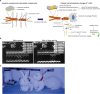 | Figure 1(A) Changes in cellular level in T-CMP. (B and C) Echocardiographic and gross change in pacing induced HF animal model. (A) Following sustained tachycardia, intracellular and extracellular remodeling leads to LV remodeling and worsening contractility. Decrease in L-type Ca2+ channel causes abnormal excitation-contraction coupling. Myocardial fibrosis persists even after recovery of LV function. (B) Echography in animal with pacing-induced HF (from Ryu et al.,29) Ross et al.38)). (C) Gross change in pacing-induced heart failure in animal (from Ryu et al.,29) Ross et al.38)): (a) normal rabbit before pacing, (b) low cardiac output status of rabbit after pacing with skin color change on ear and decreased activity, and (c) pacing induced HF rabbit showing edematous change and lethargic activity.HF = heart failure; LV = left ventricular; T-CMP = tachycardia-induced cardiomyopathy.
|
CLINICAL PRESENTATION AND DIAGNOSIS
The most common symptoms of T-CMP include palpitations, dyspnea, and presyncope/syncope; although some patients with T-CMP may not present symptoms.27) Development of T-CMP can take one month to years from the presentation of initial arrhythmia symptoms.3) In a series of T-CMP patients, T-CMP developed over 1 week after the onset of symptoms due to tachyarrhythmia.8) T-CMP should be suspected in all patients with newly diagnosed LV dysfunction without obvious etiologies and prior, persistent, or frequent paroxysmal tachycardia, because even severe LV dysfunction, so-called terminal LV dysfunction, can be normalized with appropriate treatment in T-CMP.42) Patients with underlying structural heart diseases and tachyarrhythmia should not be excluded because they may have a co-existent component of T-CMP. Previous normal LV function and LV dysfunction, disproportional to the degree of underlying heart disease, raise the possibility of T-CMP.42) The culprit tachycardia may not be documented at the time of presentation and underlying tachyarrhythmias such as AF or atrial flutter may not be the causative tachycardia.3)42) Therefore, monitoring for arrhythmia should be performed in patients with DCMP of unknown etiology or possible T-CMP and ambulatory electrocardiographic monitoring can be a useful tool for detecting the culprit tachyarrhythmia and for investigating the cause of the tachyarrhythmia in this setting.1)43) Evaluation of patients with a potential T-CMP includes common diagnostic strategies recommended by contemporary guidelines for HF.3)44)45) Thyroid function tests should be included in the initial diagnostic test because hyperthyroidism can facilitate T-CMP with tachycardias such as AF or sinus tachycardia.3) Plasma concentration of brain natriuretic peptides (BNPs) and NT pro-BNP are usually elevated in the setting of LV dysfunction due to T-CMP1) and rapid fall of plasma natriuretic peptide level following elimination of tachycardia suggests that LV dysfunction is due to T-CMP.46) As LV wall thickness is usually preserved in T-CMP, absence of LV hypertrophy pattern on electrocardiography may suggest T-CMP as a cause of heart failure and can facilitate further imaging studies focusing on differential points of T-CMP. On echocardiography, T-CMP is characterized by dilated LV dimension with a lack of ventricular hypertrophy.1)5)43) In a study comparing T-CMP and idiopathic DCMP, LV dimension and LV mass index were significantly smaller in the T-CMP group compared to the idiopathic DCMP group, and LV end-diastolic dimension ≤61 mm was a predictor of T-CMP.47) In addition to LV end-diastolic dimension, post-extra systolic potentiation of LV wall motion was more frequently observed in T-CMP rather than in DCMP from our previous work.26)
Cardiac magnetic resonance imaging (CMRI) can help differentiate T-CMP from idiopathic DCMP. In a study of patients with premature ventricular contractions-induced CMP or VT-induced CMP, absence of late gadolinium enhancement suggests an underlying scar in the LV wall which can be used as a predictor of T-CMP among patients with new onset DCMP of unknown etiology.43)48) During the early stages, T-CMP cannot easily be differentiated from HF of other etiologies and the final diagnosis of T-CMP can only be made by observation of improvement in LV systolic function. In case of improvement, recovery can be achieved within 1 to 6 months after appropriate treatment for tachycardia and HF.1)
TIME COURSE OF RECOVERY AND PROGNOSIS
In animal models of T-CMP, recovery of cardiac systolic dysfunction after cessation of rapid pacing is a unique feature of this model.5) LV systolic function improves within 1 to 2 weeks after the rapid pacing stops. Most of the hemodynamic variables return to normal by 4 weeks.5)49) On the other hand, cessation of rapid pacing does not guarantee a complete recovery of the enlarged LV volume and LV diastolic dysfunction and LV hypertrophy did develop within 4 weeks after the cessation of rapid pacing in animal models. This phenomenon can be explained by compensatory remodeling following rapid pacing or by the inability of the hypertrophic changes in response to signals inducing hypertrophy during the pacing phase.5)15)
In clinical studies, improvement of LV systolic function was observed within a month following the treatment for tachyarrhythmia.42) Recovery of LV function can take 2–3 months and usually does not take longer than 6 months.1)26)42) It is thought that the recovery of LV function can be shortened with adequate heart rate reduction treatment, below 100 beats/min in our experience. A study reported that patients who had recovered from T-CMP had a significantly higher LV volume when compared to age-, sex-, and ejection fraction-matched controls.50) This result is consistent with the previous findings in animal models and suggests the persistence of negative LV remodeling in patients with T-CMP even after normalization of LV ejection fraction.1)15)50) Patients who recovered from T-CMP are vulnerable to the recurrence of tachycardia.1) In cases where tachycardia reoccurred, deterioration of LV systolic function was induced more rapidly than that in previous T-CMP events8)51) and persistence of microstructural changes following normalization of LV dysfunction seemed to be the cause of this vulnerability.50) In this context, some studies reported sudden cardiac deaths after restoration of LV dysfunction due to T-CMP, and the prevalence of sudden cardiac death was not uncommon even after appropriate treatment and clinical improvement.8)51)
TREATMENT
Suppression of the culprit tachycardia is a key to the restoration of LV dysfunction for T-CMP. If T-CMP is suspected, treatment focusing on eliminating or controlling the culprit tachycardia should be pursued.1)3)43) This approach includes radiofrequency catheter ablation (RFCA) of culprit arrhythmia or antiarrhythmic drugs. As discussed earlier, recurrence of arrhythmia has a deleterious effect on LV systolic function. RFCA should be considered as the first therapy for arrhythmias which can be managed with a high success rate such as atrial flutter, focal AT, AVNRT and AVRT.43)
In case of decompensated HF with AF, it is generally recommended to begin with a heart rate-control treatment because recurrence rate of AF following cardioversion is common.42) Cardioversion should be performed following the acute management with optimal HF medication.42)52) In our experience, digoxin is a preferred choice for the initial rate control after excluding coronary artery disease. After appropriate rate control, heart failure management and anticoagulation therapy, chemical cardioversion with amiodarone can be performed without a concern for procedure related complications and with a moderate success rate.11) Both rate and rhythm control strategies are reasonable choices for patients with AF with compensated HF.9)10) It has been proposed that lenient rate control is as effective and feasible as strict rate control in patient with AF.53) However, the RACE II trial included a limited number of patients with reduced LV systolic function, and more data is required to set up an ideal heart rate target for these patients.10)43) Until then, a resting heart rate of up to 80 beats/min and up to 115 beats/min during moderate exercise should be an acceptable rate control target for AF patients with possible diagnosis of T-CMP.9)10)
Initial treatment of T-CMP should include standard HF therapy such as angiotensin-converting enzyme inhibitors or angiotensin receptor blockers, aldosterone blockers, beta-blockers, and diuretics to attenuate negative remodeling and relieve symptoms related to HF.1)3)42)43)44) Negative remodeling, including myocardial fibrosis and sustained LV volume enlargement is persistent even after normalization of LV systolic function.50)54) Hence, continuing standard HF management is advised for favorable remodeling. However, the duration for such therapy is not well defined.43)
FUTURE DIRECTION
Since the first reported case of T-CMP in early 1900s, T-CMP has been recognized as a distinct disease entity and we have extended our knowledge on T-CMP with multiples studies of animal models and clinical trials. Animal models contributed to a better understanding of the pathophysiology by sustained tachycardias. However, there is limited data on the pathophysiology of T-CMP in humans. With development of cardiac imaging including CMRI, our knowledge on the pathophysiology of T-CMP from animal models will be translated into the clinical practice and lead to a better understating of the pathophysiology of T-CMP in humans. We still have limited data on the risk factors of T-CMP. Further studies on genetic and clinical characteristics guided risk prediction models for T-CMP are necessary for early detection of high-risk patients with tachyarrhythmias and to prevent development of HF. Recent AF ablation trials suggested that AF alone can lead to CMP independently of rapid ventricular response55) and a more comprehensive term of AF induced CMP has been introduced. But there are gaps in our understanding of AF-induced CMP. Further studies are necessary to reveal the mechanism of AF induced CMP and to suggest appropriate treatment.
CONCLUSION
T-CMP is a reversible cause of HF. Imaging studies may be helpful for the diagnosis of T-CMP but diagnosing T-CMP requires the demonstration of improved LV function with suppression of tachycardia. T-CMP should be suspected in all patients with DCMP of undetermined etiology and tachycardia faster than 100 or 110 beats/min for early detection and treatment for these patients. T-CMP should be considered as a possible diagnosis even in patients with HF of other established causes because they may have the superimposed reversible component of T-CMP. Strategies of tachyarrhythmia management can be antiarrhythmic drugs, catheter ablation or rate control. Catheter ablation should be considered for tachyarrhythmias which can be curable with ablation. Recent studies on premature ventricular contractions-induced CMP and AF ablation trials in HF patients suggested arrhythmias besides T-CMP. In this regard, our focus should move from T-CMP to arrhythmia induced CMP.
 XML Download