

 PDF
PDF ePub
ePub Citation
 Print
Print


INTRODUCTION
A variety of materials for bone reconstruction have been used to treat bone defects caused by various diseases or trauma [1]. Autogenous bone is recognized to be the gold standard as a graft material due to its osteogenic, osteoinductive, and osteoconductive properties [2]. The disadvantage of autogenous bone lies in its quick resorption after transplantation, lower space maintenance, and the need for an additional surgical procedure during harvest [34]. Conversely, synthetic bone functions well as a space maintainer and scaffold, but has no or limited osteogenic and osteoinductive ability [5].
Recently, several studies have explored the effects of mesenchymal stem cells, which have the capability to differentiate into bone, cartilage, and muscle, on bone regeneration [67]. The use of stem cells with a synthetic or xenogenic bone graft enhanced bone regeneration when compared with a bone graft alone, possibly due to the osteogenic and osteoinductive properties of stem cells [89]. Previous studies have shown that a combination of stem cells with bone graft material improved bone regeneration [1011]. In addition, successful bone regeneration was achieved in participants with large bony defects by applying stem cells [1213]. It cannot be overlooked that a significant amount of stem cells will migrate into bone defects over time [14]. It can be assumed that the adhesion of transplanted cells and/or migrated cells may vary depending on the type and nature of the scaffold [15]. Collagen, a component of the extracellular matrix of bone tissue, has been shown to promote osteogenic differentiation in stem cells and to enhance cell adhesion at the early stage of bone graft transplantation [16]. Moreover, cell adhesion and osteogenesis were found to be improved when collagen was applied in combination with hydroxyapatite [17]. The purpose of this study was to evaluate the viability of transplanted stem cells at an early time point (24 hours) based on the type and form of the scaffold used, including type I collagen membrane and synthetic bone.
MATERIALS AND METHODS
Obtaining the stem cells
Approximately 1 cm2 of periosteum was removed during surgical procedures, including tooth extraction and periodontal surgery, from otherwise healthy patients visiting the dental clinic of Uijeongbu St. Mary's Hospital. The participants did not have any systemic diseases. We obtained the approval of the Institutional Review Board at Uijeongbu St. Mary's Hospital's College of Medicine and the Catholic University of Korea (UC12TISI0046).
Isolation and culture of stem cells derived from the periosteum were performed as follows. The periosteum was placed in sterile phosphate-buffered saline (PBS; Gibco BRL, Grand Island, NY, USA) containing 1% antibiotic and antimycotic (Gibco BRL). The periosteum was then washed with PBS (Gibco BRL) and cut into 1–2 mm2 fragments in sterile conditions. The tissues were digested with 0.06% collagenase type II (Invitrogen Corporation, Carlsbad, CA, USA) and incubated at 37°C in a humidified incubator (Forma Scientific Inc, Marietta, OH, USA) with 5% CO2 and 95% air for 4 hours. The periosteum-derived cells were filtered through a 70-μm cell strainer (BD Bioscience, Two Oak Park, Bedford, MA, USA), and centrifuged at 1,500 rpm for 5 minutes. The precipitates were suspended in Dulbecco's modified Eagle medium (Gibco BRL) containing 10% fetal bovine serum and 1% penicillin-streptomycin (Gibco BRL) and cultured in an incubator. Each day, the morphological characteristics of cells were observed under a microscope. When necessary, the culture medium was exchanged. Cells not attached to the culture dish were removed, and only the attached cells were cultured. Upon reaching 90% confluence, cells were subcultured with a coenzyme solution composed of trypsin (Gibco BRL) and ethylenediaminetetraacetic acid (EDTA; Gibco BRL). Stem cells were identified by the surface markers STRO-1 (R&D Systems, Minneapolis, MN, USA) and CD146 (R&D Systems) using a fluorescence-activated cell sorter (FACS, BD Biosciences, Bedford, MA, USA). Stem cells were subcultured several times to obtain a sufficient number of cells (Figure 1A). Fluorescent staining was performed to confirm the survival of transplanted stem cells (Figure 1B). In short, the viability of cell spheroids was qualitatively analyzed using a Live/Dead Viability/Cytotoxicity Kit for Mammalian Cells (Thermo Fisher Scientific, Waltham, MA, USA). The cells were washed and suspended in 10 mL of phosphate-buffered saline, followed by addition of 5 μL of 50 mM calcein acetoxymethyl ester solution and 20 μL of ethidium homodimer-1 for 20 minutes at room temperature.

Animal experiments
New Zealand white rabbits weighing between 2.5 and 3.0 kg were used as experimental animals. All animal experiments were strictly carried out following the approval of the Ethics Committee for Animal Experiment of Uijeongbu St. Mary's Hospital, the Catholic University of Korea (UJA2017-05A).
General anesthesia was induced with tiletamine and zolazepam (15 mg/kg, Zoletil 50; Virbac Laboratories, Carros, France) and xylazine (5 mg/kg, Kepro BV, Deventer, Netherlands) intramuscularly and hair removal was performed. After disinfection with povidone-iodine (Sung Kwang Pharm, Seoul, Korea), local anesthesia was achieved with a 2% lidocaine solution containing epinephrine (Yuhan Co., Seoul, Korea).
A skin incision was made from the nasofrontal area to the external occipital area, and a periosteal flap was raised to expose the cranium. Four symmetrical circular defects 6 mm in diameter were made using a trephine drill (Figure 2A). The defects were grafted with 1) synthetic bone (β-tricalcium phosphate/hydroxyapatite [β-TCP/HA] with a particle size of 410–1,000 μm and a β-TCP-to-HA ratio of 7:3; Cowellmedi Co., Ltd, Seoul, Korea) and 1×10⁵ MSCs, 2) a collagen membrane (type I collagen membrane, CollaTape®, Zimmer Dental Co., Carlsbad, CA, USA) and 1×10⁵ MSCs, 3) β-TCP/HA+a collagen membrane and 1×10⁵ MSCs, or 4) β-TCP/HA, a chipped collagen membrane, and 1×10⁵ MSCs (Figure 2B). Efforts were made to apply the same amount of synthetic bone and/or collagen membrane in each group. The stem cells were incubated with the graft material for 1 hour before transplantation. The defects were then covered with a Teflon sheet (W.L. Gore & Associates Inc., Flagstaff, AZ, USA) and titanium screws (Figure 2C). The periosteum was released at its base, and the wound margins were reapproximated and closed with a non-absorbable suture (3-0 black silk; Ailee Co. Ltd, Busan, Korea).
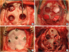 | Figure 2Surgical procedure. (A) Bone defects were made in the rabbit calvarium. A circular defect with a diameter of 6 mm was formed in the skull. (B) The groups were designated as follows; group 1: β-TCP/HA+1×105 MSCs, group 2: type I collagen membrane+1×10⁵ MSCs, group3: β-TCP/HA+type I collagen membrane+1×10⁵ MSCs and group 4: β-TCP/HA+type I collagen chipped membrane+1×10⁵ MSCs. (C) A Teflon sheet was applied and titanium screws were placed before suturing. The periosteum and scalp were replaced with non-absorbable sutures. (D) Harvesting of the grafted tissue was done at 24 hours after surgery.β-TCP/HA: β-tricalcium phosphate/hydroxyapatite, MSCs: mesenchymal stem cells.
|
After surgery, all rabbits received subcutaneous injections of antibiotics (4 mg/kg gentamicin) and anti-inflammatory analgesics (1 mg/kg ketoprofen) once per day. An E-collar (Saf-T-Shield, EJAY International Inc., Glendora, CA, USA), was placed to prevent injuries to the surgical field and fresh vegetables, including cabbage, were supplied to promote the animals' recovery. The transplanted cells and scaffold were retrieved 24 hours later using various instruments, including curettes, and the remaining portion was retrieved by pipetting (Figure 2D).
Evaluation of the cellular experiments
The cells were retrieved through the following procedure. Tissues from the rabbit skulls were placed in a 1.5 mL tube and centrifuged. After removing the supernatant, the cells were washed with phosphate-buffered saline and then treated with collagenase type II (Gibco BRL) at 1 mg/mL for groups 2, 3, and 4, which contained collagen membranes. The cells were incubated for 30 minutes to 1 hour to dissolve the collagen. All tissues were centrifuged twice at 1,200 rpm for 5 minutes. The cells were then mixed with 9 mL of distilled water and 1 mL of ×10 PBS (Gibco BRL) for 15 seconds and then centrifuged at 1,700 rpm for 6 minutes to hemolyze the red blood cells. After removing the supernatant, 1 mL of PBS and 1 mL of trypan blue solution (T-8154, Merck KGaA, Darmstadt, Germany) were mixed. Next, the number of total cells, the number of live cells, and the ratio of live cells were analyzed using an automated cell counter (TC 20TM, Bio-Rad Laboratories, Berkeley, CA, USA). Before transplantation, the fluorescent-stained stem cells were observed with an inverted microscope (IX71; Olympus Co., Tokyo, Japan) and were photographed with a digital camera (DP70; Olympus Co.). A quantitative evaluation was performed by comparing and analyzing the measured values for each defect.
Statistical analysis
Data were presented as mean ± standard deviation. Statistical analysis was performed using SPSS version 12 for Windows (SPSS Inc., Chicago, IL, USA). The normality of the distribution of the data was tested, and one-way analysis of variance with the Tukey B post hoc test was used to evaluate between-group differences. The significance level of this study was set to P<0.05.
RESULTS
Cells were extracted from the collected tissues and the number of total cells and number of viable cells were measured. Cells were easily separated from the collagen membrane, but cells attached to the synthetic bone were not easily separated (Figure 3). The number of stem cells attached to the synthetic bone in groups 1, 3, and 4 seemed to be similar.
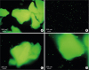 | Figure 3Fluorescent microscopic evaluation of the grafted tissue. (A) Group 1 (β-TCP/HA+MSCs): Viable MSCs were attached to β-TCP/HA. (B) Group 2 (collagen membrane+MSCs): The surviving MSCs that had been transplanted were easily separated from the collagen membrane. (C) Group 3 (β-TCP/HA+collagen membrane+MSCs). The surviving MSCs that had been transplanted were abundantly attached to the β-TCP/HA, and a small amount of MSCs were scattered around the β-TCP/HA. (D) Group 4 (β-TCP/HA+chipped collagen membrane+MSCs). The results were similar to those of group 3.β-TCP/HA: β-tricalcium phosphate/hydroxyapatite, MSCs: mesenchymal stem cells.
|
Cellular viability in tissue
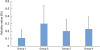
Cell migration rate
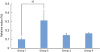
The rate of cell migration was calculated by dividing the number of total cells collected by the number of total cells transplanted [19]. The number of total cells reflected the sum of the number of transplanted cells that were not lost and the number of cells that migrated from neighboring areas. The cell migration rate was highest in group 2, but a statistically significant difference was not found (P>0.05) (Figure 5).
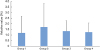
Cell survival rate
DISCUSSION
This study showed that the highest survival rate was found when the stem cells were applied to a membrane. When synthetic bone was used, utilizing a chipped membrane enhanced cell survival.
The survival of transplanted stem cells remains a major concern [2122]. Human cord blood-derived stem cells were transplanted into a critically sized femoral defect with a combined collagen/β-TCP scaffold, and the transplanted cells were reported to survive for up to 4 weeks [23]. Amniotic fluid-derived stem cells were applied to a collagen/hydroxyapatite scaffold in sinus augmentation procedures, and survival of stem cells was observed 90 days after surgery [24]. An important consideration is that it was easy to separate cells from collagen scaffolds, but the cells attached to synthetic bone were not easily separated. However, a previous study suggested that the absolute value of viability of the transplanted cells may decrease significantly, even if their survival is maintained [25]. Unlike the stable environment of the laboratory, in vivo bone defects have the possibility of leakage or loss of transplanted cells due to active bleeding or blood circulation [26].
Collagen is known to be involved in the enhancement of adhesion, proliferation, and survival of stem cells [27]. It was shown that cell attachment ability in the early phase and osteogenic differentiation were significantly enhanced in collagen-grafted porous biphasic calcium phosphate (BCP) when compared with BCP alone [16]. Human mesenchymal stem cells were cultured in a three-dimensional collagen scaffold, and a large number of osteoblasts were identified after a 50-day culture period [28]. However, it should be considered that when compared with group 2, the cells from the other groups (groups 1, 3, and 4) were securely attached to the synthetic bone and not easily separated, and direct comparison between the groups may not be feasible. It can be assumed that a direct comparison between groups 1, 3, and 4 can be made because the same amount of synthetic bone was loaded and the number of attached cells was similar. Comparing groups 1, 3, and 4, in which synthetic bone was loaded, the cell viability, cell migration, and survival rates were increased by the addition of collagen membrane. Moreover, applying a chipped membrane enhanced the survival rate, but this finding did not reach statistical significance. Cells that do not adhere to the scaffold may die prematurely, and addition of collagen may decrease the loss of the transplanted cells and enhance their viability [29]. The transplanted cells may undergo differentiation into bone cells under the influence of the surrounding environment, and stem cells may have paracrine effects by producing various factors, including growth factors [303132]. In this regard, higher cell viability and survival may have beneficial effects. In this study, the stem cells were incubated for 1 hour before transplantation to allow cells to adhere to the scaffold [3334].
Various methods have been suggested to enhance functionality [223536]. A previous report showed that three-dimensional stem cell spheroids exhibited greater resistance to apoptosis than dissociated stem cells in fibrin gels [37]. A self-assembling peptide nanofiber was applied to enhance the survival and osteogenic differentiation of peripheral blood mesenchymal stem cells in three-dimensional conditions [22]. A further study reported that fibrin gel properties were modified by supplementation with NaCl, which promoted the proangiogenic and osteogenic potential of stem cells [35]. An arginine-glycine-aspartic acid tripeptide (RGD)–modified alginate scaffold showed enhanced mesenchymal stem cell viability and osteogenic differentiation when compared with a non-modified scaffold [36]. Bone morphogenetic proteins (BMPs), including BMP-2, may be applied for bone regeneration because BMPs are capable of stimulating osteogenic differentiation during the healing period [38].
In conclusion, this study showed that stem cells could be applied to a membrane used as a scaffold under no or minimal pressure. When space maintenance is needed, stem cells can be loaded onto synthetic bone with a chipped membrane to enhance the survival rate.
 XML Download