

 PDF
PDF Citation
 Print
Print


INTRODUCTION
Accessory root canals create potential pathways through which bacteria can spread and remain unaffected by treatment procedures. It is a challenge to find techniques that can predictably disinfect accessory canals. Therefore, clinicians encounter problems when the canal is not properly disinfected and the consequent periradicular pathosis cannot be solved. It has been reported that failed endodontic treatments are related to a high incidence of apices with accessory canals, and the filling rate of the canals is low [1]. Therefore, surgical management might be required to treat the pathological problems which arise from accessory canals.
There have been a few case reports regarding the surgical approach of accessory canals in anterior teeth [23]. The incidences of anatomical complexity, including accessory canals and apical ramifications, in maxillary central incisors has been reported to be 46% [4]. Meanwhile, according to one study, 38% of maxillary first premolars had accessory canals, 12.3% had apical deltas, and 16.0% had isthmi [5]. However, few cases in the maxillary first premolars have been reported thus far, even though a ramification in the maxillary first premolar is not a rare feature, as stated above. This case report describes the surgical approach for the management of an infected accessory canal which caused periradicular pathosis on the maxillary first premolar. In addition, the application of calcium silicate-based material is highlighted.
CASE REPORT
A 55-year-old female patient presented with a gumboil on her left upper premolar area. Her medical history was noncontributory. The diagnostic radiograph showed a radiolucent lesion on the distal side of the root of the upper first premolar. The tracing radiograph using a gutta-percha cone indicated that the sinus tract originated from the distal aspect of the left upper first premolar (Figure 1A). Chronic periapical abscess was diagnosed, and nonsurgical root canal treatment was performed, but the sinus tract did not disappear (Figure 1B). Consequently, periradicular surgery was planned. With the exceptions of the incision, flap elevation, and suturing, all surgical procedures were performed using a dental operating microscope (DOM) (Global Surgical Co., Saint Louis, MO, USA).
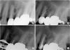 | Figure 1(A) Diagnostic radiograph showing the lateral lesion; (B) gutta-percha (GP) cone tracing indicates the origin of the sinus tract; (C and D) X-ray images showing working length measurement and canal obturation status, respectively.
|
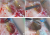
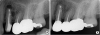
After infiltration of local anesthesia, the full mucoperiosteal flap was reflected and the lateral lesion was exposed. After the bony window was prepared, the accessory canal orifice was detected (Figure 2A). The cavity was prepared using a KiS ultrasonic tip (ObturaSpartan, Fenton, MO, USA) with a 3-mm depth driven by a piezoelectric ultrasonic unit (P5, Satelec, Merignac, France) (Figure 2B). Then, the cavity was dried and filled with a fast-setting calcium silicate cement (Endocem MTA, Maruchi, Wonju, Korea) (Figure 2C). The periapex was exposed, and 3 mm of the root-end was resected. An isthmus was found between the buccal and palatal canal, and the root-end cavity was prepared, including the isthmus (Figure 2D). Next, the cavity was also filled with Endocem MTA. The wound site was sutured with 5-0 monofilament sutures. The post-operative radiograph indicated that the lateral and main canal were adequately filled (Figure 3A). The patient was provided with post-operative care instructions. The 9-month follow-up radiograph demonstrated bony healing (Figure 3B).
DISCUSSION
Nonsurgical endodontic treatment can be difficult due to complex main root canal morphologies from root canal variations as well as complex anatomy, including accessory canals, furcation canals, isthmi, and apical deltas. According to Vertucci's [6] classification, the tooth reported in the present case had 2 root canals, with type IV (2-2) being the most common canal configuration in maxillary first premolars. After performing meticulous nonsurgical endodontic treatment, the sinus tract on the buccal gingiva was not resolved. Presumably, the chemo-mechanical preparation partially removed necrotic tissue from the entrance of the accessory canal, whereas the adjacent tissue remained inflamed, sometimes infected, and was associated with periradicular disease [7]. In other words, an insufficiently-treated large lateral canal acts as a 2-way passage for bacteria and tissue degradation products between the root canal space and periodontal tissue during conventional endodontic treatment [8]. Therefore, the surgical approach was chosen to manage the residual post-treatment infection in the accessory canal area.
The use of DOM has been highly recommended for several decades since it enhances and facilitates each phase of endodontic surgery [91011]. Notably, DOM is considered as essential equipment, especially when micro-anatomical structures, such as accessory canals and isthmi, must be appropriately managed. Kumar and Khambete [12] suggested the use of high magnification (18× to 30×) to observe and evaluate fine details during endodontic surgery. In this case, after removal of the granulation tissue from the lateral lesion, we carefully inspected the distal root surface under high magnification (19×), and we were easily able to detect the orifice. Furthermore, after the root-end resection, we could detect and effectively manage the isthmus. Floratos and Kim [13] reported that over 80% of premolars have isthmi at the 3 mm level from the apex. The isthmus can be identified and treated when DOM is used. Therefore, we strongly suggest the use of DOM with a high level of magnification for the successful surgical management of microstructures, including accessory canals, isthmi, microfractures, etc.
The type of root-end filling material is one variable that may have an impact on the outcome of periradicular surgery. Among the various types of root-end filling materials, calcium silicate cement is a good choice for root-end filling because of its biocompatibility, excellent sealing ability, hard tissue induction and conduction, and a high rate of success. Endocem MTA, one of the various calcium silicate cements, is considered to be suitable material for an accessory canal, as used in this present case. It has a superior fast-setting property which minimizes the risk of washout or exfoliation of the retrofilling material from the relatively shallow cavity. Endocem MTA exhibited a significantly shorter setting time (15.3 ± 0.5 minutes) than ProRoot MTA and OrthoMTA (318.0 ± 56.0 and 324.3 ± 2.1 minutes, respectively) [14]. Recently, Kim et al. [15] suggested that fast-setting calcium silicate cement could also be considered for use as root-end filling materials in endodontic microsurgery, particularly in complicated clinical situations which require rapid initial setting of the materials. Although the direction for lateral canal filling is different from the conventional apical root-end filling which is vertical, root-end filling material compacted densely inside the accessory canal without any voids (Figure 3A), which is reported to be one of the advantages of Endocem MTA [16]. Moreover, refractory endodontic lesions are closely associated with Enterococcus faecalis (E. faecalis) biofilm [17]. Beside the fast-setting ability of Endocem MTA, its superior inhibitory effect against E. faecalis is also assumed to have contributed to the favorable and rapid outcome in the present retreatment case [18].
Several investigators conducted short-term follow-up cases that received root-end fillings with new calcium silicate cements [19]. However, the results from studies with short-term follow-up may not indicate real treatment outcomes that will occur over an extended period, as noted in the literature [20]. Therefore, further follow-up is scheduled to investigate the long-term outcome of Endocem MTA as root-end fillings on the lateral lesion in the present case.
 XML Download