Journal List > Pediatr Infect Vaccine > v.26(2) > 1130684

Kwon, Cho, Kim, and Lee: Single or Dual Infection with Respiratory Syncytial Virus and Human Rhinovirus: Epidemiology and Clinical Characteristics in Hospitalized Children in a Rural Area of South Korea

Abstract

Purpose

Respiratory syncytial virus (RSV) and human rhinovirus (hRV) are the most common causes of child respiratory viral infections. We aimed to investigate epidemiological and clinical characteristics of RSV and hRV single infections and coinfections.

Methods

Nasopharyngeal aspirates of hospitalized children aged <5 years were tested using multiplex reverse transcription polymerase chain reaction (RT-PCR) from October 2014 to April 2017. Their medical records were retrospectively reviewed.

Results

RSV or hRV was detected in 384 patients who divided into 3 groups: patients with RSV (R group, n=258); patients with hRV (H group, n=99); and patients with both (RH group, n=27). The R group (median age, 6 months) consisted of 248 (96.1%) patients with lower respiratory tract infection (LRTI), and 14 (5.4%) needed oxygen inhalation. Infants aged <12 months (63.2%) had respiratory difficulty and were supplied oxygen more often. The H group (median age, 16 months) consisted of 56 (56.6%) patients with LRTI, 4 (4%) required oxygen inhalation, and 1 (1.0%) required mechanical ventilation. Infants (40.4%) showed longer hospitalization compared to patients aged ≥12 months (5 vs. 4 days, P<0.05). The RH group consisted of 24 (88.9%) patients with LRTI, and 2 (7.4%) needed oxygen inhalation. Hospitalization days and oxygen inhalation and mechanical ventilation rates did not differ between single infections (R and H groups) and coinfections (RH group).

Conclusions

RSV was detected more often in younger patients and showed higher LRTI rates compared to hRV. Single infections and coinfections of RSV and hRV showed no difference in severity.

References

1. Tregoning JS, Schwarze J. Respiratory viral infections in infants: causes, clinical symptoms, virology, and immunology. Clin Microbiol Rev. 2010; 23:74–98.
crossref
2. Brini I, Guerrero A, Hannachi N, Bouguila J, Orth-Höller D, Bouhlel A, et al. Epidemiology and clinical profile of pathogens responsible for the hospitalization of children in Sousse area, Tunisia. PLoS One. 2017; 12:e0188325.
crossref
3. Borchers AT, Chang C, Gershwin ME, Gershwin LJ. Respiratory syncytial virus–a comprehensive review. Clin Rev Allergy Immunol. 2013; 45:331–79.
crossref
4. Drysdale SB, Mejias A, Ramilo O. Rhinovirus – not just the common cold. J Infect. 2017; 74(Suppl 1):S41–6.
crossref
5. Asner SA, Rose W, Petrich A, Richardson S, Tran DJ. Is virus coinfection a predictor of severity in children with viral respiratory infections? Clin Microbiol Infect. 2015; 21:264.e1–6.
crossref
6. Choi EH, Lee HJ, Kim SJ, Eun BW, Kim NH, Lee JA, et al. The association of newly identified respiratory viruses with lower respiratory tract infections in Korean children, 2000–2005. Clin Infect Dis. 2006; 43:585–92.
crossref
7. Choi E, Ha KS, Song DJ, Lee JH, Lee KC. Clinical and laboratory profiles of hospitalized children with acute respiratory virus infection. Korean J Pediatr. 2018; 61:180–6.
crossref
8. Kim JK, Jeon JS, Kim JW, Rheem I. Epidemiology of respiratory viral infection using multiplex rt-PCR in Cheonan, Korea (2006–2010). J Microbiol Biotechnol. 2013; 23:267–73.
crossref
9. Eem YJ, Bae EY, Lee JH, Jeong DC. Risk factors associated with respiratory virus detection in infants younger than 90 days of age. Korean J Pediatr Infect Dis. 2014; 21:22–8. CROSSREF.
crossref
10. Lim JS, Woo SI, Kwon HI, Baek YH, Choi YK, Hahn YS. Clinical characteristics of acute lower respiratory tract infections due to 13 respiratory viruses detected by multiplex PCR in children. Korean J Pediatr. 2010; 53:373–9. CROSSREF.
crossref
11. Kim HY, Kim KM, Kim SH, Son SK, Park HJ. Clinical manifestations of respiratory viruses in hospitalized children with acute viral lower respiratory tract infections from 2010 to 2011 in Busan and Gyeongsangnam-do, Korea. Peidatr Allergy Respir Dis. 2012; 22:265–72. CROSSREF.
crossref
12. Shin HW, Cho HL, You JH, You EJ, Kim EY, Kim KS, et al. Comparison of clinical manifestations of RSV, rhinovirus and bocavirus infections in children with acute wheezing. Pediatr Allergy Respir Dis. 2011; 21:334–43. CROSSREF.
crossref
13. Calvo C, García-García ML, Blanco C, Pozo F, Flecha IC, Pérez-Breña P. Role of rhinovirus in hospitalized infants with respiratory tract infections in Spain. Pediatr Infect Dis J. 2007; 26:904–8.
crossref
14. Calvo C, García-García ML, Pozo F, Paula G, Molinero M, Calderón A, et al. Respiratory syncytial virus coinfections with rhinovirus and human bocavirus in hospitalized children. Medicine (Baltimore). 2015; 94:e1788.
crossref
15. Bicer S, Giray T, Çöl D, Erdağ GÇ, Vitrinel A, Gürol Y, et al. Virological and clinical characterizations of respiratory infections in hospitalized children. Ital J Pediatr. 2013; 39:22.
crossref
16. Liu T, Li Z, Zhang S, Song S, Julong W, Lin Y, et al. Viral etiology of acute respiratory tract infections in hospitalized children and adults in Shandong Province, China. Virol J. 2015; 12:168.
crossref
17. Chen J, Hu P, Zhou T, Zheng T, Zhou L, Jiang C, et al. Epidemiology and clinical characteristics of acute respiratory tract infections among hospitalized infants and young children in Chengdu, West China, 2009–2014. BMC Pediatr. 2018; 18:216.
crossref
18. Papenburg J, Hamelin MÈ, Ouhoummane N, Carbonneau J, Ouakki M, Raymond F, et al. Comparison of risk factors for human metapneumovirus and respiratory syncytial virus disease severity in young children. J Infect Dis. 2012; 206:178–89.
crossref
19. Shi T, Balsells E, Wastnedge E, Singleton R, Rasmussen ZA, Zar HJ, et al. Risk factors for respiratory syncytial virus associated with acute lower respiratory infection in children under five years: Systematic review and metaanalysis. J Glob Health. 2015; 5:020416.
crossref
20. Jartti T, Korppi M. Rhinovirus-induced bronchiolitis and asthma development. Pediatr Allergy Immunol. 2011; 22:350–5.
crossref
21. Jartti T, Gern JE. Role of viral infections in the development and exacerbation of asthma in children. J Allergy Clin Immunol. 2017; 140:895–906.
crossref
22. Coates BM, Camarda LE, Goodman DM. Wheezing, bronchiolitis, and bronchitis. Kliegman RM, Stanton BF, St Geme JW III, Schor NF, Behrman RE, editors. editors.Nelson textbook of pediatrics. 20th Ed.Philadelphia: Elsevier, Inc.;2016. p. 2044–49.
23. Kim KH, Kim JH, Kim KH, Kang C, Kim KS, Chung HM, et al. Identification of viral pathogens for lower respiratory tract infection in children at Seoul during autumn and winter seasons of the year of 2008–2009. Korean J Pediatr Infect Dis. 2010; 17:49–55. CROSSREF.
crossref
24. Luchsinger V, Ampuero S, Palomino MA, Chnaiderman J, Levican J, Gaggero A, et al. Comparison of virological profiles of respiratory syncytial virus and rhinovirus in acute lower tract respiratory infections in very young Chilean infants, according to their clinical outcome. J Clin Virol. 2014; 61:138–44.
crossref
25. Achten NB, Wu P, Bont L, Blanken MO, Gebretsadik T, Chappell JD, et al. Interference between respiratory syncytial virus and human rhinovirus infection in infancy. J Infect Dis. 2017; 215:1102–6.
crossref
26. Mazur NI, Bont L, Cohen AL, Cohen C, von Gottberg A, Groome MJ, et al. Severity of respiratory syncytial virus lower respiratory tract infection with viral coinfection in HIV-uninfected children. Clin Infect Dis. 2017; 64:443–50.
crossref
27. Liu W, Chen D, Tan W, Xu D, Qiu S, Zeng Z, et al. Epidemiology and clinical presentations of respiratory syncytial virus subgroups A and B detected with multiplex real-time PCR. PLoS One. 2016; 11:e0165108.
crossref

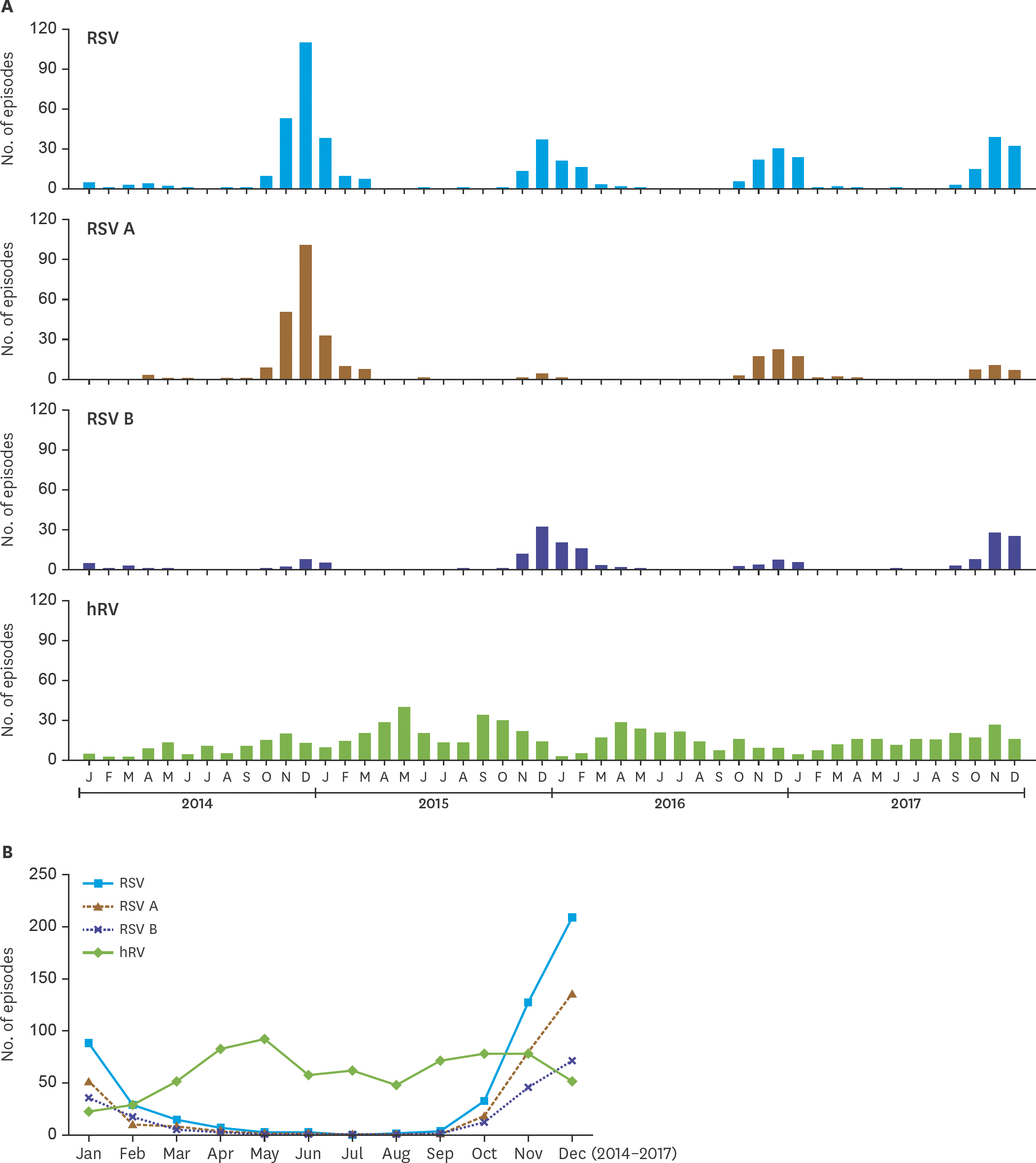
Fig. 1.
Monthly incidence of total RSV, RSV A, RSV B and hRV Infections from 2014 to 2017. Abbreviations: RSV, respiratory syncytial virus; hRV, human rhinovirus.
piv-26-99f1.tif
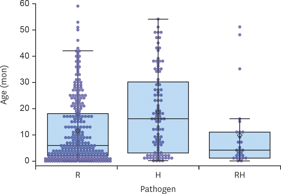
Fig. 2.
Age distribution in R, H, and RH group. Abbreviations: R, single infection of respiratory syncytial virus; H, single infection of human rhinovirus; RH, dual infection of respiratory syncytial virus and human rhinovirus.
piv-26-99f2.tif
Table 1.
Demographic findings of subjects
Variables R (n=258) H (n=99) RH (n=27) P-value Adjusted OR (95% CI)
Age (mon) 6 (0–59) 16 (0–54) 4 (0–51) <0.05  
        R<H (<0.05)  
        RH<H (<0.05)  
Sex (M:F) 1.4:1 1.3:1 2.4:1 0.41  
Past history          
  Chronic disease 12 (4.7) 6 (6.1) 2 (7.4) 0.64  
  Allergic disease 8 (3.1) 12 (12.1) 1 (3.7) <0.05  
          R<H (<0.05) 0.32 (0.12–0.84)
  Gestational age (<37 wk) 37 (14.3) 21 (21.2) 3 (11.1) 0.22  
  Birth weight (<2,500 g) 24 (9.3) 16 (16.2) 3 (11.1) 0.18  
Siblings 152 (59.1) 58 (58.6) 19 (70.4) 0.51  
Daycare center 62 (25.0) 33 (34.4) 5 (19.2) 0.14  

Values are presented as median (range) or number (%). Bold values are statistically significant.

Abbreviations: R, single infection of respiratory syncytial virus; H, single Infection of human rhinovirus; RH, dual infection of respiratory syncytial virus and human rhinovirus; OR, odds ratio; CI, confidence interval.

Post hoc analysis was performed by Dunn procedure, χ2 test and Fisher's exact test with Bonferroni correction;

Adjusted with age and sex by multivariable logistic regression;

Congenital heart diseases, chronic lung diseases, neurologic disorders.

Table 2.
Clinical findings of subjects
Variables R (n=258) H (n=99) RH (n=27) P-value Adjusted OR (95% CI)
Days of hospitalization 5 (2–12) 4 (2–15) 4 (3–13) 0.06  
LRTI 248 (96.1) 56 (56.6) 24 (88.9) <0.05  
        R>H (<0.05) 19.80 (9.19–42.68)
        RH>H (<0.05) 6.79 (1.87–24.70)
Signs of respiratory difficulty 55 (21.3) 11 (11.1) 9 (33.3) <0.05  
        RH>H (<0.05) 3.22 (1.14–9.10)
Fever 157 (60.9) 70 (70.7) 14 (51.9) 0.11  
Treatment
  Oxygen inhalation 14 (5.4) 4 (4.0) 2 (7.4) 0.76  
  Ventilator care 0 (0.0) 1 (1.0) 0 (0.0) 0.24  

Values are presented as median (range) or number (%). Bold values are statistically significant.

Abbreviations: R, single infection of respiratory syncytial virus; H, single infection of human rhinovirus; RH, dual infection of respiratory syncytial virus and human rhinovirus; OR, odds ratio; CI, confidence interval; LRTI, lower respiratory tract infection.

Post hoc analysis was performed by Dunn procedure, χ2 test and Fisher's exact test with Bonferroni correction;

Adjusted with age and sex by multivariable logistic regression.

Table 3.
Laboratory findings of subjects
Variables R (n=258) H (n=99) RH (n=27) P-value Adjusted β (SE)
WBC (×109/L) 9.2 (3.0–22.6) 11.4 (2.3–32.2) 9.6 (4.8–16.1) <0.05  
        R<H (<0.05) −2.53 (0.49)
PLT (×109/L) 349.5 (143.0–815.0) 385 (102.0–875.0) 415 (175.0–653.0) 0.13  
ANC (×109/L) 3.0 (0.4–17.9) 4.6 (0.5–29.1) 2.8 (0.9–9.6) <0.05  
        R<H (<0.05) −2.24 (0.37)
        RH<H (<0.05) −1.80 (0.66)
NEU (%) 34.4 (6.7–80.2) 47.9 (3.4–90.3) 33.1 (14.9–75.0) <0.05  
        R<H (<0.05) −7.59 (1.72)
LUC (%) 5.1 (1.7–12.9) 3.6 (0.9–8.4) 5.0 (1.9–7.0) <0.05  
        R>H (<0.05) 1.41 (0.20)
        RH>H (<0.05) 0.79 (0.36)
DNI (%) 0.0 (0.0–6.8) 0.0 (0.0–7.3) 0.0 (0.0–2.4) 0.05  
CRP (mg/dL) 0.4 (0.0–9.4) 0.7 (0.3–15.5) 0.3 (0.3–4.7) <0.05  
        R<H (<0.05) −0.65 (0.20)

Values are presented as median (range) or number (%). Bold values are statistically significant.

Abbreviations: R, single infection of respiratory syncytial virus; H, single infection of human rhinovirus; RH, dual infection of respiratory syncytial virus and human rhinovirus; β, regression coefficient; SE, standard error; WBC, white blood cell count; PLT, platelet count; ANC, absolute neutrophil count; NEU, neutrophil; LUC; large unstained cell; DNI, delta neutrophil index; CRP, C-reactive protein.

Post hoc analysis was performed by Dunn procedure with Bonferroni correction;

Adjusted with age and sex by multivariable linear regression;

Statistically significant (P<0.05).

Table 4.
Comparison of clinical and laboratory findings according to the age
Variables R (n=258) P-value H (n=99) P-value
<12 mon (n=163) ≥12 mon (n=95) <12 mon (n=40) ≥12 mon (n=59)
Days of hospitalization 5 (2–12) 4 (3–11) 0.08 5 (2–9) 4 (2–15) <0.05
LRTI 158 (96.9) 90 (94.7) 0.51 22 (55.0) 34 (57.6) 0.80
Respiratory difficulty 47 (28.8) 8 (8.4) <0.05 5 (12.5) 6 (10.2) 0.72
Fever 72 (44.2) 85 (89.5) <0.05 25 (62.5) 45 (76.3) 0.14
Severity            
  Oxygen inhalation 14 (8.6) 0 (0) <0.05 1 (2.5) 3 (5.1) 0.65
  Ventilator care 0 0   0 (0) 1 (1.7) 1.00
Siblings 107 (66.1) 45 (47.4) <0.05 29 (72.5) 29 (49.2) <0.05
Daycare center 9 (5.8) 53 (56.4) <0.05 0 (0) 33 (57.9) <0.05

Values are presented as median (range) or number (%). Bold values are statistically significant.

Abbreviations: R, single infection of respiratory syncytial virus; H, single infection of human rhinovirus; LRTI, lower respiratory tract infection.

TOOLS
Similar articles