

 PDF
PDF ePub
ePub Citation
 Print
Print


INTRODUCTION
Pathogens and the pathogenesis of healthcare-associated ventriculitis and meningitis (HAVM) differ from those in the community.1) Bacillus cereus, a gram-positive, spore-forming bacillus, causes serious central nervous system (CNS) infections that are challenging to manage because of difficulties with early detection and poor penetration of systemic antimicrobials.23) Direct antimicrobial injection into the cerebrospinal fluid (CSF) space may be an alternative option for intractable HAVM.1) We report on a patient with B. cereus HAVM treated with intraventricular (IVT) vancomycin who had not responded well to systemic antimicrobials.
CASE
A 7-year-old girl was referred to our center for newly diagnosed medulloblastoma. Two weeks before referral, she developed severe headache and drowsiness. Brain magnetic resonance imaging revealed a 3.5-cm-sized lobulated mass in the cerebellum with hydrocephalus of the lateral and third ventricles. She underwent endoscopic third ventriculostomy; however, her consciousness level continued to deteriorate. Thus, she underwent tumor resection surgery 2 days later. Pathologic biopsy revealed medulloblastoma; there was no evidence of metastasis to other sites. Fever (39.7°C) started on postoperative day 2; CSF examination on postoperative day 3 revealed white blood cell (WBC) counts of 10,240/mm3 (neutrophils 95%); glucose level, 40 mg/dL; and protein level, 242 mg/dL. The extraventricular drain (EVD) was immediately removed; empiric antimicrobial treatment, including vancomycin and gentamycin, was initiated. B. cereus was isolated from CSF, peripheral blood, and EVD tip cultures; it was sensitive to vancomycin, teicoplanin, clindamycin, levofloxacin, and trimethoprim/sulfamethoxazole and resistant to penicillin, oxacillin, and erythromycin. Treatment comprised intravenous vancomycin (15 mg/kg/dose every 6 hours; trough level, 7.6 µg/mL) and gentamycin (7.5 mg/kg/day); she was referred to our center on postoperative day 15 with persistent fever. On physical examination, she was alert with no sign of infection or inflammation at the surgical site. Regarding the laboratory work-up, complete peripheral blood counts showed the following: WBC count, 12,760/µL (normal, 6,000–15,000/µL); hemoglobin, 10.6 g/dL (normal, 10.5–14.0 g/dL); and platelets, 431 K/µL (normal, 150–450 K/µL). C-reactive protein (CRP) and procalcitonin levels were 0.05 mg/dL (normal, 0–0.3 mg/dL) and 0.11 ng/mL (normal, 0–0.49 ng/mL), respectively. CSF examination revealed the following: WBC counts, 23/mm3 (neutrophils 12%); glucose level, 33 mg/dL (serum level, 83 mg/dL); and protein level, 40 mg/dL. After admission, she was treated with intravenous vancomycin alone (22.6 mg/kg/dose every 6 hours; trough level, 14.3 µg/mL on hospitalization day [HD] 4) with a target trough level of 15 µg/mL. Following this, no fever >38.0°C was observed.
On HD9, fever developed again; cefepime was added as the empiric antimicrobial treatment for HAVM but there was no response (Fig. 1A). Further CSF examination revealed the following: WBC counts, 4/mm3 (neutrophils 16%); glucose level, 28 mg/dL (serum level, 78 mg/dL); and protein level, 47 mg/dL. No pathogens were isolated from the peripheral blood, CSF, or urine specimens. After confirming all negative culture results, antimicrobials were discontinued for 2 days (HD13–HD14). CSF was again examined on HD15 and revealed the following: WBC count 230/mm3 (neutrophils 56%); glucose level, 28 mg/dL (serum level, 95 mg/dL); and protein level, 42 mg/dL. Magnetic resonance imaging showed that the previously enhanced lesion at the EVD insertion site had decreased in size; however, an enhanced lesion in the parietal horn of the left lateral ventricle was newly observed in T1-enhanced images (Supplementary Fig. 1). However, no pathogens were isolated in subsequent CSF cultures. Suspecting treatment failure for B. cereus HAVM and HAVM due to other pathogens, broad spectrum antimicrobial treatment, including linezolid, meropenem, levofloxacin, and rifampin, was initiated. However, her fever persisted, and response of the CSF profile appeared to fluctuate (Fig. 1A). A decision was made to change linezolid to vancomycin with EVD insertion to monitor vancomycin concentration in the CSF. Consequently, her fever improved slightly; the CSF WBC count gradually improved from 115–440/mm3 to 40–50/mm3, but the response rate was very slow. The CSF vancomycin trough level was below the reference level (<3.0 µg/mL); the median serum vancomycin concentration was 14.1 µg/mL (range, 8.5–14.5 µg/mL). The CSF cytology identified previously unseen malignant cells; there was concern about delaying further anti-cancer therapy, especially proton therapy. Therefore, after confirming that the draining amount from the EVD was constant, IVT vancomycin was administered from HD34. The initial vancomycin dose was 5 mg, but the trough levels at 24 hours after infusion were often below the reference level. Therefore, we increased the dose to 6 mg from HD41. IVT vancomycin (5–6 mg/dose every 24 hours, clamped for 30 minutes) was administered with systemic antimicrobials for 48 days, followed by IVT only for 2 weeks. The median CSF volume from the drain was 289 mL/day (interquartile range [IQR], 234–326 mL/day; range, 148–336 mL/day); the median trough level of 61 CSF samples was 5.5 µg/mL (IQR, 4.25–6.4 µg/mL; range, 3–9.7 µg/mL). Defervescence occurred on day 6 (HD39) of IVT treatment; normalization of CSF parameters was achieved on day 18 (HD51). During EVD change on HD63, there was only one fever episode, which resolved spontaneously without treatment (Fig. 1A). CSF vancomycin concentrations were monitored on IVT days 1 and 2; vancomycin concentrations at 30 minutes, 2 hours, 4 hours, 8 hours, 16 hours, and 24 hours after IVT administration were 110.7, 29.6, 16.8, 8.9, 4.8, and 4.1 µg/mL, respectively (Fig. 1B). She received proton therapy for 6 weeks starting on day 13 after IVT treatment (HD46). The only adverse event was an EVD obstruction on HD83; vancomycin clamping was maintained for 3.5 hours until the function was restored after EVD irrigation. CSF vancomycin concentration at the time of irrigation was 44.5 µg/mL. Other toxicities, including CSF eosinophilia, drug fever, and CNS toxicity, were not observed. Interestingly, until discharge from hospitalization, peripheral blood CRP levels remained within the normal range and never increased. She recovered well and is currently undergoing chemotherapy.
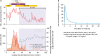 | Fig. 1Clinical course and CSF vancomycin pharmacokinetic data of our patient. (A) Clinical course, (B) CSF vancomycin levels during IVT infusion on days 1 and 2. The peak level was 110.7 µg/mL 1 hour after infusion with 5 mg vancomycin with 30-minute clamping. The triangle indicates the CSF vancomycin level; at time points of 2 hours, 8 hours, and 24 hours, we used an average CSF vancomycin level for plotting.Abbreviations: VAN, vancomycin; CFP, cefepime; Levo, levofloxacin; LZD, linezolid; MER, meropenem; RIF, rifampin; IVT, intraventricular; CSF, cerebrospinal fluid; WBC, white blood cell.
|
The patient's parents provided consent. The study was approved by the Institutional Review Board of the National Cancer Center (NCC 2018-0175).
DISCUSSION
To our knowledge, this is the first case of B. cereus HAVM treated using IVT vancomycin with CSF vancomycin pharmacokinetics data. A recently published guideline for HAVM included recommendations for IVT antimicrobial treatment, but most recommendations were based on studies concerning common pathogens of HAVM including methicillin-resistant Staphylococcus aureus, vancomycin-resistant Enterococcus, and multidrug-resistant gram-negative bacilli; evidence for B. cereus is scarce.1)
B. cereus causes serious infections with high mortality, especially in immunocompromised patients.4) Gaur et al.2) reported 12 pediatric cases of B. cereus bacteremia and meningitis. Among 4 patients with CNS disease (3 proven and 1 possible), two died and one had severe sequelae. They also reported a low CSF vancomycin concentration (2.2 µg/mL 9 hours post-injection) with a vancomycin dose of 15 mg/kg every 8 hours in one deceased patient. Stevens et al.5) reviewed cases of B. cereus CNS infections and reported 33 CNS infection cases (20 children) with a high crude mortality (66%) in 22 case reports. Conversely, our patient responded well to treatment without sequelae despite serious CNS infection, possibly due to the effect of IVT vancomycin therapy; however, we note that our patient was not severely immunocompromised and the infected EVD was removed immediately when infection was suspected.
She initially responded well to systemic antimicrobial treatment but became unresponsive 22 days after systemic treatment (HD10), possibly owing to changes in CNS penetration of vancomycin during treatment. At the time of first infection, the meninges might have been severely inflamed, resulting in a high CNS permeability to vancomycin. Subsequently, the meninges were no longer inflamed as the infection was controlled; therefore, the permeability to vancomycin might have been decreased. In a review of CNS penetration of antimicrobials, vancomycin permeability in inflamed meninges was 0.3 (serum/CSF)—approximately 2 times higher than that of uninflamed meninges (0.14–0.18; serum/CSF).3) For our patient, serum CRP was very high at the time of first infection (15.8 mg/dL; normal, 0–0.5 mg/dL), but during hospitalization in our center, peripheral blood CRP levels remained within the normal range; thus, CNS permeability to vancomycin may have been low because of uninflamed meninges.
There is no specific data suggesting an exact optimal dose of IVT vancomycin. However, based on limited data, a recent guideline suggested that IVT doses of 5–20 mg vancomycin may be given every 1 to 3 days in adults, taking into account the amount of EVD and the size of the ventricles.1) In our patient, we aimed to maintain a trough level at approximately 5–10 µg/mL to exceed an inhibitory quotient of 10–20 under the assumption that B. cereus in our patient was susceptible to vancomycin (minimal inhibitory concentration <0.5 mg/dL). Having collected detailed pharmacokinetic data regarding CSF vancomycin levels for only the first 2 days of IVT treatment was a limitation of the study. However, due to issues concerning contamination and infection, it was difficult to continue frequent sampling.
To our knowledge, this was the first study on the successful treatment with IVT vancomycin in combination with systemic antimicrobial treatment in a patient with B. cereus HAVM, which is known for its high morbidity and mortality. We also reported CSF pharmacokinetic data for vancomycin in the pediatric population. In conclusion, IVT vancomycin with systemic antimicrobials may be considered for patients with intractable B. cereus ventriculitis.
 XML Download