

 PDF
PDF ePub
ePub Citation
 Print
Print


Abstract
Purpose:
The purpose of this study was to document the sonographic morphology of the subscapularis footprint, particularly the 1st facet, of the non-pathologic subscapularis tendon and footprint, and analyze the correlation between the size of the 1st facet and the demographic variables.
Materials and Methods:
Between March 2015 and December 2017, retrospectively data analysis was performed for the ultrasound (US) scans of 115 consecutive shoulder (mean age 53.4 years, range 23–74 years) with non-pathologic subscapularis tendon and footprint. The sonographic findings of the 1st facet of the subscapularis footprint was a very unique, flat, broad, and plane angle in the upward direction, which were distinguished from the other facets. On US, the transverse (medio-lateral) and longitudinal (superior-inferior) length of the 1st facet on axis of the humerus shaft were recorded. The demographic variables, including age, site, body height, weight, body mass index (BMI), and arm length, were reviewed.
Results:
On US, the mean transverse length of the 1st facet was 12.75 mm (range 10.54–14.50 mm, standard deviation [SD] 0.712) and the mean longitudinal length was 12.22 mm (range 9.20–13.30 mm, SD 0.888). The transverse and longitudinal length of the size of the 1st facet were significantly greater in males than in females (p<0.001, p=0.001). Of the demographic data (body height, weight, BMI, arm length) that showed a significant positive linear correlation, the correlation with body height (transverse r=0.749, p<0.001; longitudinal r=0.642, p<0.001) showed the strongest relationship, and the correlation with the BMI was weakly related. The relationships between the size of the 1st facet to site/age were not statistically significant or appeared to have no linear correlation.
Go to : 
References
1. Dyrna F, Kumar NS, Obopilwe E. . Relationship between deltoid and rotator cuff muscles during dynamic shoulder abduction: a biomechanical study of rotator cuff tear progression. Am J Sports Med. 2018. 46:1919–26.

2. Gamulin A, Pizzolato G, Stern R, Hoffmeyer P. Anterior shoulder instability: histomorphometric study of the subscapularis and deltoid muscles. Clin Orthop Relat Res. 2002. 398:121–6.
3. Keating JF, Waterworth P, Shaw-Dunn J, Crossan J. The relative strengths of the rotator cuff muscles. A cadaver study. J Bone Joint Surg Br. 1993. 75:137–40.
4. Adams CR, Schoolfield JD, Burkhart SS. The results of arthroscopic subscapularis tendon repairs. Arthroscopy. 2008. 24:1381–9.
5. Flury MP, John M, Goldhahn J, Schwyzer HK, Simmen BR. Rupture of the subscapularis tendon (isolated or in combination with supraspinatus tear): when is a repair indicated? J Shoulder Elbow Surg. 2006. 15:659–64.
6. Jeong JY, Pan HL, Song SY, Lee SM, Yoo JC. Arthroscopic subscapularis repair using single-row mattress suture technique: clinical results and structural integrity. J Shoulder Elbow Surg. 2018. 27:711–9.
7. Kim SJ, Choi YR, Jung M, Lee W, Chun YM. Arthroscopic repair of anterosuperior massive rotator cuff tears: does repair integrity affect outcomes? Am J Sports Med. 2017. 45:1762–8.
8. Kim YK, Kim DW, Noh YT, Lee SB. Arthroscopic repair of combined rotator cuff tears involving the subscapularis tendon. J Korean Orthop Assoc. 2010. 45:392–8.
9. Warner JJ, Higgins L, Parsons IM 4th, Dowdy P. Diagnosis and treatment of anterosuperior rotator cuff tears. J Shoulder Elbow Surg. 2001. 10:37–46.
10. Denard PJ, Burkhart SS. Arthroscopic recognition and repair of the torn subscapularis tendon. Arthrosc Tech. 2013. 2:e373–9.
11. Gerhardt C, Bartl C, Voigt C. . Recovery of subscapularis and shoulder function following arthroscopic treatment of isolated anterior and combined anterosuperior rotator cuff lesions. Arch Orthop Trauma Surg. 2016. 136:75–81.
12. Lafosse L, Jost B, Reiland Y, Audebert S, Toussaint B, Gobezie R. Structural integrity and clinical outcomes after arthroscop-ic repair of isolated subscapularis tears. J Bone Joint Surg Am. 2007. 89:1184–93.
13. Lo IK, Burkhart SS. The comma sign: an arthroscopic guide to the torn subscapularis tendon. Arthroscopy. 2003. 19:334–7.
14. Adams CR, Schoolfield JD, Burkhart SS. Accuracy of preoperative magnetic resonance imaging in predicting a subscapularis tendon tear based on arthroscopy. Arthroscopy. 2010. 26:1427–33.
15. Alilet M, Behr J, Nueffer JP, Barbier-Brion B, Aubry S. Multi-modal imaging of the subscapularis muscle. Insights Imaging. 2016. 7:779–91.
16. Ryu HY, Song SY, Yoo JC, Yun JY, Yoon YC. Accuracy of sagittal oblique view in preoperative indirect magnetic resonance arthrography for diagnosis of tears involving the upper third of the subscapularis tendon. J Shoulder Elbow Surg. 2016. 25:1944–53.
17. Narasimhan R, Shamse K, Nash C, Dhingra D, Kennedy S. Prevalence of subscapularis tears and accuracy of shoulder ultrasound in pre-operative diagnosis. Int Orthop. 2016. 40:975–9.
18. Roy JS, Braën C, Leblond J. . Diagnostic accuracy of ultrasonography, MRI and MR arthrography in the characterisation of rotator cuff disorders: a systematic review and meta-analysis. Br J Sports Med. 2015. 49:1316–28.
19. Rutten MJ, Jager GJ, Kiemeney LA. Ultrasound detection of rotator cuff tears: observer agreement related to increasing experience. AJR Am J Roentgenol. 2010. 195:W440–6.
20. Teefey SA, Middleton WD, Payne WT, Yamaguchi K. Detection and measurement of rotator cuff tears with sonography: analysis of diagnostic errors. AJR Am J Roentgenol. 2005. 184:1768–73.
21. Yoo JC, Rhee YG, Shin SJ. . Subscapularis tendon tear classification based on 3-dimensional anatomic footprint: a cadaveric and prospective clinical observational study. Ar-throscopy. 2015. 31:19–28.
22. Clark JM, Harryman DT 2nd. Tendons, ligaments, and capsule of the rotator cuff. Gross and microscopic anatomy. J Bone Joint Surg Am. 1992. 74:713–25.
23. Klapper RC, Jobe FW, Matsuura P. The subscapularis muscle and its glenohumeral ligament-like bands. A histomorphologic study. Am J Sports Med. 1992. 20:307–10.
24. Arai R, Sugaya H, Mochizuki T, Nimura A, Moriishi J, Akita K. Subscapularis tendon tear: an anatomic and clinical investigation. Arthroscopy. 2008. 24:997–1004.
25. D’Addesi LL, Anbari A, Reish MW, Brahmabhatt S, Kelly JD. The subscapularis footprint: an anatomic study of the subscapularis tendon insertion. Arthroscopy. 2006. 22:937–40.
26. Ide J, Tokiyoshi A, Hirose J, Mizuta H. An anatomic study of the subscapularis insertion to the humerus: the subscapularis footprint. Arthroscopy. 2008. 24:749–53.
27. Richards DP, Burkhart SS, Tehrany AM, Wirth MA. The subscapularis footprint: an anatomic description of its insertion site. Arthroscopy. 2007. 23:251–4.
28. Halder A, Zobitz ME, Schultz E, An KN. Structural properties of the subscapularis tendon. J Orthop Res. 2000. 18:829–34.
29. Gausden EB, McCarthy MM, Kontaxis A, Corpus KT, Gulot-ta LV, Kelly AM. Subscapularis tendon loading during activities of daily living. J Shoulder Elbow Surg. 2017. 26:331–6.
30. Omi R, Sano H, Ohnuma M. . Function of the shoulder muscles during arm elevation: an assessment using positron emission tomography. J Anat. 2010. 216:643–9.
31. Jang HS, Kong DH, Jang SH. Correlation between subscapularis tears and the outcomes of physical tests and isokinetic muscle strength tests. Clin Should Elbow. 2016. 19:90–5.
Go to :
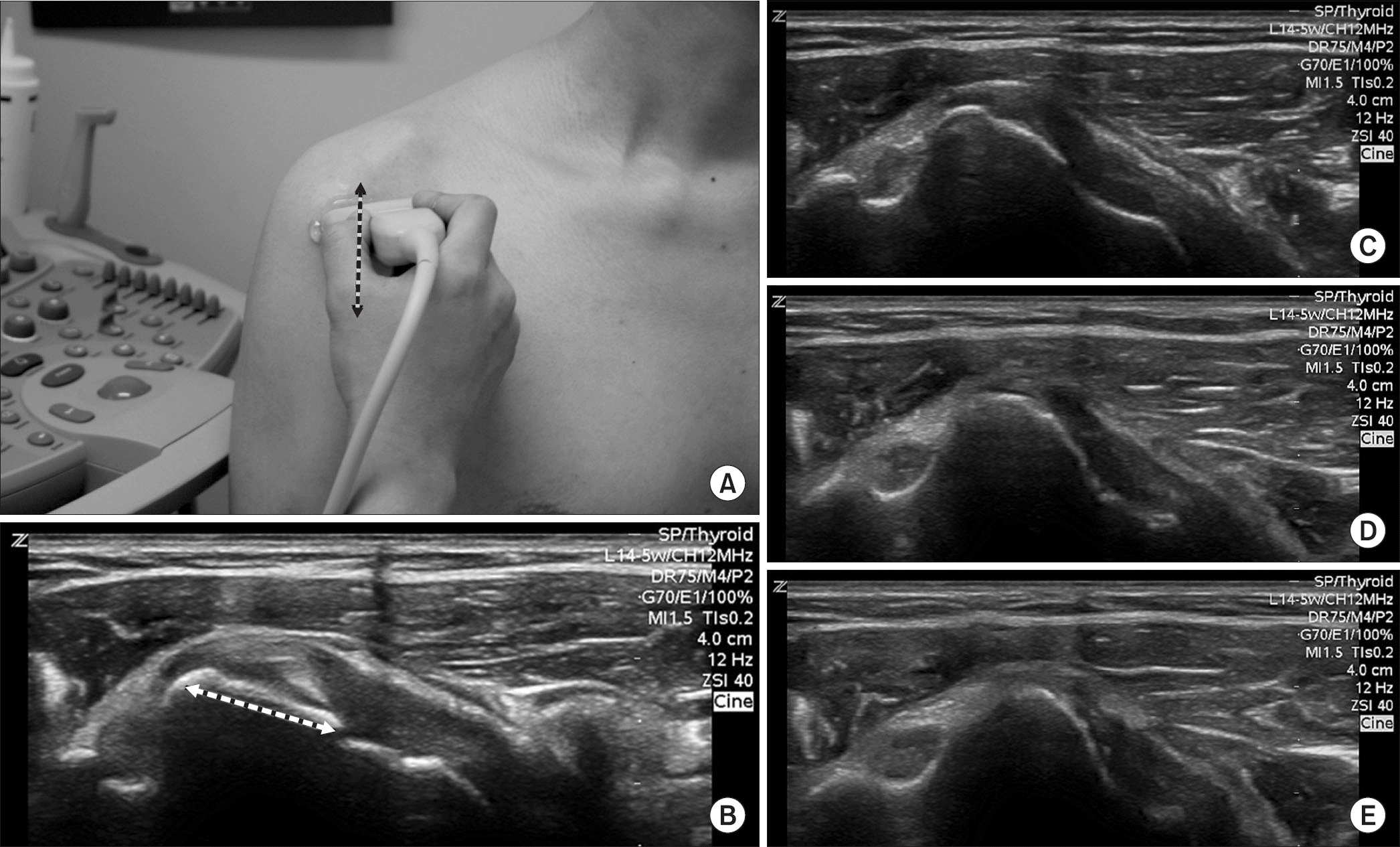 | Figure 1.(A) Transverse scans of the subscapularis footprint (same as longitudinal scan of subscapularis tendon) tracing from superior to inferior (B-E) on the axis of humerus shaft. The sonographic morphology of the 1st facet of subscapularis footprint was unique with its flat and broad shape, which were distinguished from the other facets. The longest length was measured on a transverse scan of the 1st facet (transverse or medio-lateral length). |
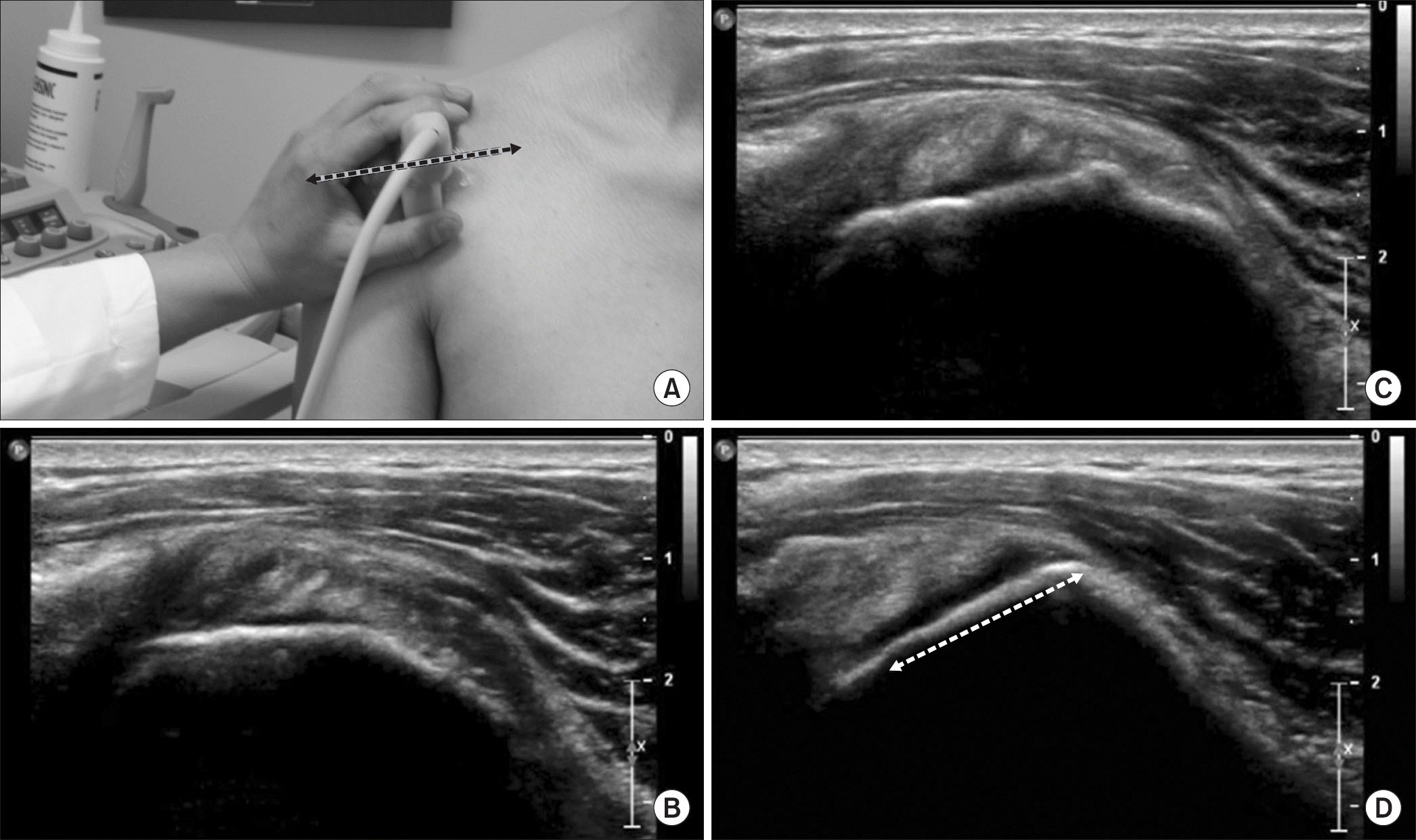 | Figure 2.(A) Longitudinal scans of the subscapularis footprint (same as transverse scan of subscapularis tendon) tracing from medial to lateral (B-D) on axis of humerus shaft. The shapes of the tendinous portion of the subscapularis have undergone gradual echogenic and morphologic changes. On the medial portion (A, B), the corresponding ultrasound images of the subscapularis tendon revealed its multipennate appearance showing areas of hyper- and hypo-echogenicity. The echogenicity of the subscapularis tendon became denser and more hyperechoic as the tendon was closer to the 1st facet of the subscapularis footprint. The 1st facet of the subscapularis footprint also has a plane angle in the upward direction, which was distinguished from other facets with the plane angle in the anterior direction. |
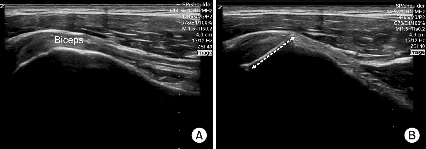 | Figure 3.Longitudinal scans of subscapularis 1st facet tracing from longitudinal scan of biceps long the head tendon (A) to just medial on the axis of humerus shaft (B). |
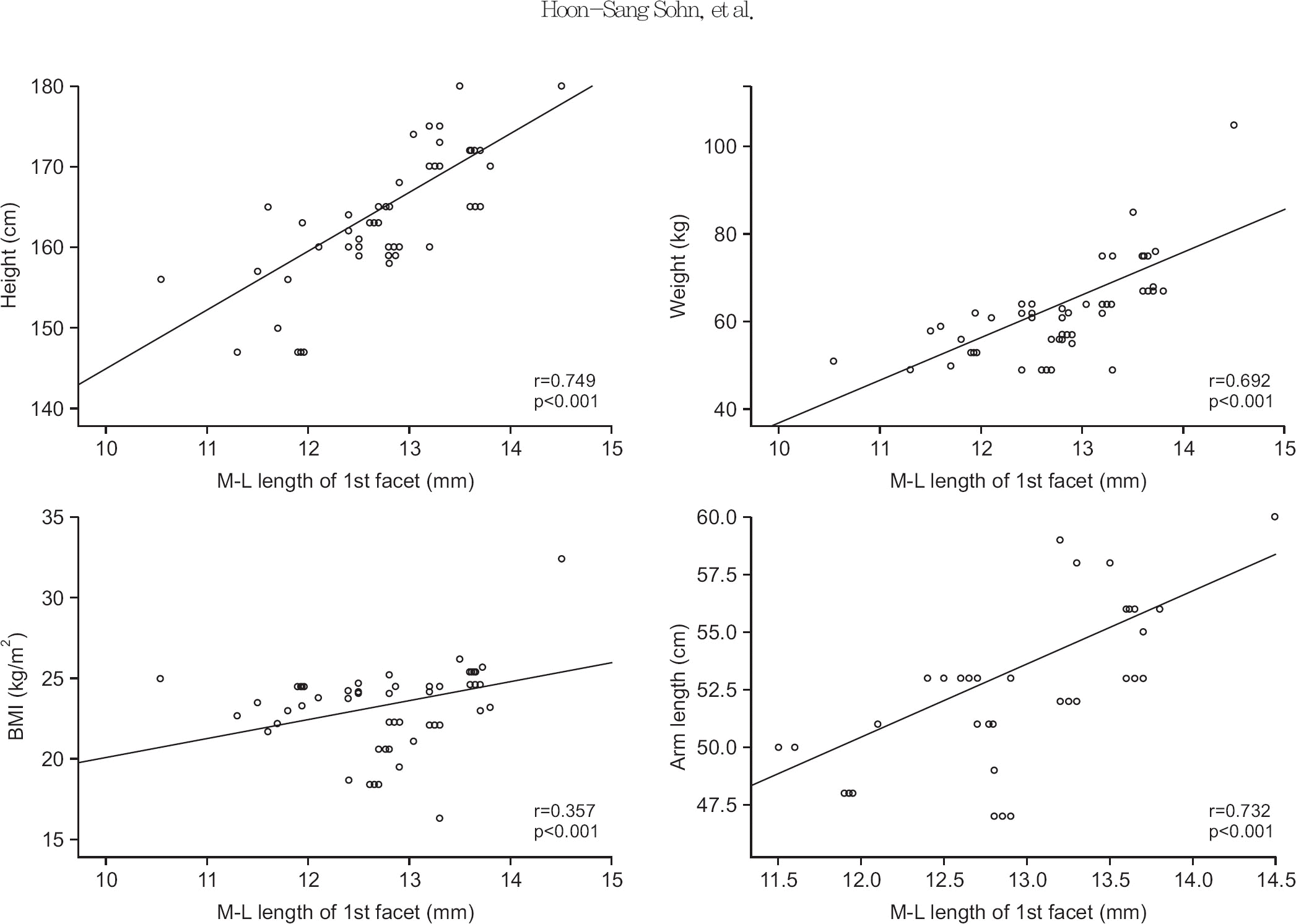 | Figure 4.Correlation analysis between the demographic data and the medio-lateral (M-L) length of the subscapularis 1st facet on ultrasound. r, Pearson correlation coefficient; BMI, body mass index. |
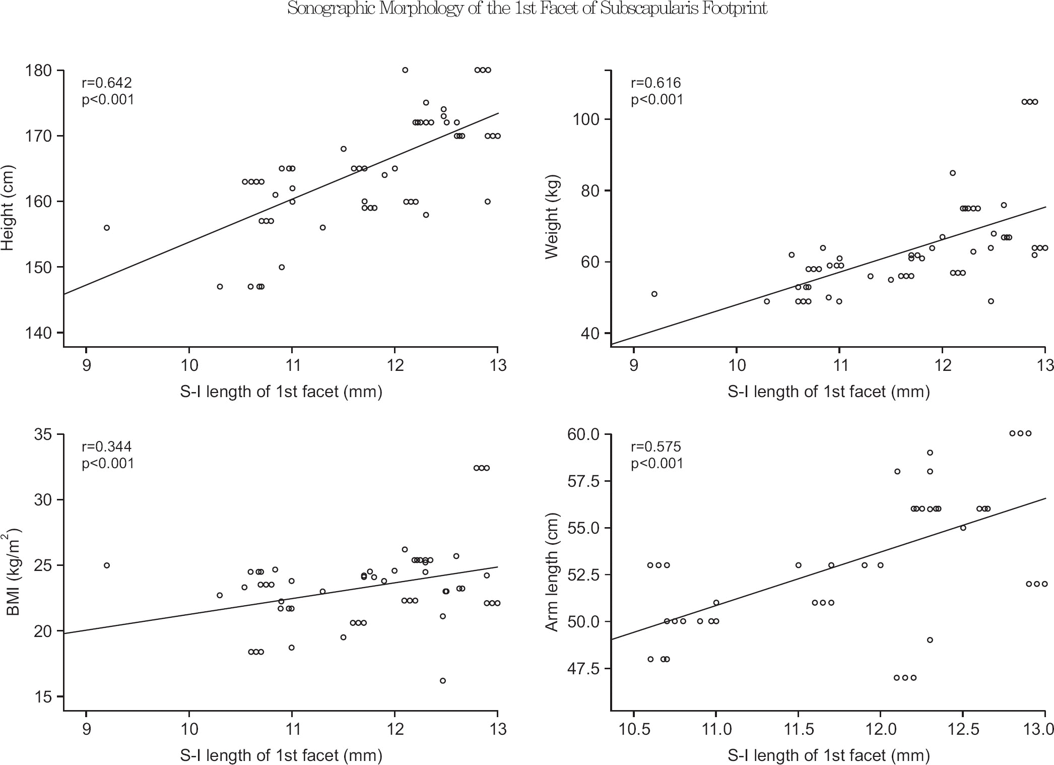 | Figure 5.Correlation analysis between the demographic data and the superior-inferior (S-I) length of the subscapularis 1st facet on ultrasound. r, Pearson correlation coefficient; BMI, body mass index. |
Table 1.
Patient Demography (n=115)
| Variable | Value |
|---|---|
| Age (yr) | 53.4±13.26 (23–74) |
| Sex (male:female) | 71:44 |
| Site (right:left) | 68:47 |
| Height (cm) | 165.50±7.69 (147–180) |
| Weight (kg) | 64.31±11.14 (49–105) |
| Body mass index (kg/m2) | 23.29±2.60 (16.3–32.4) |
| Arm length (cm)∗ | 53.27±3.31 (47–60) |
 XML Download