

 PDF
PDF ePub
ePub Citation
 Print
Print


Abstract
Purpose
To report a case of choroidal neovascularization in a Best disease patient treated with intravitreal bevacizumab injection and followed up with optical coherence tomography angiography (OCTA).
Case summary
A 20-year-old female visited our clinic with decreased visual acuity of the left eye for 6 months. On optical coherence tomography (OCT), subretinal fluid and hyperreflective subretinal clumps were observed in the macula of the right eye. Subretinal hemorrhage and subretinal fluid were observed in the left eye. Choroidal neovascularization in the left eye was observed using OCTA, fluorescein angiography, and indocyanine green angiography. A full-field electroretinogram was normal in both eyes, but an electrooculogram revealed that the Arden ratio was 1.564 in the right eye and 1.081 in the left eye. Intravitreal bevacizumab injection was performed in the left eye. At 6 months after the intravitreal injection, the best-corrected visual acuity of the left eye had recovered to 20/20. OCT revealed that subretinal fluid reduced and choroidal neovascularization was stable. After 12 months, visual acuity of the left eye was maintained at 20/20, but OCTA revealed that choroidal neovascularization had increased.
Figures and Tables
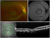 | Figure 1Multimodal imaging findings of the right eye. (A) Fundus photography shows a pale yellowish lesion surrounding the foveola and dark brown spot on the parafovea. (B) Fundus autofluorescence shows ring-shaped hyperautofluorescence around the fovea and hypoautofluorescent spot in the inferior macula. (C) Optical coherence tomography shows subretinal fluid and hyper-reflective subretinal clumps, and retinal pigment epithelium hyperplasia at the dark brown spot.
|
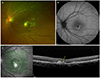 | Figure 2Multimodal imaging findings of the left eye. (A) Fundus photography shows juxtafoveal retinal hemorrhage, whitish subfoveal fibrous membrane and yellowish white-colored spots around fovea. (B) Fundus autofluorescence shows hyperautofluorescent ring around the hypoautofluorescence at the fovea. (C) Optical coherence tomography shows subretinal fluid, choroidal neovascular membrane and subretinal hyperreflective material due to subretinal hemorrhage (yellow arrow).
|
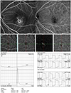 | Figure 3Multimodal imaging findings of the left eye. (A) Fluorescein angiography and indocyanine green angiography of the left eye shows choroidal neovascularization. (B) Optical coherence tomography angiography shows lacy choroidal neovascularization in the outer retina and choriocapillaris slab. (C) Electrooculography shows that the Arden ratio is 1.564 in the right eye and 1.081 in the left eye.
|
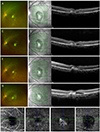 | Figure 4Multimodal imaging findings of the left eye. Fundus photography of left eye at 1 month (A), at 3 months (C), at 6 months (E), and at 12 months (G) after intravitreal bevacizumab injection show disappeared juxtafoveal retinal hemorrhage and whitish fibrotic scar. Optical coherence tomography at 1 month (B) and at 3 months (D) show that the subretinal fluid and choroidal neovascularization membrane decreased. (F) Optical coherence tomography at 6 months shows more decreased subretinal fluid and stable choroidal neovascular membrane. (H) Optical coherence tomography at 12 months shows increased subretinal fluid and choroidal neovascular membrane. (I) Optical coherence tomography angiography at 12 months shows fan shaped choroidal neovascularization in the outer retina slab. Choroidal neovascularization is also seen in the choriocapillaris slab.
|
References
1. Marmorstein AD, Marmorstein LY, Rayborn M, et al. Bestrophin, the product of the Best vitelliform macular dystrophy gene (VMD2), localizes to the basolateral plasma membrane of the retinal pigment epithelium. Proc Natl Acad Sci U S A. 2000; 97:12758–12763.

2. Deutman AF, Hoyng CB, van Lith-Verhoeven JJC. Macular dystrophies. In : Ryan SJ, Schachat AP, Wilkinson C, editors. Retina. 4th ed. Baltimore: Elsevier Mosby;2006. v. 2:chap. 63.
3. Shahzad R, Siddiqui MA. Choroidal neovascularization secondary to Best vitelliform macular dystrophy detected by optical coherence tomography angiography. J AAPOS. 2017; 21:68–70.
4. Patel RC, Gao SS, Zhang M, et al. Optical coherence tomography angiography of choroidal neovascularization in four inherited retinal dystrophies. Retina. 2016; 36:2339–2347.
5. Stattin M, Ahmed D, Glittenberg C, et al. Optical coherence tomography angiography for the detection of secondary choroidal neovascularization in vitelliform macular dystrophy. Retin Cases Brief Rep. 2017; 08. 16. DOI: 10.1097/ICB.0000000000000626.
6. Guduru A, Gupta A, Tyagi M, et al. Optical coherence tomography angiography characterisation of Best disease and associated choroidal neovascularisation. Br J Ophthalmol. 2018; 102:444–447.
7. Wang XN, You QS, Li Q, et al. Findings of optical coherence tomography angiography in Best vitelliform macular dystrophy. Ophthalmic Res. 2018; 60:214–220.
8. Andrade RE, Farah ME, Cardillo JA, et al. Optical coherence tomography in choroidal neovascular membrane associated with Best's vitelliform dystrophy. Acta Ophthalmol Scand. 2002; 80:216–218.
9. Sodi A, Murro V, Caporossi O, et al. Long-term results of photodynamic therapy for choroidal neovascularization in pediatric patients with Best vitelliform macular dystrophy. Ophthalmic Genet. 2015; 36:168–174.
10. Leu J, Schrage NF, Degenring RF. Choroidal neovascularisation secondary to Best's disease in a 13-year-old boy treated by intravitreal bevacizumab. Graefes Arch Clin Exp Ophthalmol. 2007; 245:1723–1725.
11. Heidary F, Hitam WH, Ngah NF, et al. Intravitreal ranibizumab for choroidal neovascularization in Best's vitelliform macular dystrophy in a 6-year-old boy. J Pediatr Ophthalmol Strabismus. 2011; 48:e19–e22.
12. Ruiz-Moreno O, Calvo P, Ferrández B, Torrón C. Long-term outcomes of intravitreal ranibizumab for choroidal neovascularization secondary to Best's disease: 3-year follow-up. Acta Ophthalmol. 2012; 90:e574–e575.
13. Khan KN, Mahroo OA, Islam F, et al. Functional and anatomical outcomes of choroidal neovascularization complicating BEST1-related retinopathy. Retina. 2017; 37:1360–1370.
14. O'Gorman S, Flaherty WA, Fishman GA, Berson EL. Histopathologic findings in Best's vitelliform macular dystrophy. Arch Ophthalmol. 1988; 106:1261–1268.
15. Kim HK, Rho CR, Lee WK, Kim KS. Optical coherence tomography findings in Best disease. J Korean Ophthalmol Soc. 2008; 49:845–852.
 XML Download