

 PDF
PDF ePub
ePub Citation
 Print
Print


Abstract
Purpose
We report a case of infectious keratitis caused by Shewanella putrefaciens in a patient after fishing.
Case summary
A 75-year-old male with no underlying disease other than hypertension was admitted to our hospital because of decreased visual acuity and congestion in his left eye for 2 weeks. At the first ophthalmic examination, the best-corrected visual acuity (BCVA) of the left eye was counting fingers. Slit lamp examination showed stromal infiltrates with 2.0 × 2.0 mm corneal epithelial defects, endothelial inflammatory plaques and 1 mm height hypopyon with severe inflammation in the anterior chamber. Bacterial culture tests were performed by corneal scraping, which were positive for Shewanella putrefaciens, followed by treatment with moxifloxacin and ceftazidime topical antibiotics. After 2 months of treatment, the BCVA of the left eye improved to 0.4 and the corneal lesion clinically improved with residual mild stromal opacity.
Figures and Tables
Figure 1
Slit lamp photograph and anterior segment optical coherence tomography at the first ophthalmic examination. (A) Slit lamp photograph at the first ophthalmic examination showing infectious keratitis with central epithelial defects, stromal infiltrates, perilesional stromal edema and severe chamber reaction with linear hypopyon. (B) Anterior segment optical coherence tomography showing central stromal infiltrates, endothelial inflammatory plaques and edema.
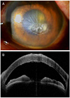
Figure 2
Bacterial culture tests by corneal scraping. On blood agar plates, the Shewanella putrefaciens colonies are convex and large, with a brown pigment.
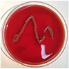
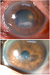
Figure 3
Slit lamp photographs at 1 week and 3 weeks after antibacterial treatments. (A) One week after antibacterial treatments, epithelial defects size, stromal infiltration and hypopyon decreased. (B) Three weeks of antibacterial treatments, the corneal lesions healed with a remaining stromal opacity.
References
1. Janda JM. Shewanella: a marine pathogen as an emerging cause of human disease. Clin Microbiol Newsl. 2014; 36:25–29.

2. Vignier N, Barreau M, Olive C, et al. Human infection with Shewanella putrefaciens and S. algae: report of 16 cases in Martinique and review of the literature. Am J Trop Med Hyg. 2013; 89:151–156.
3. To KK, Wong SS, Cheng VC, et al. Epidemiology and clinical features of Shewanella infection over an eight-year period. Scand J Infect Dis. 2010; 42:757–762.
4. Tsai MS, You HL, Tang YF, Liu JW. Shewanella soft tissue infection: case report and literature review. Int J Infect Dis. 2008; 12:e119–e124.
5. Mohan N, Sharma S, Padhi TR, et al. Traumatic endophthalmitis caused by Shewanella putrefaciens associated with an open globe fishhook injury. Eye (Lond). 2014; 28:235.
6. Park HJ, Tuli SS, Downer DM, et al. Shewanella putrefaciens keratitis in the lamellar bed 6 years after LASIK. J Refract Surg. 2007; 23:830–832.
7. Holt HM, Søgaard P, Gahrn-Hansen B. Ear infections with Shewanella alga: a bacteriologic, clinical and epidemiologic study of 67 cases. Clin Microbiol Infect. 1997; 3:329–334.
8. Holt HM, Gahrn-Hansen B, Bruun B. Shewanella alga and Shewanella putrefaciens: clinical and microbiological characteristics. Clin Microbiol Infect. 2005; 11:347–352.
9. Khashe S, Janda JM. Biochemical and pathogenic properties of Shewanella alga and Shewanella putrefaciens. J Clin Microbiol. 1998; 36:783–787.
10. Oh SY, Lee SJ, Park JM. A case of endophthalmitis caused by Shewanella algae after trauma. J Korean Ophthalmol Soc. 2013; 54:365–369.
11. Bravenec CA, Pandit RT, Beaver HA. Shewanella algae keratitis. Indian J Ophthalmol. 2019; 67:148–150.
12. Holt HM, Gahrn-Hansen B, Bruun B. Shewanella species: infections in Denmark and phenotypic characterisation. Clin Microbiol Infect. 2004; 10:348–349.
13. Héritier C, Poirel L, Nordmann P. Genetic and biochemical characterization of a chromosome-encoded carbapenem-hydrolyzing ambler class D beta-lactamase from Shewanella algae. Antimicrob Agents Chemother. 2004; 48:1670–1675.
14. Willcox MD. Pseudomonas aeruginosa infection and inflammation during contact lens wear: a review. Optom Vis Sci. 2007; 84:273–278.
15. Massey EL, Weston BC. Vibrio vulnificus corneal ulcer: rapid resolution of a virulent pathogen. Cornea. 2000; 19:108–109.
 XML Download