

 PDF
PDF ePub
ePub Citation
 Print
Print


Abstract
Purpose
To determine changes in subfoveal choroidal thickness (SCT) after intravitreal injection of bevacizumab in eyes with macular edema secondary to retinal vein occlusion (RVO).
Methods
Forty-four patients treated with intravitreal bevacizumab for unilateral macular edema due to RVO were retrospectively reviewed. Before injection, patients underwent best-corrected visual acuity (BCVA) assessment, dilated fundus examination, fluorescein angiography, and enhanced depth imaging optical coherence tomography. Changes in BCVA, SCT, and central macular thickness (CMT) of the RVO eyes were evaluated and compared with those of the normal contralateral eyes at baseline and at 1, 3, and 6 months after injection.
Results
The mean SCT in RVO eyes (265.41 ± 43.02 µm) was significantly thicker than that in the fellow eyes (244.77 ± 30.35 µm) at baseline (p < 0.001). The mean SCT was significantly reduced at 1, 3, and 6 months after intravitreal bevacizumab injection (all p < 0.001), and the change in SCT was significantly correlated with the change in CMT (r = 0.327, p = 0.030). While there was an improvement in BCVA together with a reduction in SCT (p < 0.001), no significant correlation was found (p = 0.126).
Figures and Tables
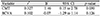
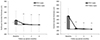 | Figure 1Changes and comparisons of the subfoveal choroidal thickness and the central macular thickness between RVO eyes and normal fellow eyes. Vertical lines indicate 1 standard error of the means. RVO = retinal vein occlusion. *p < 0.05 for the difference between the two groups. **p < 0.05 for the change from the baseline.
|
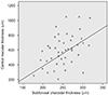 | Figure 2Scatter plot shows correlation between subfoveal choroidal thickness and central macular thickness. At baseline, a significant correlation between the two is shown in eyes with macular edema due to retinal vein occlusion (*r = 0.468, p = 0.049). *Pearson's correlation coefficient.
|
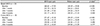
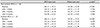
Table 2
Changes in SCT, CMT, and BCVA after intravitreal bevacizumab
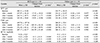
Continuous variables are presented as mean ± SD.
SCT = subfoveal choroidal thickness; CMT = central macular thickness; BCVA = best-corrected visual acuity; RVO = retinal vein occlusion; logMAR = the logarithm of the minimum angle of resolution; SD = standard deviation.
*p for the change from the baseline; †p for the difference between RVO eyes and the fellow eyes.
![]()
References
1. Klein R, Klein BE, Moss SE, Meuer SM. The epidemiology of retinal vein occlusion: the Beaver Dam Eye Study. Trans Am Ophthalmol Soc. 2000; 98:133–141. discussion 141-3.
2. Rogers S, McIntosh RL, Cheung N, et al. The prevalence of retinal vein occlusion: pooled data from population studies from the United States, Europe, Asia, and Australia. Ophthalmology. 2010; 117:313–319.e1.

3. Ehlers JP, Fekrat S. Retinal vein occlusion: beyond the acute event. Surv Ophthalmol. 2011; 56:281–299.
4. Haller JA, Bandello F, Belfort R Jr, et al. Dexamethasone intravitreal implant in patients with macular edema related to branch or central retinal vein occlusion twelve-month study results. Ophthalmology. 2011; 118:2453–2460.
5. Moon J, Kim M, Sagong M. Combination therapy of intravitreal bevacizumab with single simultaneous posterior subtenon triamcinolone acetonide for macular edema due to branch retinal vein occlusion. Eye (Lond). 2016; 30:1084–1090.
6. Imamura Y, Fujiwara T, Margolis R, Spaide RF. Enhanced depth imaging optical coherence tomography of the choroid in central serous chorioretinopathy. Retina. 2009; 29:1469–1473.
7. Maruko I, Iida T, Sugano Y, et al. Subfoveal choroidal thickness after treatment of Vogt-Koyanagi-Harada disease. Retina. 2011; 31:510–517.
8. Maruko I, Iida T, Sugano Y, et al. Subfoveal retinal and choroidal thickness after verteporfin photodynamic therapy for polypoidal choroidal vasculopathy. Am J Ophthalmol. 2011; 151:594–603.e1.
9. Regatieri CV, Branchini L, Carmody J, et al. Choroidal thickness in patients with diabetic retinopathy analyzed by spectral-domain optical coherence tomography. Retina. 2012; 32:563–568.
10. Du KF, Xu L, Shao L, et al. Subfoveal choroidal thickness in retinal vein occlusion. Ophthalmology. 2013; 120:2749–2750.
11. Tsuiki E, Suzuma K, Ueki R, et al. Enhanced depth imaging optical coherence tomography of the choroid in central retinal vein occlusion. Am J Ophthalmol. 2013; 156:543–547.e1.
12. Esen E, Sizmaz S, Demircan N. Choroidal thickness changes after intravitreal dexamethasone implant injection for the treatment of macular edema due to retinal vein occlusion. Retina. 2016; 36:2297–2303.
13. Willoughby AS, Vuong VS, Cunefare D, et al. Choroidal changes after suprachoroidal injection of triamcinolone acetonide in eyes with macular edema secondary to retinal vein occlusion. Am J Ophthalmol. 2018; 186:144–151.
14. Yumusak E, Ornek K, Dikel NH. Comparison of choroidal thickness changes following intravitreal dexamethasone, ranibizumab, and triamcinolone in eyes with retinal vein occlusion. Eur J Ophthalmol. 2016; 26:627–632.
15. Hidayat AA, Fine BS. Diabetic choroidopathy. Light and electron microscopic observations of seven cases. Ophthalmology. 1985; 92:512–522.
16. Langham ME, Grebe R, Hopkins S, et al. Choroidal blood flow in diabetic retinopathy. Exp Eye Res. 1991; 52:167–173.
17. Rayess N, Rahimy E, Ying GS, et al. Baseline choroidal thickness as a predictor for response to anti-vascular endothelial growth factor therapy in diabetic macular edema. Am J Ophthalmol. 2015; 159:85–91. 91.e1–91.e3.
18. Lee EK, Han JM, Hyon JY, Yu HG. Changes in choroidal thickness after intravitreal dexamethasone implant injection in retinal vein occlusion. Br J Ophthalmol. 2015; 99:1543–1549.
19. Kim KH, Lee DH, Lee JJ, et al. Regional choroidal thickness changes in branch retinal vein occlusion with macular edema. Ophthalmologica. 2015; 234:109–118.
20. Noma H, Mimura T, Eguchi S. Association of inflammatory factors with macular edema in branch retinal vein occlusion. JAMA Ophthalmol. 2013; 131:160–165.
21. Aiello LP, Northrup JM, Keyt BA, et al. Hypoxic regulation of vascular endothelial growth factor in retinal cells. Arch Ophthalmol. 1995; 113:1538–1544.
22. Tilton RG, Chang KC, LeJeune WS, et al. Role for nitric oxide in the hyperpermeability and hemodynamic changes induced by intravenous VEGF. Invest Ophthalmol Vis Sci. 1999; 40:689–696.
23. Chung YK, Shin JA, Park YH. Choroidal volume in branch retinal vein occlusion before and after intravitreal anti-VEGF injection. Retina. 2015; 35:1234–1239.
24. Park J, Lee S, Son Y. Effects of two different doses of intravitreal bevacizumab on subfoveal choroidal thickness and retinal vessel diameter in branch retinal vein occlusion. Int J Ophthalmol. 2016; 9:999–1005.
25. Rayess N, Rahimy E, Ying GS, et al. Baseline choroidal thickness as a predictor for treatment outcomes in central retinal vein occlusion. Am J Ophthalmol. 2016; 171:47–52.
26. Heiduschka P, Fietz H, Hofmeister S, et al. Penetration of bevacizumab through the retina after intravitreal injection in the monkey. Invest Ophthalmol Vis Sci. 2007; 48:2814–2823.
 XML Download