

 PDF
PDF ePub
ePub Citation
 Print
Print


INTRODUCTION
The skin is the largest organ in mammals. Skin has many important roles, including protection, maintenance of homeostasis, thermoregulation, vitamin D synthesis, and sensory perception. Damage to the skin by trauma or surgery activates a series of interdependent and sequential physiological events involving inflammation, cell proliferation, matrix deposition, and remodeling for tissue repair. Excessive collagen production and deposition during wound healing cause abnormal dermal proliferation and the formation of hypertrophic scars. Hypertrophic scars often present with redness and itching as firm, rubbery lesions or shiny, fibrous nodules on the surface of the skin.12
Various mechanisms have been proposed to explain the pathogenesis of scars.3 However, treatment is difficult due in part to the lack of an accurate, practical, reproducible, and economical animal model with which to systematically study scar remodeling. Animal models are important for studying the complex interactions that occur in living tissue during wound healing without the limitations of in vitro techniques. Currently, many researchers use rodent models to study wound healing due to low costs, small animal size, and relative ease of handling and care. However, hypertrophic scars do not develop in these species. In addition, the dermal composition of rodents does not resemble that of humans. In contrast, porcine skin is a good model for studying human dermal repair due to a number of similarities with human skin. Porcine and human skin has remarkably similar anatomies and physiologies, similar thicknesses of dermal and epidermal layers, and the presence of similar dermal appendages.4567 In addition, juvenile domestic pigs can be used instead of adult swine to address difficult handling and housing issues. Although juvenile animals tend to heal quicker than their older counterparts, the healing process takes a few months in juvenile pigs. Thus, sufficient time is available to assess the physiology and cellular biology of the healing process in a juvenile porcine model.8
The 6-kDa polypeptide hormone relaxin (RLX) is a member of the insulin family of peptides. RLX is primarily produced by the ovaries and/or placenta during pregnancy in mammals. RLX has multiple pregnancy-related functions, including connective tissue remodeling in the reproductive tract and cervix during pregnancy and softening of the cervix and vagina at the time of delivery.9 Recent studies have demonstrated that RLX also has biological effects on the brain, heart, kidneys, lungs, and connective tissues independent of its roles in reproduction.101112 These findings suggest that RLX may have therapeutic applications for non-reproductive conditions, such as fibrosis. RLX may reduce fibrosis by suppressing collagen synthesis and increasing collagenase activity with subsequent collagen degradation.131415
During the past 20 years, the field of virus-mediated gene therapy has grown tremendously. Adenoviruses (Ads)-based vectors, in particular, have attracted attention as gene-delivery vehicles due to their high titer capability and transduction efficiency in both dividing and non-dividing cells.1617 In this study, we introduced a RLX-expressing Ad vector (dE1-RGD/LacZ/RLX) into scar tissue on the skin of a pig model. A previous investigation from our lab demonstrated that RLX-expressing Ad has potential therapeutic effects on keloid and hypertrophic scars due to the reversal of pathological fibrosis.181920 However, a replication-incompetent Ad that expressed relaxin (Ad-RLX) vector system was limited by immunogenicity, local inflammation, a short half-life, enzymatic inactivation, and transient effects, which resulted in short-term expression of RLX.21 To address this problem, we tested a matrix-based depot system with a biodegradable alginate gel, which is a natural polymer frequently used in biomedical applications. Alginate gel can be used as a depot system for sustained release of Ad and maintenance of Ad viral activity due to its biocompatible environment.2223 Thus, the use of alginate gel should ultimately increase therapeutic efficacy of RLX.
MATERIALS AND METHODS
Scar formation
The scar model was created with three Yorkshire pigs (XP Bio, Anseong, Korea; four months of age, 40 kg). Anesthesia was induced in each pig via intramuscular injection of Zoletil (5 mg/kg, Virbac, Carros, France) and Rompun (2 mg/kg, Bayer, Seoul, Korea). Hair was then completely removed from the backs and bellies of each pig. Inhalation anesthesia was achieved with isoflurane (IsoFlo, Abbott Laboratories, Abbott Park, IL, USA). After anesthetizing the pigs, 36 full-thickness skin defects (3×3 cm2) were created symmetrically on the back of each Yorkshire pig with 18 wounds on each side of the midline. Each wound reached the depth of the muscle fascia to mimic scar formation. The distance of each wound was maintained 5 cm in the horizontal direction and 3 cm in the vertical direction to control the interval of each scar (Supplementary Fig. 1, only online). Intravenous antibiotics and wound dressings with TegaDerm® (3M, St. Paul, MN, USA) were administered postoperatively for 5 days, after which open dressings were maintained. Scar tissue, which was distinct in appearance from that of the peripheral normal tissue, formed at 50 days after creating the initial skin defect (Supplementary Fig. 2, only online).7 The Animal Care and Experiment Committee of Yonsei University approved the experimental protocol (2011-0181).
Preparation of Ad and alginate gel
A replication-incompetent Ad that expressed relaxin (Ad-RLX) and control Ad (Ad-LacZ)18 was used in this study. The propagation, purification, and titration of Ad were performed as previously described.2425 An alginate (alginic acid sodium salt, Sigma, St. Louis, MO, USA) solution of 5 wt.% was prepared with 0.5 g of alginate and 0.09 g of NaCl (Sigma) in 10 mL of phosphate-buffered saline. The alginate solution was stirred for 24 h at room temperature and gelated at various concentrations of CaCl2 (Sigma), ranging from 20 to 200 mM, to determine the optimal conditions for Ad transduction.
Injection of Ad into the pig scar model
Wound epithelialization and scar formation occurred on postoperative day 50. Anesthesia was induced in each pig by intramuscular injection of Zoletil (5 mg/kg; Virbac) and Rompun (2 mg/kg; Bayer). Hair was then completely removed from the backs and bellies of each pig. Scars were divided into three groups (gel, gel/Ad-LacZ and gel/Ad-RLX). We injected 1 mL of alginate gel, the same amount of alginate gel encapsulating dE1-RGD/LacZ [5×10⁷ plaque forming unit (PFU)], or alginate gel encapsulating dE1-RGD/LacZ/RLX in each group respectively. Injections were performed with a 27-gauge needle and 1-mL syringe, and directly into the intradermal layers of the scar regions.
Therapeutic evaluation of scar area, color index, and pliability
The surface areas of scars were imaged with a digital camera. The images of the surface areas of the scars were measured with a ruler and compared to a 1-cm2 standard. Measurements were assessed with Image J software (National Institutes of Health, Bethesda, MD, USA). The color of the scar was quantitatively analyzed with a spectrophotometer (CM-700D; Konica Minolta, Inc., Tokyo, Japan). The color of each scar was examined with melanin (the degree of scar darkness) and erythema (the degree of scar redness) indices.26 Pliability was measured with a durometer (H1000 Mini-Dial, RexGauge Co., Buffalo Grove, IL, USA). Each value was measured more than three times, and the average value was calculated. All of the evaluations were performed every 10 days until the 50th day after virus injection by the same individual who was blinded to the experimental group. Scar area, color index, and pliability were measured by an independent blinded physician.
Immunofluorescence microscopy
Specimens were collected at 10 days after the administration of the Ad virus and were embedded in paraffin blocks. Paraffin-embedded tissues were sectioned, deparaffinized, hydrated, and treated with blocking buffer (20% normal goat serum, S-100, Vector Lab Inc., Burlingame, CA, USA) for one hour. After removing the blocking buffer, sections were immersed in primary antibody (anti-relaxin, Santa Cruz Biotechnology, Dallas, TX, USA) overnight at 4℃ and then incubated in secondary antibody (DyLight594, Vector Lab Inc.) at room temperature for 2 hours. The nucleus was stained with mounting solution containing DAPI. The degree of RLX expression in the tissue was evaluated by confocal microscopy (LSM700, Carl Zeiss, Oberkochen, Germany).
Arrangement of collagen fibers and mast cell count
Picrosirius red (Sigma-Aldrich Co., St. Louis, MO, USA) and toluidine blue (Sigma-Aldrich Co.) staining was performed on specimens 50 days after virus injection. Full thickness skin tissues, 1×1 cm, were obtained from the center areas of scars and embedded in paraffin: we obtained 5 µm sections. Using the stained tissues, the number of mast cells (toluidine blue staining) and the collagen arrangement (picrosirius red staining) were compared using optical microscopy (BX51, Olympus, Tokyo, Japan). The number of mast cells was counted in five different areas randomly chosen for each slide, and counting was performed by magnifying images under ×100 magnification.
Immunohistochemistry
Scar tissue sections were incubated at 4℃ overnight with one of the following primary antibodies: mouse anti-collagen type-I (ab6308; Abcam, Ltd., Cambridge, UK), mouse anti-collagen type-III (C7805; Sigma-Aldrich Co.), mouse anti-elastin (E4013; Sigma-Aldrich Co.), mouse anti-fibronectin (sc-52331; Santa Cruz Biotechnology), rabbit anti-transforming growth factor-β1 (anti-TGF-β1) (ab9758; Abcam, Ltd.), or mouse antinatural killer-1.1 (NK-1.1) antibody (108701; Biolegend, San Diego, CA, USA). Tissues were then incubated at room temperature for 20 min with the DAKO Envision™ Kit (DAKO, Glostrup, Denmark) as a secondary antibody. Expression levels of type-I and type-III collagens, elastin, fibronectin, TGF-β1, and NK-1.1 were semi-quantitatively analyzed with MetaMorph® image analysis software (Universal Image Corp., Buckinghamshire, UK). Results are expressed as the mean optical density of six different digital images. Immunohistochemistry was performed by the same individual who was blinded to the experimental group, and the tissue sections for analysis were obtained from the center areas of scar tissue.
Real-time reverse transcriptase-polymerase chain reaction for expression of TGF-β1 and TGF-β3 mRNAs
Scar tissues were treated with gel, gel/Ad-LacZ, or gel/Ad-RLX at 5×10⁷ PFU of Ad. On the injection date and 50 days posttreatment, cDNA was synthesized with AccuPower™ RT Pre-Mix (Bioneer, Daejeon, Korea) after extracting total RNA with TRIzol reagent (Invitrogen, Carlsbad, CA, USA). PCR was performed in a 20-µL reaction volume containing the synthesized cDNA and 2× Taq Premix (Solgent, Daejeon, Korea). Thermal cycling was performed initially for 5 min at 94℃, followed by 30 cycles of 30 sec at 94℃, 30 sec at 58℃, and 30 sec at 72℃, and finally 5 min at 72℃. After electrophoresis on a 1% agarose gel, each band was evaluated by densitometry (Image J, National Institutes of Health). The standard value (β-actin) was converted to 1, and analyses were performed by obtaining the ratio with the other measured values [TGF-β1 (Ss03382325, ThermoFisher Scientific, MA, USA) and TGF-β3 (Ss03394349, ThermoFisher Scientific)]. PCR was performed three times for each mRNA in each tissue, and the average value was calculated.
Statistical analysis
All test results are presented as a mean±standard error of the mean. The results of quantitative analyses were statistically analyzed in three groups by one-way ANOVA and the multiple-comparison Bonferroni correction method. A p value less than 0.05 was considered statistically significant. Statistical analyses were performed with SAS software (version 9.1.3, SAS Institute Inc., Cary, NC, USA).
RESULTS
Relaxin-expressing Ad decreases scar size, color index, and pliability in pig scar tissue
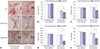
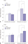
Scars with full thickness were generated on the backs of Yorkshire pigs. An RLX-expressing Ad vector entrapped in alginate gel was evaluated for effects on scar remodeling. The sizes of the initial scars were 3.15±0.15 cm2, 3.12±0.11 cm2, and 3.18±0.08 cm2 in the gel, gel/Ad-LacZ, and gel/Ad-RLX groups, respectively. Alginate gel (gel), alginate gel encapsulating dE1-RGD/LacZ (gel/Ad-LacZ), or alginate gel encapsulating dE1-RGD/LacZ/RLX (gel/Ad-RLX), respectively, was injected into the scar tissue. The sizes of the scars decreased to 1.61±0.15 cm2, 1.70±0.10 cm2, and 1.37±0.05 cm2 in each group, respectively, by 50 days after treatment (Fig. 1A and B, Supplementary Fig. 3, only online). These results indicated that RLX expression from Ad (gel/Ad-RLX) reduces the size of scars compared to those of the control groups (gel or gel/Ad-LacZ) (p<0.05; Fig. 1B).
To further examine the effect of RLX on scar remodeling, color changes and pliability were investigated by spectrophotometry in conjunction with erythema and melanin indices and a durometer. The erythema index values of the initial scars were 1.97±0.20, 2.01±0.10, and 2.01±0.09 in the gel, gel/Ad-LacZ, and gel/Ad-RLX groups, respectively. Erythema was significantly reduced (1.52±0.15; p<0.05) in the gel/Ad-RLX group at 50 days after treatment, compared to those in the gel (2.07±0.35) and gel/Ad-LacZ (2.05±0.08) groups (Fig. 1C). The initial melanin index values were 0.23±0.06, 0.23±0.02, and 0.23±0.03 in the gel, gel/Ad-LacZ, and gel/Ad-RLX groups, respectively (Fig. 1D). This index was also significantly (p<0.05) decreased to 0.08±0.02 in the gel/Ad-RLX group, compared to 0.21±0.05 and 0.21±0.04 in the gel and gel/Ad-LacZ control groups, respectively, at 50 days after treatment. Similarly, pliability was significantly (p<0.05) reduced in the gel/Ad-RLX group, compared to the gel and gel/Ad-LacZ control groups (Fig. 1E). The initial scars had pliability measurements of 20.50±1.42, 19.73±0.88, and 19.08±1.06 in the gel, gel/Ad-LacZ, and gel/Ad-RLX groups, respectively. Pliability was reduced to 18.00± 0.58, 17.46±1.62, and 11.15±1.72 in the gel, gel/Ad-LacZ, and gel/Ad-RLX groups, respectively, by 50 days after treatment. Taken together, these data suggested that RLX expression from gel/Ad-RLX promotes the remodeling of scar tissue.
Relaxin-expressing Ad induces collagen rearrangement and decreases expression of major ECM components in scar tissue
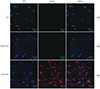
We examined RLX expression by immunofluorescence staining in pig scar tissues transduced with gel, gel/Ad-LacZ, or gel/Ad-RLX. The gel/Ad-RLX-treated groups showed markedly increased immunoreactivity for RLX, compared to the control groups (gel or gel/Ad-LacZ). These results confirmed an association between increased RLX expression and reduced scar tissue formation in tissues transduced with the gel/Ad-RLX depot system (Fig. 2).
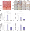
To evaluate the effects of RLX-expressing Ad on collagen-fiber arrangement, tissues were stained with picrosirius red, which binds specifically to collagen fibrils of various diameters. The gel/Ad-RLX-treated groups had closely packed collagen fibers and formed bundles of collagen (Fig. 3A), compared to those in the control groups (gel or gel/Ad-LacZ). These data suggested that RLX induces collagen rearrangement to resemble that of mature, bundle-shaped collagen fibers.
We next examined the effects of RLX overexpression on the major extracellular matrix (ECM) components of scar tissues. Immunohistochemical staining of scar sections revealed significant reductions in type-I collagen, type-III collagen, elastin, and fibronectin in the gel/Ad-RLX-treated group, compared with those in the gel/Ad-LacZ group (p<0.01) (Fig. 3B-F). These data strongly suggested that expression levels of the major ECM components were significantly decreased by RLX overexpression in pig scar tissues. Moreover, these results implied that RLX can play a prominent role in ECM remodeling during the development of scar tissue.
Relaxin-expressing Ad increases MMP-1 and decreases TIMP-1 and α-SMA expression in pig scar tissue
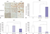
Matrix metalloproteinase-1 (MMP-1), tissue inhibitor of metalloproteinase-1 (TIMP-1), and alpha-smooth muscle actin (α-SMA) are important markers of the effects of TGF-β on wound repair. Therefore, we performed immunohistochemistry to examine expression of MMP-1, TIMP-1, and α-SMA. MMP-1 expression levels were significantly increased by 7.0-fold in pig scar tissues treated with gel/Ad-RLX versus that of the gel/Ad-LacZ-treated tissues (p<0.01) (Fig. 4). In contrast, TIMP-1 and α-SMA expression levels were significantly decreased by 1.9- and 9.9-fold, respectively, in pig scar tissues treated with gel/Ad-RLX transduction (p<0.05). These results suggested that RLX upregulates MMP-1 and downregulates TIMP-1 and α-SMA, which are major players in collagen breakdown.
Relaxin-expressing Ad downregulates TGF-β1 and upregulates TGF-β3 expression in pig scar tissue
To examine the mechanism by which RLX-expressing Ad suppresses expression of the major ECM components, TGF-β1 and TGF-β3 mRNA levels were examined in scar tissues. The gel/Ad-RLX group showed decreased TGF-β1 mRNA expression and increased TGF-β3 mRNA expression in scar tissues, compared with those in the gel or gel/Ad-LacZ group (Fig. 5) (p<0.05), suggesting that the reduced expression of ECM components by RLX overexpression is associated with decreased expression of TGF-β1 and increased expression of TGF-β3.
We also checked TGF-β1 expression via immunohistochemical staining of the scar sections. The results revealed significant reductions in TGF-β1 in the gel/Ad-RLX-treated group, compared to the gel/Ad-LacZ group (p<0.05) (Supplementary Fig. 4, only online).
Relaxin-expressing Ad decreases mast cell counts and NK-1.1 expression in pig scar tissue
Mast cells are reported to be involved in the proliferation and contraction of fibroblasts, and the synthesis of ECM. They also play a key role in scar formation. The amount of mast cells was increased by four times in hypertrophic scars than in normal skin.272829 To examine the local inflammatory effects of RLX-expressing Ad, mast cell counts and NK-1.1 immunohistochemistry were assessed. Immediately after scar formation, the mean numbers of mast cells were 6.20±1.30, 7.00±0.80, and 6.30±0.70 in the gel, gel/Ad-LacZ, and gel/Ad-RLX groups, respectively, with no significant differences. Fifty days after injection of the virus, the mean numbers of observed mast cells were 6.80±0.42 in the gel group and 7.09±0.68 in the gel/Ad-LacZ group. A significant decrease was seen in the mean number of mast cells in the gel/Ad-RLX group (4.89±0.40; p<0.01; Supplementary Fig. 5, only online). NK-1.1 expression was significantly lower in the gel/Ad-LacZ and gel/Ad-RLX-treated groups, compared to the gel group (p<0.05) (Supplementary Fig. 6, only online).
DISCUSSION
Scars, which are caused by collagen deposition, confer a large number of functional problems. Nowadays, many patients want to minimize scar formation, and many treatment modalities, such as scar revision, laser therapy, and medications, have been applied to prevent scar formation. However, the successful treatment of scars has not been fully resolved.303132
This study was performed by adopting a pig scar model used our previous study. The aim of this study was to verify the antifibrotic effect of RLX on scars. Recent studies supported our data and suggested that RLX exerts alternative biological effects mediated directly through TGF-β-dependent signaling.141819303132 These effects elicit downregulation of TGF-β1 and upregulation of TGF-β3 to inhibit myofibroblast accumulation, collagen synthesis/secretion, and ultimately, fibrosis progression in scar tissue. Other studies have suggested that TGF-β1 is a potent fibrogenic growth factor, whereas TGF-β3 reduces scar tissue by decreasing synthesis and increasing degradation of type-I collagen.33 TGF-β plays a key role in the pathophysiology of fibroplasia by decreasing the expression of MMPs and increasing the expression of TIMPs.19
Furthermore, TGF-β induces α-SMA expression, implying that α-SMA expression is a marker of TGF-β activity.323334353637 Thus, the increased expression of MMP-1 and decreased levels of TIMP and α-SMA suggest that dE1-RGD/LacZ/RLX plays a prominent role in ECM remodeling in hypertrophic scars. MMP-1 promotes degradation and ECM remodeling, whereas TIMP-1 decreases the activities of plasminogen activators, such as MMPs, and shifts the ECM equilibrium toward degradation. Thus, increased MMP-1 production and decreased TIMP-1 expression should reduce the amount of abnormal or unfolded collagen during wound healing. In addition, reduced levels of α-SMA may be a positive indicator of TGF-β activity, which was decreased in the gel/Ad-RLX group in this study. Taken together, these studies suggest that dE1-RGD/LacZ/RLX is a potential therapeutic intervention for hypertrophic scars.
Ad has been used extensively as a gene-delivery system in experimental models of cancer and cardiovascular diseases.383940 However, one of the chief concerns with translating these experimental results to the clinical setting is the large and repeated dose that is usually required to generate a modest clinical effect or to achieve the desired therapeutic concentration at the target. To address this concern, we utilized an Arg-Gly-Asp-modified (RGD) replication-incompetent Ad vector as a strategy to allow direct delivery of the RLX gene to wounds. Previous studies from our lab showed that RGD-modified Ad markedly increases gene transfer efficiency in primary keloid fibroblasts. Thus, we believe that RGD-modified Ad is a highly efficient gene-transfer vehicle that does not adversely affect the replication potential of target cells.1417
To further advance this potential therapy for hypertrophic scars, we generated and characterized an alginate gel-matrix system to entrap RLX-expressing Ad as a delivery vehicle.2223 Previous studies have indicated that the biological activity of Ad loaded in alginate gel is prolonged, compared with that of naked Ad, over an extended period of time. Cells that were transduced with gel-released Ad expressed green fluorescence protein at a 13-fold greater intensity at seven days post-incubation compared to that of naked Ad. Because long-term transduction is needed for in vivo applications, the Ad/alginate gel may be very useful as a depot system. The Ad/alginate gel acts as a reservoir that releases Ad in a sustained manner and maintains the biological activity of Ad. Alginate gel has also been shown to limit the mobility of Ad by entrapping Ad within its scaffold, further minimizing the spread of the replication-incompetent Ad outside of the scar tissue. These results show that sustained and controlled delivery of Ad in alginate gel markedly augments therapeutic effects, prolongs maintenance of Ad activity, and provides specificity for local delivery of replication-incompetent Ad at high concentrations. These advantages of the Ad/alginate system will allow for potent local viral therapy to a target site.
In conclusion, we provide strong evidence that RLX promotes the remodeling of scar tissue. Moreover, sustained and controlled delivery of Ad in alginate gel markedly augments the therapeutic effects of RLX. Our data support further evaluation of gel/Ad-RLX as a novel gene-therapy system for treating human scars.
 XML Download