

 PDF
PDF ePub
ePub Citation
 Print
Print


INTRODUCTION
Ameloblastoma occurs rarely in long bones and is slightly more common in the tibia.1 Most reports comprise individual cases, among which the treatment methods have differed.2345678 Since 1993, many scholars have used customized tibia prostheses to reconstruct limbs after tumor resection.91011121314 However, no cases of ameloblastoma were reported among these cases. Herein, we report a case of IB stage ameloblastoma in the middistal tibia and distal fibula of the right limb that was treated with tumor radical resection combined with implanted composite of mid-distal tibial three-dimensional (3D)-printing prosthesis and allogenic tibial segment implantation. The study was approved by the institutional review board of the Affiliated Wuxi People's Hospital of Nanjing Medical University [IRB No: 2017-(01)]. Informed consent was obtained from the patient for the operation.
CASE REPORT
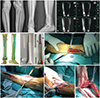
A 40-year-old man presented with right crus pain for a month. Magnetic resonance imaging showed mixed signals in the mid-distal segment of the right tibia and the distal part of the fibula, with low and high signals on T1- and T2-weighted images, respectively (Fig. 1). Tibial lesions penetrated the cortex, and puncture biopsy revealed ameloblastoma. Enneking stage was IB (G1, T2, and M0).
Implant customization
In a computer, 3D images of the tibia were reconstructed with reverse data of computed tomography (CT) of 0.625-mm slice thickness. The digital image of the prosthesis was designed according to the predicted range of radical resection. Then, the resinous tibia and prosthesis were printed with Lite 450 HD (Shanghai Union 3D Technology Co., Ltd., Shanghai, China) at a 1:1 size for the installation test. After debugging, a titanium alloy (Ti-6Al-4V; AP&C Advanced Powder and Coatings Inc., Boisbriand, Quebec, Canada) prosthesis was printed in one step with its porous coating with Arcam Q10 (Arcam AB, Mölndal, Sweden) by Beijing Chunlizhengda Medical Instruments Company. The aperture, wire diameter, and porosity of the porous coating were 0.40–0.50, 0.40–0.45, and 68–78%, respectively. The prosthetic cylindrical body was 2.6 cm in diameter and 23.5 cm in length, and could be split in the middle. The stem was 1.8 cm in diameter and 9.5 cm in length. The keyhole was equipped with an interlocking screw with a steel plate.
Surgical procedures
After general endotracheal anesthesia, a 12.0-cm long incision (I) was performed on the surface of the distal fibula. The fibula was amputated at 3.0 cm above the lesion. Then, a 18.0-cm long incision (II) was made on the surface of the mid-distal tibia, which included a needle path for biopsy. At last, a 15.0-cm long incision (III) on the anterior surface of the middle tibia was performed, and the tibia was amputated with stepsection at 3.0 cm above the lesion. The diseased bone and surrounding soft tissues were removed completely at its margin, and no residual tumor tissue was found in the frozen-section examination during the operation. The tibial stump was repaired with allogenic tibia to restore its intact structure. After directional reaming with a 1.6-cm diameter reamer, the prosthetic stem with a porous surface was inserted into the medullary cavity, and then fixed with a plate and interlocking screw. Next, the metal ankle mortise and the osteotomic cancellous bone of the talus were also compacted closely and fixed with a number of screws. Finally, two parts of the prosthesis were connected with screws.
Surgical outcome
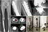
There were no injuries to vessels and nerves. The patient was able to walk with a single crutch after 2 weeks, and he was told to wear a brace when walking after 8 weeks. At 12 weeks, the brace was removed. The limb function was excellent at 1 year after surgery, and the MSTS 93 score was 86.7% (26/30).15 The ranges of movement were 0° (extension) to 130° (flexion) in the knee joint, and 15° (dorsal extension) to 10° (plantar flexion) in the mid-tarsal joint (Table 1). CT and radiography showed no signs of prosthesis loosening (partial regenerated bone ingrowth around the prosthesis and no progressive radiolucent zone) (Fig. 2). No tumor recurrence and metastasis were found.
DISCUSSION
The first type of tibial prostheses with an ankle joint was hinge-restricted or semi-constrained.910111214 The main complications were talus collapse, prosthesis loosening, fibula impact, incision necrosis, deep infection, and tumor recurrence. Although these scholars all believed that a tibial prosthesis with an ankle joint can achieve better short-term function with limb reconstruction than with other methods, such as bone transplantation and tumor bone replantation after inactivation, they also emphasized the need for strict selection of cases before operaoperation in view of more complications and poor late outcomes in some cases.910111214
Another type of prosthesis has no joints. Economopoulos, et al.13 reported a case of giant cell tumor of the distal tibia that was repaired with porous tantalum metal prosthesis without a joint. At 5 years after the operation, the patient had no pain when walking, and the mid-tarsal joint had 5° of dorsal extension and 10° of plantar flexion. They believed that the life expectancy of jointless prosthesis is better, although the tibiotalar joint will have lost its mobility. Also, the simpler surgical process in the operation is another advantage of jointless prosthesis.
In this case, ameloblastoma has a wide range of lesions, accounting for more than 75% of the total length of the tibia. If the bone of the diseased limb is reconstructed with a large segment of allograft, the bone regeneration will be slow, and the possibility of complications, such as allograft rejection, fracture, and nonunion, will increase. As mentioned above, local stress to a hinged prosthesis will be more intense and complications, such as loosening and talus collapse, will occur more readily.1116 There are still complications, such as talus prosthesis loosening, after the use of a semi-constrained artificial ankle.101112 As in the present case, the distal fibular tumor segment often cannot be retained, and it can be very difficult to reconstruct the peri-ankle ligaments between the metal prosthesis and the host skeleton. Therefore, a mid-distal tibial prosthesis without an ankle joint should be used for limb reconstruction. Also, the initial stability of the prosthesis was beneficial for early initiation of rehabilitation, which is an advantage of this type of limb salvage, compared with allograft bone transplantation.
It has been reported that postoperative incision necrosis and infection are not uncommon during the operation of tibial tumors.91011121314 In the present case, the lesion was extensive. To prevent skin necrosis after the operation, three incisions were designed. All were within 20 cm in length and staggered with each other. Incision I was 7.0 cm away from incision II. Likewise, the distal end of incision III and the proximal end of incision II were also 4.5 cm apart.
In the present case, a more intuitive understanding and accurate data of the diseased tibia could be obtained using a computer and resin model before the operation. Thereby, the most feasible schemes were devised during the manufacturing and implantation of the personalized prosthesis. When using the computer to simulate the surgery, we discovered that the remaining tibia may not be able to grasp the prosthetic stem according to the criteria of osteotomy. Thus, an allogenic tibia was prefabricated for bone grafting.
Hollander, et al.17 reported that after co-culture of porous titanium alloy scaffolds with human osteoblasts in vitro for 2 weeks, a large number of osteoblasts adhered to and proliferated in the surface and pores of the scaffolds. In this case, CT scan showed that the regenerated bone of the host had grown into the porous surface of the prosthetic stem and ankle mortise at 9 months after operation. The new extra-cortical bone bridges connected the tibial stump to the bone-prosthesis junction. Meanwhile, the new bone bridge spanned the junction of the talus and prosthesis in the anterior side of the talus. For 12 consecutive months after operation, radiography showed no progressive radiolucent zones around the prosthetic stem and metal ankle mortise, suggesting no obvious loosening of the prosthesis.
In this case, the application of 3D-printing technology to construct a prosthesis replacement achieved good short-term efficacy. We believe that the trabecular porous structure design will result in a good outcome for the long-term biological fixation of the prosthesis.

 XML Download