

 PDF
PDF Citation
 Print
Print


Abstract
Objectives
We report a case of Ogilvie's syndrome following posterior decompression surgery in a spinal stenosis patient who presented with acute abdominal distension, nausea, and vomiting.
Summary of Literature Review
Ogilvie's syndrome is a rare and potentially fatal disease that can easily be mistaken for postoperative ileus, and is also known as acute colonic pseudo-obstruction. Early recognition and diagnosis enable treatment prior to bowel perforation and requisite abdominal surgery.
Materials and Methods
An 82-year-old woman presented with 6 months of worsening back pain with walking intolerance due to weakness in both legs. She had hypertension, asthma, and Cushing syndrome without bowel or bladder symptoms. Further workup demonstrated the presence of central spinal stenosis on magnetic resonance imaging. The patient underwent an L2-3 laminectomy and posterior decompression. Surgery was uneventful.
Results
The patient presented with acute abdominal distension, nausea, and vomiting on postoperative day 1. The patient was initially diagnosed with adynamic ileus and treated conservatively with bowel rest, reduction in narcotic dosage, and a regimen of stool softeners, laxatives, and enemas. Despite this treatment, her clinical course failed to improve, and she demonstrated significant colonic distension radiographically. Intravenous neostigmine was administered as a bolus with a rapid and dramatic response.
Conclusion
Ogilvie's syndrome should be included in the differential diagnosis of postoperative ileus in patients developing prolonged unexplained abdominal distension and pain after lumbar spinal surgery. Early diagnosis and initiation of conservative management can prevent major morbidity and mortality due to bowel ischemia and perforation.
REFERENCES
1. Maloney N, Vargas HD. Acute intestinal pseudo-obstruction (Ogilvie's syndrome). Clin Colon Rectal Surg. 2005 May; 18(2):96–101. DOI: 10.1055/s-2005-870890.

2. Ogilvie WH. William Heneage Ogilvie 1887–1971. Large-intestine colic due to sympathetic deprivation. A new clinical syndrome. Dis Colon Rectum. 1987 Dec; 30(12):984–7. DOI: 10.1007/bf02554291.
3. Tsirikos AI, Sud A. Ogilvie's syndrome following posterior spinal arthrodesis for scoliosis. Indian J Orthop. 2013 Jul; 47(4):408–12. DOI: 10.4103/0019-5413.114934.
4. Vanek VW, Al-Salti M. Acute pseudo-obstruction of the colon (Ogilvie's syndrome). An analysis of 400 cases. Dis Colon Rectum. 1986 Mar; 29(3):203–10. DOI: 10.1007/bf02555027.
5. Anuras S, Baker CR Jr. The colon in the pseudoobstructive syndrome. Clin Gastroenterol. 1986 Oct; 15(4):745–62.
6. Bachulis BL, Smith PE. Pseudoobstruction of the colon. Am J Surg. 1978 Jul; 136(1):66–72. DOI: 10.1016/0002-9610 (78)90202-7.
7. Feldman RA, Karl RC. Diagnosis and treatment of Ogilvie's syndrome after lumbar spinal surgery. Report of three cases. J Neurosurg. 1992 Jun; 76(6):1012–6. DOI: 10.3171/jns.1992.76.6.1012.
8. Ozkurt H, Yilmaz F, Bas N, et al. Acute colonic pseudo-obstruction (Ogilvie's syndrome): radiologic diagnosis and medical treatment with neostigmine. Report of 4 cases. Am J Emerg Med. 2009 Jul; 27(6):757. e1-4. DOI: 10.1016/j.ajem.2008.10.020.
9. Singleton AO Jr., Wilson M. Air-fluid obstruction of the colon. South Med J South Med J. 1967 Sep; 60(9):909–13. DOI: 10.1097/00007611-196709000-00001.
10. Livingston EH, Passaro EP Jr. Postoperative ileus. Dig Dis Sci. 1990 Jan; 35(1):121–32. DOI: 10.1007/bf01537233.
Fig. 1.
(A, B) Preoperative anteroposterior and lateral radiographs of an 82-year-old woman show sclerotic changes and disc space narrowing at L2-3. (C, D) Postoperative anteroposterior and lateral radiographs show L2-3 decompression.

Fig. 2.
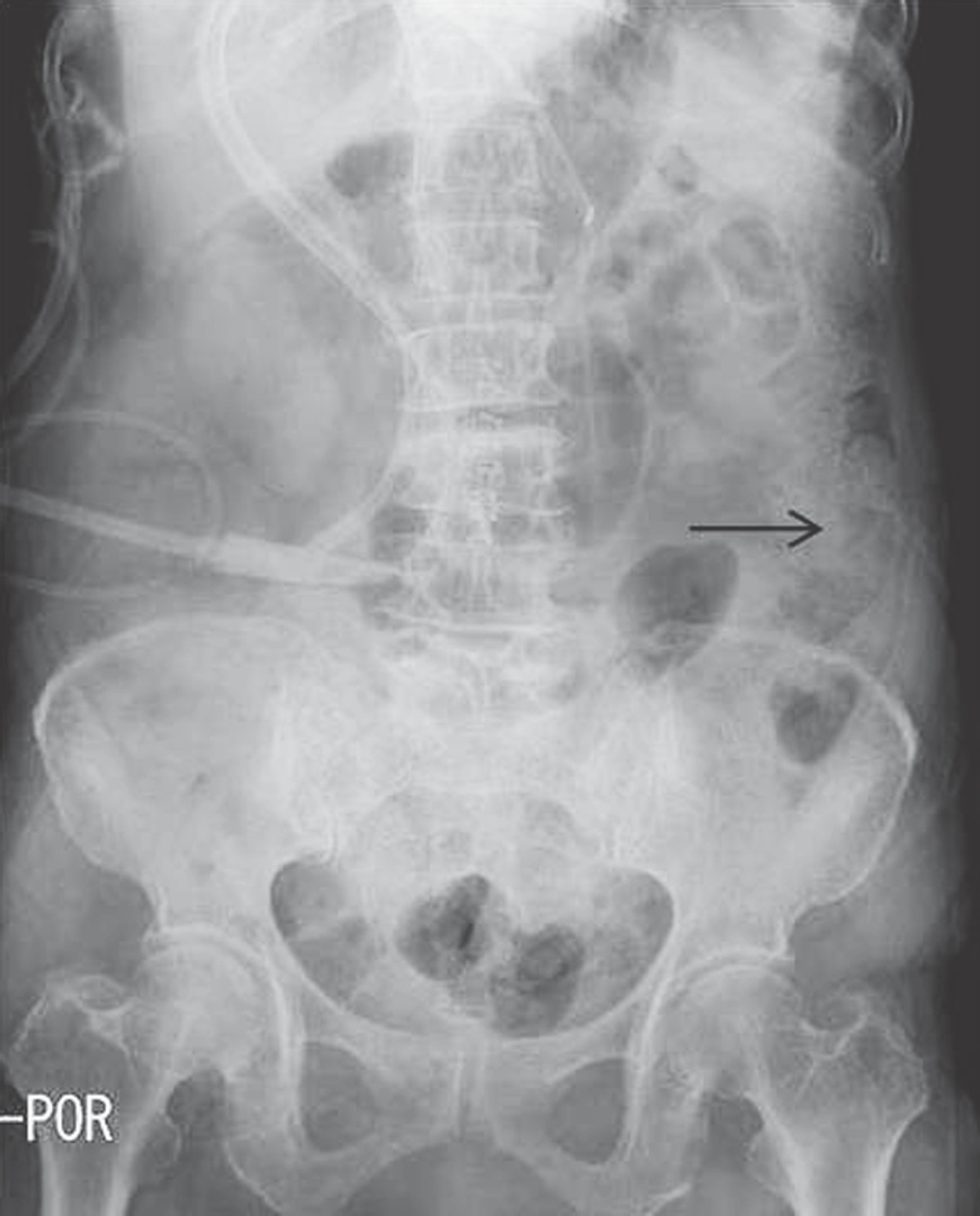
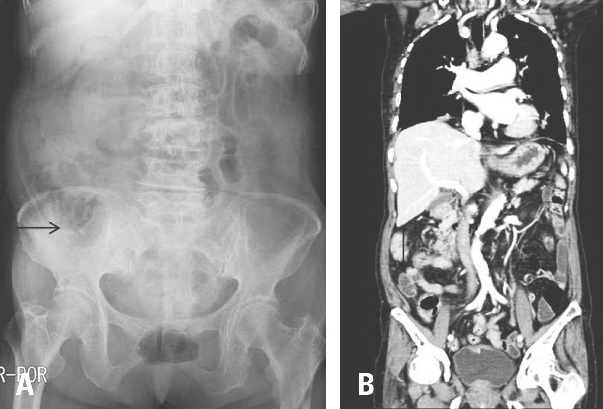
An abdominal radiograph on postoperative day 1 shows abdominal distension and fecal impaction with gas retention.

 XML Download