

 PDF
PDF Citation
 Print
Print


Abstract
Summary of Literature Review
There have been no reports on the prevalence of thoracic scoliosis in Korea.
Materials and Methods
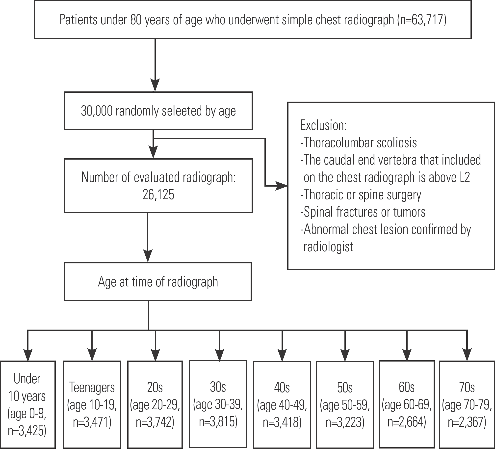
From August 2011 to October 2012, radiographs of patients under 80 years of age who underwent routine chest radiographs were retrospectively reviewed. Based on their age when the chest radiographs were obtained, the patients were divided into 8 groups. The prevalence and angle of the curve of thoracic scoliosis were investigated in each age group, and the prevalence of thoracic scoliosis according to sex, the direction of the curve, number of vertebrae in the major curve, the location and rotation of the apical vertebrae, and osteophyte location were examined.
Results
The prevalence of thoracic scoliosis was 2.4% (621 patients), and female patients (3.0%, 375 of 12471) showed a higher prevalence than male patients (1.8%, 246 of 13654) (p<0.001). Right curvature was present in 445 patients and left curvature in 176 patients. In each age group, the prevalence and degree of thoracic scoliosis were 1.1% (14.2°±3.2°), 2.3% (17.4°±7.7°), 2.5% (17.0°±8.9°), 1.9% (15.8°±5.9°), 1.3% (15.5°±6.6°), 2.1% (18.0°±13.6°), 2.9% (14.3°±3.6°), and 6.1% (16.2°±4.8°), respectively. The mean curvature in all scoliosis patients was 16.0°±7.0°. The angle of the curve was significantly different by sex (15.4°±7.1° for males, 16.8°±7.6° for females). The average curve angle of patients with thoracic scoliosis was 16.0°±7.0°, among whom it was 10°-20° in 533 patients, 20°-30° in 64, 30°-40° in 11, and over 40° in 13.
Go to : 
REFERENCES
1. Kostuik JP, Bentivoglio J. The incidence of low back pain in adult scoliosis. Acta Orthop Belg. 1981 May; 6(3):268–73. DOI: 10.1097/00007632-198105000-00009.
2. Kebaish KM, Neubauer PR, Voros GD, et al. Scoliosis in adults aged forty years and older: prevalence and relationship to age, race, and gender. Spine (Phila Pa 1976). 2011 Apr; 36(9):731–6. DOI: 10.1097/BRS.0b013e3181e9f120.
3. Hong JY, Suh SW, Modi HN, et al. The prevalence and radiological findings in 1347 elderly patients with scoliosis. J Bone Joint Surg Br. 2010 Jul; 92(7):980–3. DOI: 10.1302/0301-620X.92B7.23331.

4. Schwab F, Dubey A, Gamez L, et al. Adult scoliosis: prevalence, SF-36, and nutritional parameters in an elderly volunteer population. Spine (Phila Pa 1976). 2005 May; 30(9):1082–5. DOI: 10.1097/01.brs.0000160842. 43482.cd.
5. Nash CL Jr., Moe JH. A study of vertebral rotation. J Bone Joint Surg Am. 1969 Mar; 51(2):223–9. DOI: 10.2106/00004623-196951020-00002.
6. Kim JH Suk SI, Chung ER, et al. Epidemiologic study of lumbar scoliosis with plain abdominal X-ray. J Korean Soc Spine Surg. 2004 Dec; 11(4):246–52. DOI: 10.4184/jkss.2004.11.4.246.
7. Carter OD, Haynes SG. Prevalence rates for scoliosis in US adults: results from the first National Health and Nutrition Examination Survey. Int J Epidemiol. 1987 (Dec); 16(4):537–44. DOI: 10.1093/ije/16.4.537.
8. Urrutia J, Besa P, Bengoa F. A prevalence study of thoracic scoliosis in Chilean patients aged 10-20 years using chest radiographs as a screening tool. J Pediatr Orthop B. 2018 (Mar); 27(2):159–62. DOI: 10.1097/BPB.0000000000000466.
9. Urrutia J, Zamora T, Klaber I. Thoracic scoliosis prevalence in patients 50 years or older and its relationship with age, sex, and thoracic kyphosis. Spine (Phila Pa 1976). 2014 (Jan); 39(2):149–52. DOI: 10.1097/BRS. 0000000000000095.
10. Chen JB, Kim AD, Allan-Blitz L, et al. Prevalence of thoracic scoliosis in adults 25 to 64 years of age de-tected during routine chest radiographs. Eur Spine J. 2016 Oct; 25(10):3082–7. DOI: 10.1007/s00586-015-4215-4.
11. Pritchett JW, Bortel DT. Degenerative symptomatic lumbar scoliosis. Spine (Phila Pa 1976). 1993 May; 18(6):700–3. DOI: 10.1097/00007632-199305000-00004.
12. Grubb SA, Lipscomb HJ, Coonrad RW. Degenerative adult onset scoliosis. Spine (Phila Pa 1976). 1988 (Mar); 13(3):241–5. DOI: 10.1097/00007632-198803000-00004.
13. Kirkaldy-Willis WH, Farfan HF. Instability of the lumbar spine. Clin Orthop Relat Res. 1982 May; 165:110–23. DOI: 10.1097/00003086-198205000-00015.
14. Konieczny MR, Senyurt H, Krauspe R. Epidemiology of adolescent idiopathic scoliosis. J Child Orthop. 2013 Feb; 7(1):3–9. DOI: 10.1007/s11832-012-0457-4.
Go to :
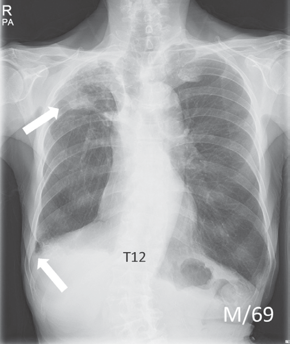 | Fig. 2.A chest radiograph of a 69-year-old man revealed a focal nodular opacity in the right upper lobe and right costophrenic angle blunting. In addition, thoracolumbar scoliosis was also observed on the chest radiograph, and the patient was ultimately excluded from this study. |
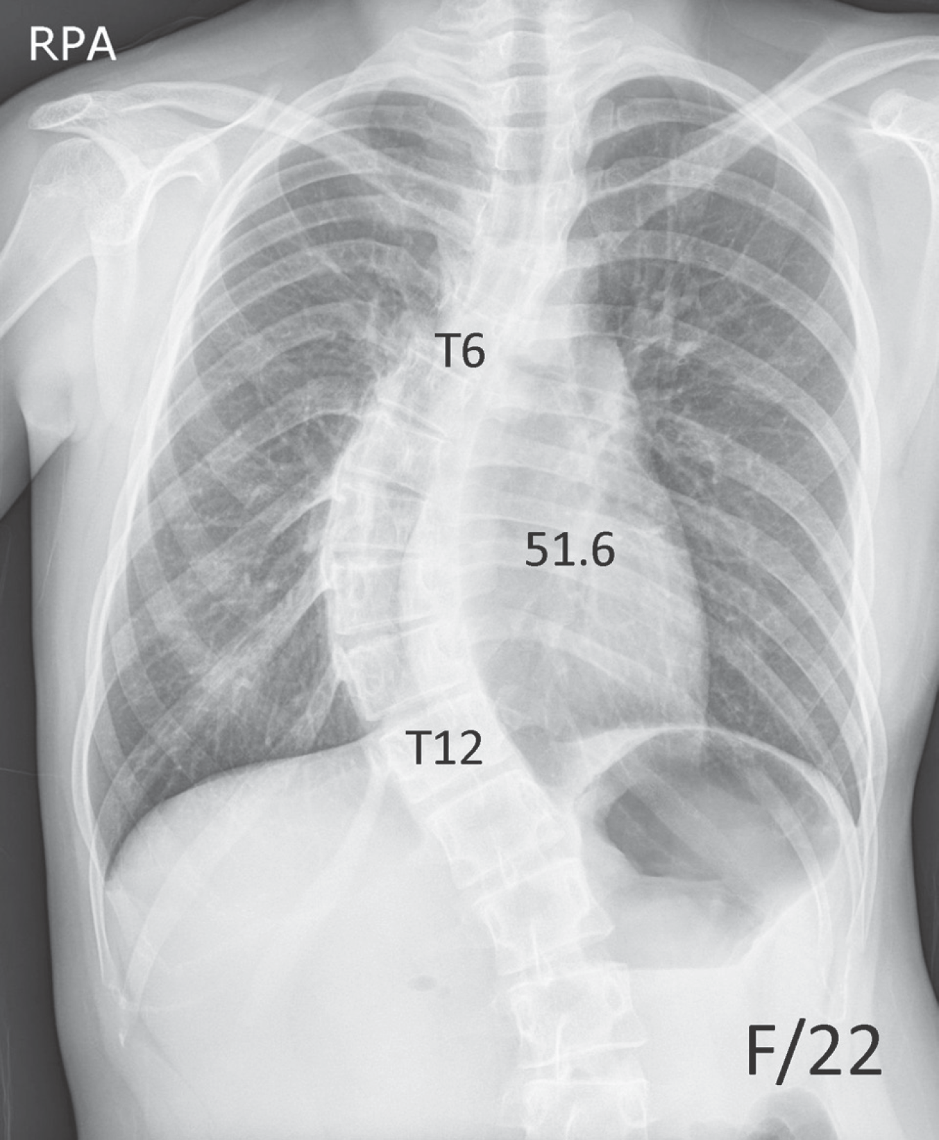 | Fig. 3.A 22-year-old woman presented with thoracic scoliosis with a Cobb angle of 51.6° from T6 to T12 on her chest radiograph. |
Table 1.
Prevalence of thoracic scoliosis according to age and its distribution by gender and curve direction
| Age | 0-9 | 10-19 | 20-29 | 30-39 | 40-49 | 50-59 | 60-69 | 70-79 | total |
|---|---|---|---|---|---|---|---|---|---|
| Population of checked X-ray | 3425 | 3471 | 3742 | 3815 | 3418 | 3223 | 2664 | 2367 | 26125 |
| Number of thoracic scoliosis | 38 (1.1%) | 81 (2.3%) | 95 (2.5%) | 74 (1.9%) | 44 (1.3%) | 68 (2.1%) | 77 (2.9%) | 144 (6.1%) | 621 (2.4%) |
| Gender (male/female) | 17/21 | 35/46 | 26/69 | 29/45 | 15/29 | 36/32 | 32/45 | 56/88 | 246/375 |
| Direction (Rt/Lt) | 16/22 | 55/26 | 70/25 | 51/23 | 28/16 | 47/21 | 57/20 | 121/23 | 445/176 |
| Cobb angle (˚)∗ | 14.2±3.2 | 17.4±7.7 | 17.1±8.9 | 15.9±5.9 | 15.5±6.6 | 18.0±13.6 | 14.3±3.6 | 16.2±4.8 | 16.0±7.0 |
Table 2.
Number of involved segments
Table 3.
Location of apex of curve
| Age | 0-9 | 10-19 | 20-29 | 30-39 | 40-49 | 50-59 | 60-69 | 70-79 | total |
|---|---|---|---|---|---|---|---|---|---|
| Location of Apex of curve∗ | |||||||||
| T3 | 0 | 2 | 1 | 3 | 0 | 1 | 3 | 0 | 10 |
| T4 | 1 | 16 | 11 | 5 | 7 | 2 | 10 | 7 | 59 |
| T5 | 1 | 6 | 7 | 5 | 5 | 3 | 8 | 16 | 51 |
| T6 | 3 | 6 | 6 | 7 | 4 | 12 | 8 | 29 | 75 |
| T7 | 4 | 10 | 24 | 21 | 12 | 22 | 9 | 32 | 134 |
| T8 | 9 | 20 | 25 | 16 | 11 | 18 | 9 | 25 | 133 |
| T9 | 11 | 17 | 14 | 7 | 3 | 5 | 10 | 13 | 80 |
| T10 | 7 | 4 | 6 | 8 | 2 | 2 | 11 | 13 | 53 |
| T11 | 2 | 0 | 1 | 2 | 0 | 3 | 9 | 9 | 26 |
Table 4.
Prevalence and degree of vertebral body rotation using Nash & Moe method
| Age | 0-9 | 10-19 | 20-29 | 30-39 | 40-49 | 50-59 | 60-69 | 70-79 | total |
|---|---|---|---|---|---|---|---|---|---|
| Rotation of apex∗ | |||||||||
| Grade 1 | 4 | 22 | 20 | 5 | 10 | 13 | 42 | 77 | 193 |
| Grade 2 | 3 | 10 | 9 | 1 | 0 | 6 | 16 | 13 | 58 |
| Grade 3 | 1 | 0 | 0 | 0 | 0 | 2 | 0 | 1 | 4 |
 XML Download