Abstract
Objectives
To evaluate preoperative factors related with spinal canal expansion after posterior decompression for the treatment of multilevel cervical myelopathy.
Summary of Literature Review
Data about preoperative factors related with spinal canal expansion after posterior cervical decompression surgery are inconsistent.
Materials and Methods
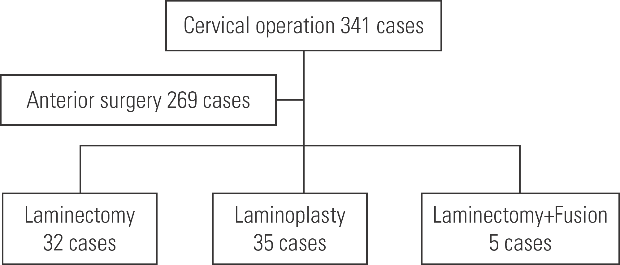
We reviewed 67 patients with cervical myelopathy who underwent posterior laminectomy or laminoplasty. Radiologically, we evaluated the C2-7 Cobb angle and range of motion using X-rays from the preoperative assessment and final follow-up. Expansion of the spinal canal at 6 weeks postoperatively was evaluated using magnetic resonance imaging and compared with the preoperative values. The preoperative factors of age, sex, number of operated levels, operation method, and radiological parameters were investigated as factors potentially related to postoperative spinal canal expansion using multivariate regression and correlation analyses. The clinical outcome was analyzed by the Neck Disability Index (NDI) and Japanese Orthopaedic Association (JOA) scores.
Results
The postoperative spinal canal expansion was 4.76 mm in sagittal images and 4.31 mm in axial images, with higher values observed in males and cases of severe preoperative cord compression. A lordotic preoperative Cobb angle was related to postoperative spinal canal expansion and JOA score improvement, but without statistical significance. The clinical outcomes of NDI (18.3→14.8) and JOA scores (10.81→14.6) showed improvement, but were not significantly related with any preoperative factors.
REFERENCES
1. Denaro V, Di Martino A. A cervical spine surgery: an historical perspective. Clin Orthop Relat Res. 2011 Mar; 469(3):639–48. DOI: 10.1007/s11999-010-1752-3.
2. Rhee JM, Basra S. Posterior surgery for cervical myelopathy: laminectomy, laminectomy with fusion, and laminoplasty. Asian Spine J. 2008; 2(2):114–26. DOI: 10.4184/asj.2008.2.2.114.

3. Aita I, Hayashi K, Wadano Y, Yabuki T. Posterior move-ment and enlargement of the spinal cord after cervical laminoplasty. J Bone Joint Surg (Br). 1998 Jan; 80(1):33–7. DOI: 10.1302/0301-620x.80b1.7919.

4. Fujimura Y, Nishi Y, Nakamura M. Dorsal shift and expansion of the spinal cord after expansive open-door laminoplasty. J Spinal Disord. 1997 Aug; 10(4):282–95. DOI: DOI:10.1097/00002517-199708000-00002.

5. Sodeyama T, Goto S, Mochizuki M, Takahashi J, Moriya H. Effect of decompression enlargement laminoplasty for posterior shifting of the spinal cord. Spine. 1999 Agu; 24(15):1527–31. DOI: DOI:10.1097/00007632-199908010-00005.

6. Tashjian VS, Kohan E, McArthur DL, Holly LT. The relationship between preoperative cervical alignment and postoperative spinal cord drift after decompressive laminectomy and arthrodesis for cervical spondylotic myelopathy. Surg Neurol. 2009 Aug; 72(2):112–7. DOI: 10.1016/j.surneu.2009.02.024.

7. Du W, Zhang P, Shen Y, Zhang YZ, Ding WY, Ren LX. Enlarged laminectomy and lateral mass screw fixation for multilevel cervical degenerative myelopathy associated with kyphosis. Spine J. 2014 Jan; 14(1):57–64. DOI: 10.1016/j.spinee.2013.06.017.

8. Denaro V, Longo UG, Berton A, Salvatore G, Denaro L. Cervical spondylotic myelopathy: the relevance of the spinal cord back shift after posterior multilevel decompression. A systematic review. Eur Spine J. 2015 Nov; 24(S7):832–41. DOI: 10.1007/s00586-015-4299-x.

9. Hirabayashi K, Satomi K. Operative procedure and results of expansive open-door laminoplasty. Spine (Phila Pa 1976). 1998; 13:870–6.

10. Song KJ, Choi BW, Kim JK. Adjacent segment pathology following anterior decompression and fusion using cage and plate for the treatment of degenerative cervical spinal diseases. Asian Spine J. 1988 Jun; 13(7):870–6. DOI: 10.1097/00007632-198807000-00032.

11. Baba H, Uchida K, Maezawa Y, Furusawa N, Azuchi M, Imura S. Lordotic alignment and posterior migration of the spinal cord following en bloc open-door laminoplasty for cervical myelopathy: a magnetic resonance imaging study. J Neurol. 1996; 243(9):626–32. DOI: 10.1007/bf00878657.

12. Hatta Y, Shiraishi T, Hase H, Yato Y, Ueda S, Mikami Y, Harada T, Ikeda T, Kubo T. Is posterior spinal cord shifting by extensive posterior decompression clinically significant for multisegmental cervical spondylotic myelopathy? Spine. 2005 Nov; 30(21):2414–9. DOI: 10.1097/01. brs.0000184751.80857.3e.

13. Lee JY, Sharan A, Baron EM, Lim MR, Grossman E, Al-bert TJ, Vaccaro AR, Hilibrand AS. Quantitative prediction of spinal cord drift after cervical laminectomy and arthrodesis. Spine. 2006 Jul; 31(16):1795–8. DOI: 10.1097/01. brs.0000225992.26154.d0.

14. Shiozaki T, Otsuka H, Nakata Y, Yokoyama T, Takeuchi K, Ono A, Numasawa T, Wada K, Toh S. Spinal cord shift on magnetic resonance imaging at 24 hours after cervical laminoplasty. Spine. 2009 Feb; 34(3):274–9. DOI: 10.1097/brs.0b013e318194e275.

15. Fujimura Y, Nishi Y, Nakamura M. Dorsal shift and expansion of the spinal cord after expansive open-door lami-noplasty. J Spinal Disord. 1997; 10:282–7.

16. Yoon ST, Raich A, Hashimoto RE, Riew KD, Shaf-frey CI, Rhee JM, Tetreault LA, Skelly AC, Fehlings MG. Predictive factors affecting outcome after cervical lami-noplasty. Spine. 2013 Oct; 38:232–52. DOI: 10.1097/brs.0b013e3182a7eb55.

17. Ranger MR, Irwin GJ, Bunbury KM, Peutrell JM. Changing body position alters the location of the spinal cord with-in the vertebral canal: a magnetic resonance imaging study. Br J Anaesth. 2008 Dec; 101(6):804–9. DOI: 10.1093/bja/aen295.

18. Kato Y, Iwasaki M, Fuji T, Yonenobu K, Ochi T. Long term follow-up results of laminectomy for cervical myelopathy caused by ossification of the posterior longitudinal ligament. J Neurosurg. 1998 Aug; 89(2):217–23. DOI: 10.3171/jns.1998.89.2.0217.
Fig. 2.
Severity of cord compression at the site of maximum compression was analyzed using T2-weighted sagittal and axial views on magnetic resonance imaging.(A) Sagittal occupy ratio was measured as B/A. (B) Axial occupy ratio was measured as B/A. (C, D) At the 6-week postoperative follow-up, the degree of spinal cord expansion was compared and analyzed with that prior to surgery as B-A.

Table 1.
Effect of demographic and radiological factors on postoperative increase of sagittal diameter (Multivariate regression analysis)
| β∗ | p-value† | |
|---|---|---|
| Age | −0.016 | 0.896 |
| Sex | −0.332 | 0.004 |
| Number of fused vertebra | 0.188 | 0.401 |
| Preoperative Cobb angle | 0.103 | 0.397 |
| Preoperative range of motion | 0.034 | 0.792 |
| Preoperative sagittal diameter | −0.530 | 0.000 |
| Preoperative axial diameter | −0.190 | 0.039 |
| Sagittal occupying ratio | −0.214 | 0.166 |
| Axial occupying ratio | −0.237 | 0.126 |
| Operation method | −0.413 | 0.075 |
Table 2.
Effect of demographic and radiological factors on postoperative increase of axial diameter (Multivariate regression analysis)
| β∗ | p-value† | |
|---|---|---|
| Age | 0.250 | 0.090 |
| Sex | −0.276 | 0.041 |
| number of fused vertebra | 0.005 | 0.985 |
| Preoperative Cobb angle | 0.024 | 0.870 |
| Preoperative range of motion | 0.029 | 0.851 |
| Preoperative sagittal diameter | −0.239 | 0.019 |
| Preoperative axial diameter | −0.297 | 0.027 |
| Sagittal occupying ratio | 0.068 | 0.709 |
| Axial occupying ratio | −0.336 | 0.070 |
| Operation method | −0.118 | 0.662 |
Table 3.
Radiological and clinical analysis according to preoperative Cobb angle (Chi square, t-test)
Table 4.
Correlation analysis between clinical outcomes and radiologica factors.


 PDF
PDF Citation
 Print
Print


 XML Download