

 PDF
PDF ePub
ePub Citation
 Print
Print


Abstract
Purpose:
To evaluate the efficacy of antibiotic-loaded cement spacers (ALCSs) for the treatment of diabetic foot infections with osteomyelitis as a salvage procedure and to analyze the risk factors of treatment failure.
Materials and Methods:
This study reviewed retrospectively 39 cases of diabetic foot infections with osteomyelitis who underwent surgical treatment from 2009 to 2017. The mean age and follow-up period were 62±13 years and 19.2±23.3 months, respectively. Wounds were graded using the Wagner and Strauss classification. X-ray, magnetic resonance imaging (or bone scan) and deep tissue cultures were taken preoperatively to diagnose osteomyelitis. The ankle-brachial index, toe-brachial index (TBI), and current perception threshold were checked. Lower extremity angiography was performed and if necessary, percutaneous transluminal angioplasty was conducted preoperatively. As a surgical treatment, meticulous debridement, bone curettage, and ALCS placement were employed in all cases. Between six and eight weeks after surgery, ALCS removal and autogenous iliac bone graft were performed. The treatment was considered successful if the wounds had healed completely within three months without signs of infection and no additional amputation within six months.
Results:
The treatment success rate was 82.1% (n=32); 12.8% (n=5) required additional amputation and 5.1% (n=2) showed delayed wound healing. Bacterial growth was confirmed in 82.1% (n=32) with methicillin-resistant Staphylococcus aureus being the most commonly identified strain (23.1%, n=9). The lesions were divided anatomically into four groups; the largest number was the toes: (1) toes (41.0%, n=16), (2) metatarsals (35.9%, n=14), (3) midfoot (5.1%, n=2), and (4) hindfoot (17.9%, n=7). A significant difference in the Strauss wound score and TBI was observed between the treatment success group and failure group.
Go to : 
REFERENCES
1.Lipsky B. Infectious problems of the foot in diabetic patients. Bowker JH, Pfeifer MA, editors. Levin and O'Neal's the diabetic foot. St. Louis (MO): Mobsy;2001. p. 467–80.

2.Lipsky BA. Medical treatment of diabetic foot infections. Clin Infect Dis. 2004. 39(Suppl 2):S104–14. DOI: doi: 10.1086/383271.
3.Frykberg RG., Wittmayer B., Zgonis T. Surgical management of diabetic foot infections and osteomyelitis. Clin Podiatr Med Surg. 2007. 24:469–82. viii-ix.DOI: doi: 10.1016/j.cpm.2007.04.001.
4.Dalla L., Faglia E., Caminiti M., Clerici G., Ninkovic S., Deanesi V. Ulcer recurrence following first ray amputation in diabetic patients: a cohort prospective study. Diabetes Care. 2003. 26:1874–8. DOI: doi: 10.2337/diacare.26.6.1874.
5.Izumi Y., Satterfield K., Lee S., Harkless LB. Risk of reamputation in diabetic patients stratified by limb and level of amputation: a 10-year observation. Diabetes Care. 2006. 29:566–70. DOI: doi: 10.2337/diacare.29.03.06.dc05-1992.
6.Sage RA. Biomechanics of ambulation after partial foot amputation: prevention and management of reulceration. J Prosthet Orthot. 2007. 19:77–9. DOI: doi: 10.1097/JPO.0b013e3180dc92fb.
7.Hsieh PH., Chang YH., Chen SH., Ueng SW., Shih CH. High concentration and bioactivity of vancomycin and aztreonam eluted from Simplex cement spacers in two-stage revision of infected hip implants: a study of 46 patients at an average follow-up of 107 days. J Orthop Res. 2006. 24:1615–21. DOI: doi: 10.1002/jor.20214.
8.Park SJ., Cho Y., Lee SW., Woo HY., Lim SE. In vitro study evaluating the antimicrobial activity of vancomycin-impregnated cement stored at room temperature in methicillin-resistant staphylococcus aureus. J Korean Foot Ankle Soc. 2018. 22:38–43. DOI: doi: 10.14193/jkfas.2018.22.1.38.
9.Buchholz HW., Engelbrecht H. [Depot effects of various antibiotics mixed with Palacos resins]. Chirurg. 1970. 41:511–5. German.
10.Ostermann PA., Seligson D., Henry SL. Local antibiotic therapy for severe open fractures. A review of 1085 consecutive cases. J Bone Joint Surg Br. 1995. 77:93–7. DOI: doi: 10.1302/0301-620x.77b1.7822405.
11.Klemm K. [Gentamicin-PMMA-beads in treating bone and soft tissue infections (author's transl)]. Zentralbl Chir. 1979. 104:934–42. German.
12.Klemm K. [Local treatment of infection with gentamicin-PMMA chains and minichains]. Aktuelle Probl Chir Orthop. 1990. 34:65–77. German.
13.Klemm K. The use of antibiotic-containing bead chains in the treatment of chronic bone infections. Clin Microbiol Infect. 2001. 7:28–31.
14.Schade VL., Roukis TS. The role of polymethylmethacrylate antibiotic-loaded cement in addition to debridement for the treatment of soft tissue and osseous infections of the foot and ankle. J Foot Ankle Surg. 2010. 49:55–62. DOI: doi: 10.1053/j.jfas.2009.06.010.
15.Nelson CL., Evans RP., Blaha JD., Calhoun J., Henry SL., Patzakis MJ. A comparison of gentamicin-impregnated polymethylmethacrylate bead implantation to conventional parenteral antibiotic therapy in infected total hip and knee arthroplasty. Clin Orthop Relat Res. 1993. 295:96–101. DOI: doi: 10.1097/00003086-199310000-00014.
16.Melamed EA., Peled E. Antibiotic impregnated cement spacer for salvage of diabetic osteomyelitis. Foot Ankle Int. 2012. 33:213–9. DOI: doi: 10.3113/fai.2012.0213.
17.Elmarsafi T., Oliver NG., Steinberg JS., Evans KK., Attinger CE., Kim PJ. Long-term outcomes of permanent cement spacers in the infected foot. J Foot Ankle Surg. 2017. 56:287–90. DOI: doi: 10.1053/j.jfas.2016.10.022.
18.Krause FG., deVries G., Meakin C., Kalla TP., Younger AS. Outcome of transmetatarsal amputations in diabetics using antibiotic beads. Foot Ankle Int. 2009. 30:486–93. DOI: doi: 10.3113/FAI.2009.0486.
19.Roukis TS., Landsman AS. Salvage of the first ray in a diabetic patient with osteomyelitis. J Am Podiatr Med Assoc. 2004. 94:492–8. DOI: doi: 10.7547/0940492.
20.Lavery LA., Armstrong DG., Peters EJ., Lipsky BA. Probe-to-bone test for diagnosing diabetic foot osteomyelitis: reliable or relic? Diabetes Care. 2007. 30:270–4. DOI: doi: 10.2337/dc06-1572.
21.Ha Van G., Siney H., Danan JP., Sachon C., Grimaldi A. Treatment of osteomyelitis in the diabetic foot. Contribution of conservative surgery. Diabetes Care. 1996. 19:1257–60. DOI: doi: 10.2337/diacare.19.11.1257.
22.Lipsky BA., Berendt AR., Deery HG., Embil JM., Joseph WS., Karchmer AW, et al. Infectious Diseases Society of America. Diagnosis and treatment of diabetic foot infections. Plast Re-constr Surg. 2006. 117(7 Suppl):212S–38S. DOI: doi: 10.1097/01. prs.0000222737.09322.77.
23.Kandemir O., Akbay E., Sahin E., Milcan A., Gen R. Risk factors for infection of the diabetic foot with multi-antibiotic resistant microorganisms. J Infect. 2007. 54:439–45. DOI: doi: 10.1016/j.jinf.2006.08.013.
24.Wong MW., Hui M. Development of gentamicin resistance after gentamicin-PMMA beads for treatment of foot osteomyelitis: report of two cases. Foot Ankle Int. 2005. 26:1093–5. DOI: doi: 10.1177/107110070502601216.
25.Park SJ., Jung HJ., Shin HK., Kim E., Lim JJ., Yoon JW. Microbiology and antibiotic selection for diabetic foot infections. J Korean Foot Ankle Soc. 2009. 13:150–5.
26.Im CS., Lee MJ., Kang JM., Cho YR., Jo JH., Lee CS. Usefulness of percutaneous transluminal angioplasty before operative treatment in diabetic foot gangrene. J Korean Foot Ankle Soc. 2018. 22:32–7. DOI: doi: 10.14193/jkfas.2018.22.1.32.
27.Chun DI., Jeon MC., Choi SW., Kim YB., Nho JH., Won SH. The amputation rate and associated risk factors within 1 year after the diagnosis of diabetic foot ulcer. J Korean Foot Ankle Soc. 2016. 20:121–5. DOI: doi: 10.14193/jkfas.2016.20.3.121.
Go to :
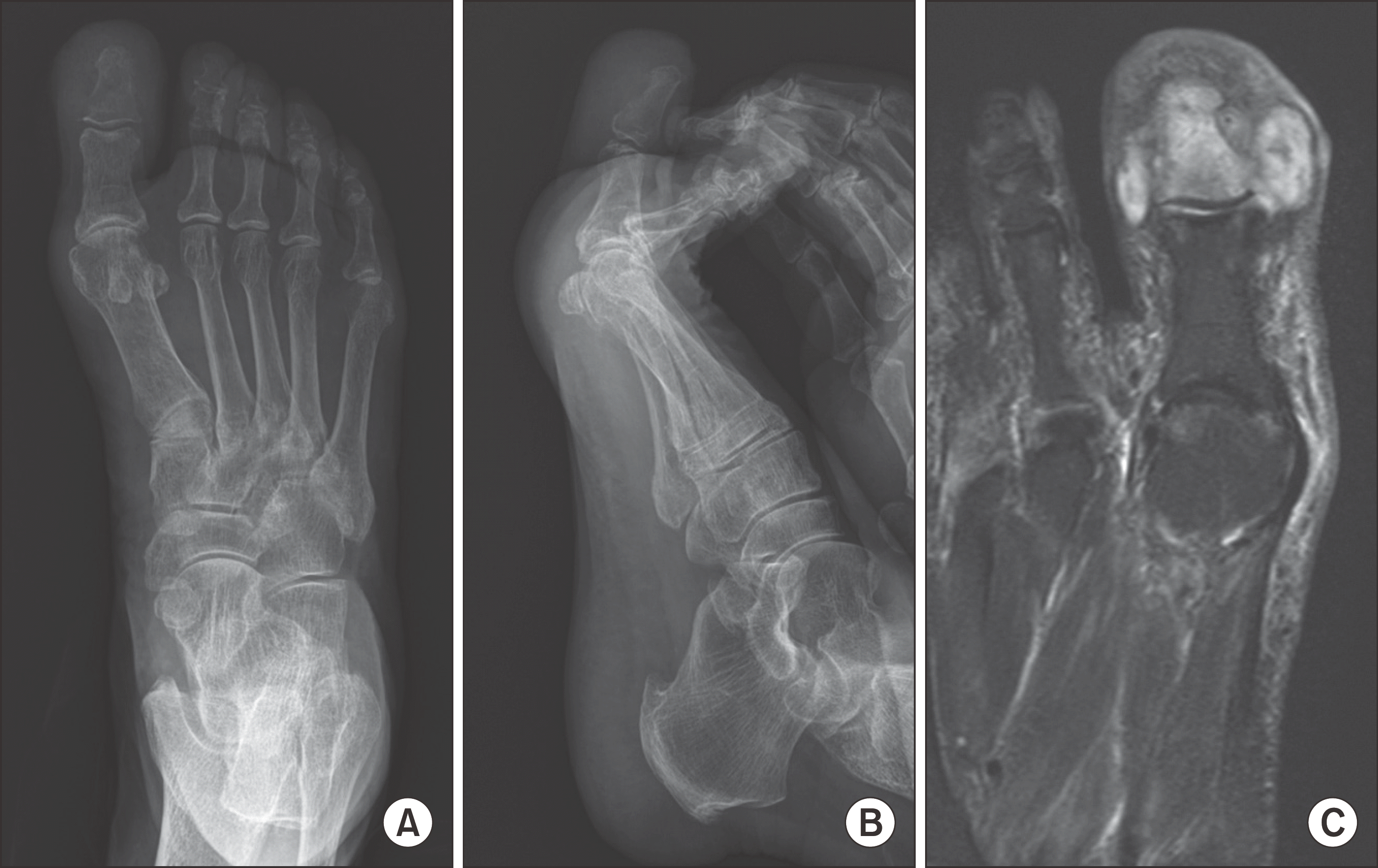 | Figure 1.Right foot anteroposterior (A) and lateral (B) radiographs show mild osteolytic lesion on the 1st distal phalanx. T2-weighted axial magnetic resonance imaging (C) shows hypersignal intensity on the 1st distal phalanx and suspicious of abscess formation around it, compatible with osteomyelitis. |
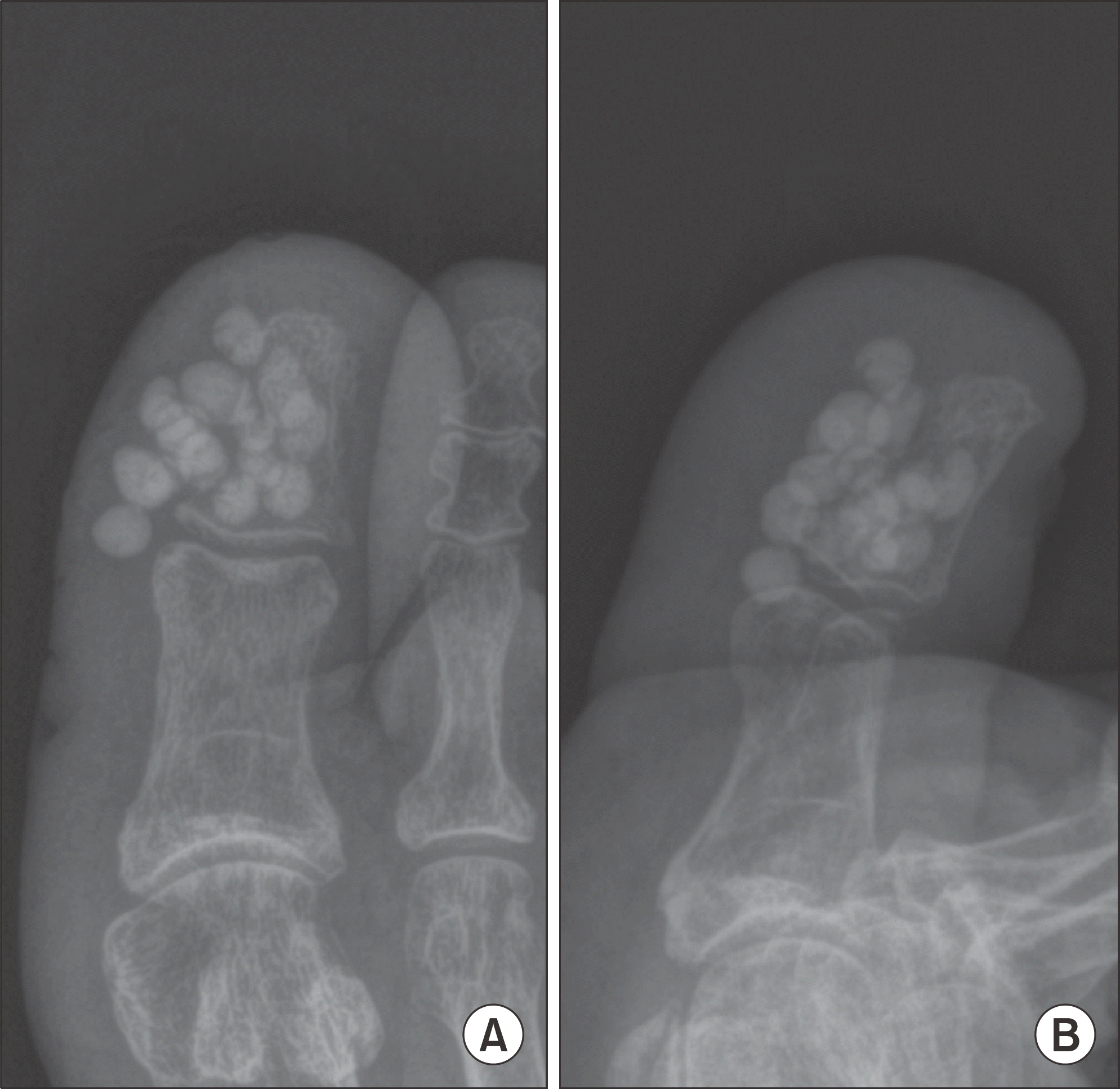 | Figure 2.Right 1st toe anteroposterior (A) and lateral (B) radiographs after debridement, bone curettage and antibiotic cement spacer insertion. |
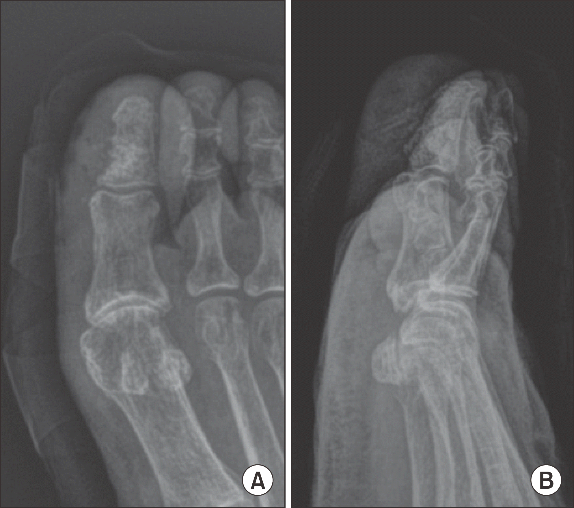 | Figure 3.Right 1st toe anteroposterior (A) and lateral (B) radiographs after antibiotic cement spacer removal and autogenous iliac bone graft. |
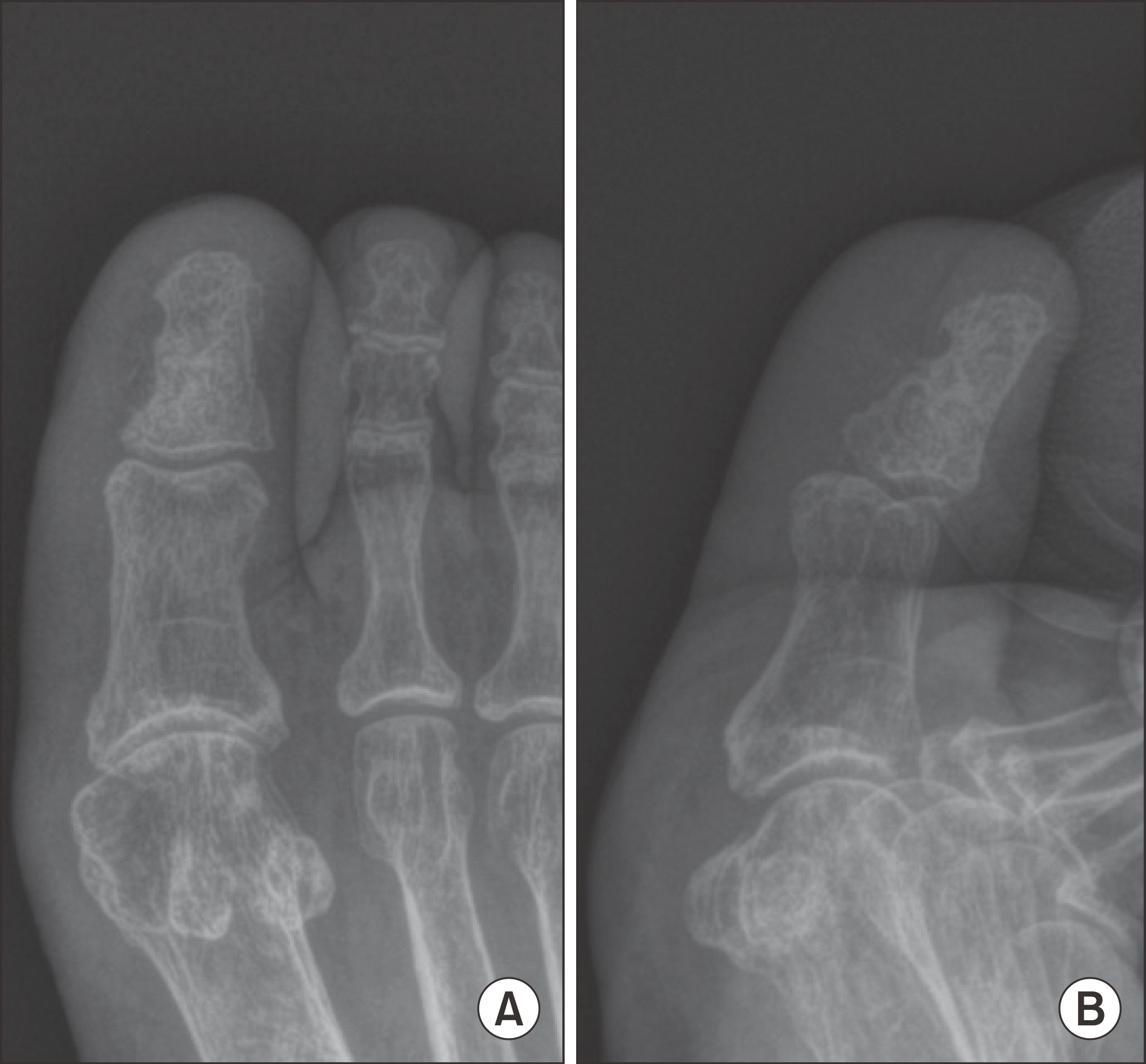 | Figure 4.Right 1st toe anteroposterior (A) and lateral (B) radiographs after 4 months of antibiotic cement spacer removal and autogenous iliac bone graft. The 1st distal phalanx healed nearly as normal, and we could prevent amputation. |
Table 1.
Strauss Wound Classification Scoring System
| Criteria* | 2 points | 1 point | 0 point |
|---|---|---|---|
| Appearance (wound base) | Red | Yellow or withe | Black |
| Size | Thumb tip or smaller | Between Thumb tip and fist | Larger than fist |
| Depth (maximum depth of probe) | Skin or subcutaneous | Muscle or tendon | Bone or joint |
| Infection (bioburden) | Colonized | Cellulitis | Septic† |
| Perfusion | Palpable pulses | Biphasic or triphasic dopplerable pulses (cool, pale or dusky, capillary refill 2~5 sec) | Monophasic or imperceptible pulses (cold, black/cyanotic/purplish, capillary refill >5 sec) |
Table 2.
Current Perception Threshold (CPT) by Neurometer
| Grade* | Sensory classification |
|---|---|
| 12 | Completely anesthetic |
| 9.9 | Severe hypoesthesia |
| 8.82 | Moderate hypoesthesia |
| 7.74 | Mild hypoesthesia |
| 6.62 | Moderate hyperesthesia |
| 5.54 | Mid hyperesthesia |
| 4.82 | Mid dysfunction |
| 3.9 | Very mild dysfunction |
| 2.78 | Extremely mild dysfunction |
| 1.66 | Slight dysfunction |
| 0 | No abnormal measures |
Table 3.
Summary of Patient Data Showing Successful Treatment Outcome
FU: follow-up, ALCS: antibiotic-loaded cement spacers, Interval: interval to secondary surgery, ABI: ankle-brachial index, TBI: Toe-brachial index, CPT: current perception threshold, Rt.: right, Lt.: left, MP: middle phalanx, DP: distal phalanx, PP: proximal phalanx, MSSA: Methicillin-sensitive Staphylococcus aureus, MRSA: Methicillin-resistant Staphylococcus aureus.
Table 4.
Summary of Patient Data Showing Non-Successful Treatment Outcome
Table 5.
Distribution of Lesions by Location
| Location | Number of patients (%) |
|---|---|
| Toes | 16 (41.0) |
| Metatarsals | 14 (35.9) |
| Midfoot | 2 (5.1) |
| Hindfoot | 7 (17.9) |
| Total | 39 (100) |
Table 6.
Comparing between Successful Group and Non-Successful Group
| Variable | Successful group (n=32) | Non-successful group (n=7) | p-value* |
|---|---|---|---|
| Age (yr) | 62.66+13.14 | 56.71+12.41 | 0.398 |
| Wagner wound grade | 3.28+0.45 | 3.43+0.49 | 0.554 |
| Strauss wound score | 5.13+0.41 | 2.86+0.23 | <0.01 |
| ABI | 1.01+0.16 | 0.98+0.16 | 0.654 |
| TBI | 0.67+0.16 | 0.52+0.16 | 0.040 |
| Neurometer | 10.69+1.52 | 11.33+0.92 | 0.359 |
 XML Download