

 PDF
PDF ePub
ePub Citation
 Print
Print


Introduction
Last couple of decades, successful vaccines against various type of pathogens have been developed to improve the human health by identification of antigens that induce immune response and elucidation of the mechanism associated with long-term memory [1]. Recent advances in immunology have expanded our knowledge about how innate immune system modulates the quantity and the quality of long-term T and B cell memories for protecting ourselves from pathogens [2]. However, there have been difficulties in developing vaccines against deadly diseases with extensive antigenic diversity, and an in-depth knowledge of the molecules and the signaling mechanisms is required for successful vaccine development against pathogens with antigenic variability [1]. As a part of an effort, imaging the vaccine components such as antigen or adjuvants and immune cell dynamics can provide important information for the development of effective vaccines.
For effective vaccination, vaccine components include sufficient danger recognition patterns, and initiate successful innate immune responses to the local cells such as stromal cells, tissue-resident monocytes, muscle cells, or mucosal cells at the injection site. Signals from these cells stimulate recruitment of circulating monocytes, leukocytes, or dendritic cells to the injection site, and amplify further inflammatory signals to activate the localized or recruited antigen presenting cells (APCs). Thus, effective vaccine candidates need to elicit an increase of antigen or adjuvant uptake, antigen processing and presenting by activated APCs. For sustained vaccine response, locally activated antigen bearing APCs (antigen-decorated, adjuvant-activated) must migrate to the T and B cell enriched region of the draining lymph nodes. In addition, soluble antigens and adjuvants also reach to the draining lymph nodes by free-fluid diffusion.
APCs display antigen peptides on MHC class I and II to activate CD8 T cells, and induce to differentiate antigen specific CD4 T cells into various types of effector cells such as Th1, Th2, Th17, Treg, and Tfh cells. With the cooperation of these cells, sufficient pool of central memory T cells in the skin and nodes, and effector memory T cells in the peripheral tissues are generated for sustain vaccine response. In addition, large pools of antigen-specific memory B cells ready to differentiate to produce antibody within a few days, and long-lived antibody secreting plasma cells in specific bone marrow niche are also required for successful vaccination. Antigen-specific B cells are generated in the B-cell follicles by binding of antigen to B cells, and co-activating signals from antigen expressing APCs and CD4 helper cells activate antigen-specific B cells to induce germinal center reaction. After a series of selections, B cells differentiate into antibody-secreting plasma cells or memory B cells.
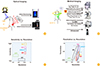
Therefore, monitoring vaccine components, such as antigen or adjuvants, and immune cell dynamics at the site of vaccination or draining lymph nodes can provide important information to understand more about the vaccine response. Immune cell dynamics represent effective activation of locally resident or attracted APCs at the site of vaccination and elicit specific T, B cells with antigen-specific memory, so it is required to develop more sensitive imaging methods in vivo. A variety of imaging modalities including bioluminescence imaging, nuclear medicine imaging (such as positron emission tomography [PET], single photon emission computed tomography [SPECT]), and magnetic resonance imaging (MRI) can provide in vivo non-invasive imaging for visualizing immune cell kinetics (Fig. 1) [3].
Imaging Strategies for Immune Cell Dynamics
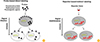
By accumulating a particular imaging signal to the background signals make it possible to visualize the localization of small molecules, proteins, or cells. Probe-based direct labeling or reporter-based indirect labeling methods are frequently used. Direct labeling requires an imaging probe such as fluorophore, radiotracer or paramagnetic nanoparticle to be internalized by the immune cells, while indirect labeling requires genetic modification for the expression of a detectable reporter protein or an enzymatic reaction for imaging (Fig. 2).
By conjugating a specific optical probe such as quantum dots and fluorescent molecules designed for ex vivo direct labeling, it is possible to study immune cell tracking in vivo. For visualization with nuclear imaging (PET/SPECT), 18F-fluorodeoxyglucose (FDG) for PET, 99mTc-hexamethyl propylene amine oxime (HMPAO), 111In-oxine for SPECT have been frequently used to label immune cells. These probes can easily label cells by entering the cytoplasm or by binding to the membrane, various magnetic nanoparticles such as iron oxides, gadolinium chelates and manganese chelates have been developed as probes for monitoring the immune cell dynamics with MRI. Despite easy labeling procedures, direct labeling has several drawbacks; limited by probe uptake or retention capacity, signal dilution from mitotic events, and changes of cellular characteristics after probe uptake.
Reporter based indirect labeling requires in vitro transfection of reporters to the immune cells or the use of isolated immune cells from reporter expressing transgenic animals. Frequently used reporter genes are as follows: fluorescence proteins (green fluorescent proteins, red fluorescent proteins, etc.) and luciferases (renilla, firefly, etc.) for optical imaging; herpes simplex virus-thymidine kinase (HSV-TK) with 18F labeled ganciclovir analogues and sodium iodide symporter (NIS) with radioiodine or 99mTc for nuclear imaging; transferrin receptor for MRI.
Various fluorescence reporter genes emitting at different wavelengths have been developed, but only some of them (red or near-infra-red fluorescence reporters) can be used for non-invasive in vivo imaging to overcome the limitation of low photon emission, signal to noise ratio and tissue absorption [4]. In the case of invasive intra-vital microscopic approaches, a broad range of fluorescence proteins is still available. However, various luciferase genes have been used to visualize immune cells because bioluminescence only needs a specific substrate for each luciferase and emits sufficient photons [356]. Although imaging costs are expensive, reporters for medical imaging (nuclear imaging, MRI) have less limitation for in vivo imaging and ready to use for clinical application compared to optical reporters such as fluorescent proteins and luciferases.
Pre-clinical Imaging of Immune Cell Dynamics for Vaccine Development
Considering the characteristics of immune cells for vaccine responses, the selection of labeling methods and modality is really important to visualize spatio-temporal immune cell kinetics in vivo. Detection and trafficking immune cells are still challengeable because of the difficulties of small number of light emitting cells and signal amplification to the detectable range. Various engineered reporter genes have been developed to improve expression and sensitivity [678]. To insert an exogenous reporter gene into the immune cells requires genetic modification, but the reporter gene can be passed on to the next generations without signal dilution, making it possible to track the fate of a given population of cells for a long term studies.
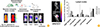
In addition, reporter transgenic mouse can be used to overcome the disadvantages of reporter transfection with a better understanding of immune cells (Fig. 3A). Instead of creating a protocol for each immune cell cluster, reporter transgenic mouse can be used to isolate specific immune cell clusters and observe the dynamics of immune cells representing the in vivo environment [8910].
Lymph node activation following vaccination in vivo is closely related to the localization of the antigens, which can be demonstrated with a variety of imaging probes and reporters. Fig. 3B shows an example of visualizing the location of 125I-labeled large hepatitis B virus antigen with different adjuvants at the vaccinated site and nearby draining lymph nodes. Depending on the type of adjuvant, time to stay at the injection site and the time to delivery to the draining lymph node were different resulting in different levels of lymph node activation.
Clinical Imaging of Immune Cell Dynamics for Vaccine Development
Since the pattern, magnitude, and duration of lymph node activation following vaccination in vivo have not been clearly defined yet, the role of vaccine adjuvants to improve the immune response should be investigated to access of vaccine efficacy. 18F-FDG PET is most commonly used as a standard option for imaging malignant tumors, but transient inflammation of lymph nodes due to vaccination can be visualized with 18F-FDG PET scans.
A recent study has been reported to visualize lymph node activation following administration of the Food and Drug Administration-approved human papillomavirus vaccines, Cervarix and Gardasil, which contain similar antigens with different adjuvants [11]. Following intramuscular vaccination, differences between duration of uptake and intensity of standard uptake value (SUV, a semi-quantitative measurement unit most commonly used to describe uptake intensity and compensates for variation in body size, injected activity and radioactive decay) were observed in the lymph nodes between Cervarix and Gardasil recipients. There were no major differences between duration of uptake and intensity of SUV between Cervarix and Gardasil recipients in ipsilateral axillary lymph nodes, but there was a significant contralateral node activation until the one month after the first vaccination only in Cervarix recipients, possibly reflecting the differences in vaccine adjuvant formulation.
Conclusion
Monitoring vaccine components and immune cell dynamics at the site of vaccination or the draining lymph nodes can provide important information to evaluate the efficacy of vaccine candidates. Considering the characteristics of immune cells to vaccine response, the labeling method and the modality should be carefully selected to visualize spatio-temporal immune cell kinetics in vivo. Detection and trafficking of immune cells are still challengeable, but various probes and reporters with higher sensitivity and specificity are being developed to improve immune cell imaging in both preclinical and clinical studies.
 XML Download