

 PDF
PDF ePub
ePub Citation
 Print
Print


Abstract
Purpose
The aim of this study was to determine the outcomes of fixation of AO/OTA type C2 fractures among IntraArticular fractures of the distal humerus using the paratricipital approach (side to side retraction of the triceps).
Materials and Methods
From June 2008 to January 2018, 12 patients underwent an open reduction and internal fixation with the paratricipital approach and were followed-up for more than 10 months after surgery. According to the AO/OTA classification, type C2 fractures were chosen among the IntraArticular distal humerus fractures. An extended posterior incision was used over the olecranon in the prone position, preserving the insertion site of the triceps brachii muscle. The fracture site was exposed by retracting the muscle side-to side through a dissection of the medial and lateral intermuscular septum of the triceps brachii muscle. The therapeutic results were assessed by the anatomical reduction of the articular surface and integrity of the metaphyseal contour in postoperative simple radiographs, complications, such as neuropathy or non-union, and the Mayo elbow performance score (MEPS) were checked to estimate the functional outcome.
Results
In the postoperative simple radiographs, no case showed more than 1 mm step-off and the disrupted contour of the distal humerus was recovered to normal alignment in most cases. The range of elbow joint motion in the last follow-up was 133.8° on average with a mean flexion contracture of 5.0°. The clinical results depending on the MEPS were excellent, except for two cases, which were good. Neuropathy of the ulnar nerve was observed in one patient, which was resolved after metal removal.
Go to : 
References
1. Yoon YC, Oh JK. Treatment of distal humeral fractures. J Korean Fract Soc. 25:223–232. 2012.

2. Sharma S, John R, Dhillon MS, Kishore K. Surgical approaches for open reduction and internal fixation of intraarticular distal humerus fractures in adults: a systematic review and metaanalysis. Injury. 49:1381–1391. 2018.
3. Ali AM, Hassanin EY, El-Ganainy AE, Abd-Elmola T. Management of intercondylar fractures of the humerus using the extensor mechanism-sparing paratricipital posterior approach. Acta Orthop Belg. 74:747–752. 2008.
4. Schildhauer TA, Nork SE, Mills WJ, Henley MB. Extensor mechanism-sparing paratricipital posterior approach to the distal humerus. J Orthop Trauma. 17:374–378. 2003.
5. Zalavras CG, Papasoulis E. IntraArticular fractures of the distal humerus-a review of the current practice. Int Orthop. 42:2653–2662. 2018.
6. Brouwer KM, Guitton TG, Doornberg JN, Kloen P, Jupiter JB, Ring D. Fractures of the medial column of the distal humerus in adults. J Hand Surg Am. 34:439–445. 2009.
7. Jupiter JB, Barnes KA, Goodman LJ, Saldaña AE. Multiplane fracture of the distal humerus. J Orthop Trauma. 7:216–220. 1993.
8. Nauth A, McKee MD, Ristevski B, Hall J, Schemitsch EH. Distal humeral fractures in adults. J Bone Joint Surg Am. 93:686–700. 2011.
10. Wong AS, Baratz ME. Elbow fractures: distal humerus. J Hand Surg Am. 34:176–190. 2009.
11. Wilkinson JM, Stanley D. Posterior surgical approaches to the elbow: a comparative anatomic study. J Shoulder Elbow Surg. 10:380–382. 2001.
12. Zlotolow DA, Catalano LW 3rd, Barron OA, Glickel SZ. Surgical exposures of the humerus. J Am Acad Orthop Surg. 14:754–765. 2006.
13. Bryan RS, Morrey BF. Extensive posterior exposure of the elbow. A triceps-sparing approach. Clin Orthop Relat Res. 166:188–192. 1982.
14. Kasser JR, Richards K, Millis M. The triceps-dividing approach to open reduction of complex distal humeral fractures in adolescents: a cybex evaluation of triceps function and motion. J Pediatr Orthop. 10:93–96. 1990.
15. Remia LF, Richards K, Waters PM. The Bryan-Morrey triceps-sparing approach to open reduction of T-condylar humeral fractures in adolescents: cybex evaluation of triceps function and elbow motion. J Pediatr Orthop. 24:615–619. 2004.
16. Erpelding JM, Mailander A, High R, Mormino MA, Fehringer EV. Outcomes following distal humeral fracture fixation with an extensor mechanism-on approach. J Bone Joint Surg Am. 94:548–553. 2012.
17. McKee MD, Jupiter JB. A contemporary approach to the management of complex fractures of the distal humerus and their sequelae. Hand Clin. 10:479–494. 1994.
18. McKee MD, Jupiter JB, Bamberger HB. Coronal shear fractures of the distal end of the humerus. J Bone Joint Surg Am. 78:49–54. 1996.
19. O'Driscoll SW. Parallel plate fixation of bicolumn distal humeral fractures. Instr Course Lect. 58:521–528. 2009.
20. O'Driscoll SW, Sanchez-Sotelo J, Torchia ME. Management of the smashed distal humerus. Orthop Clin North Am. 33:19–33. vii,. 2002.
Go to :
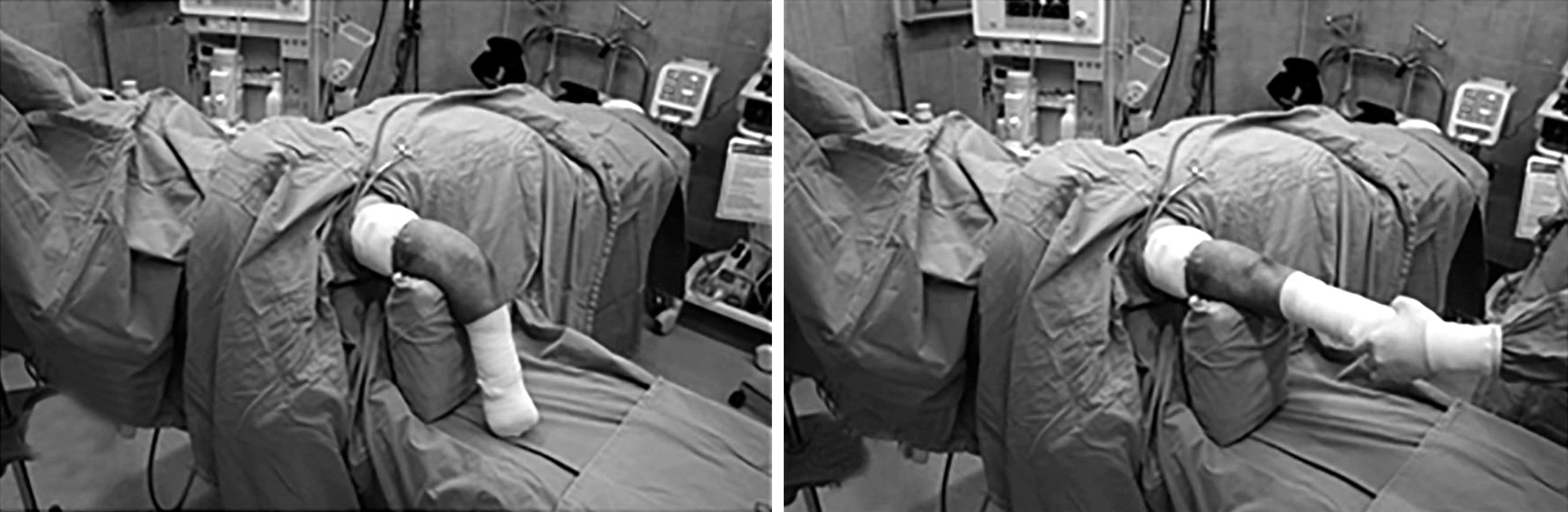 | Fig. 1.Positioning for the paratricipital approach. The approach was used over the olecranon in the prone position. The pedestal was located on the distal portion of the upper arm and by supporting the fracture site of distal humerus, allowed the operators to flex and extend the elbow joint to acquire sufficient exposure of the surgical field. |
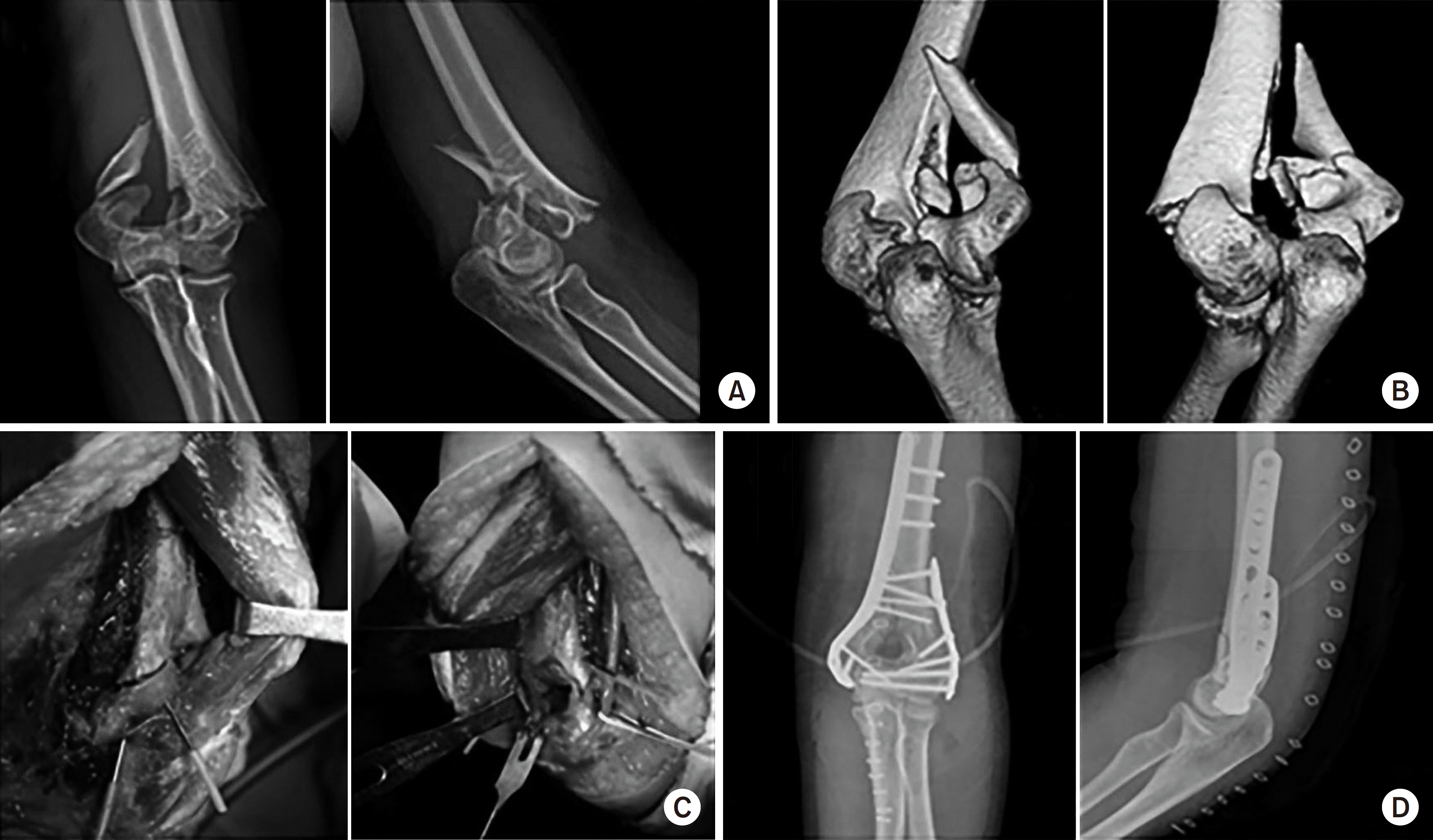
 | Fig. 2.A 58-year-old female slipped down and injured her elbow. (A, B) Anteroposterior X-ray and lateral X-ray and computed tomographies of a type C2 IntraArticular fracture of the distal humerus. (C) Intraoperative photos of the paratricipital approach. (D) Postoperative anteroposterior and lateral X-rays of the patient fixed with a plate and screws and maintained anatomical reduction. The patient had an excellent result. |
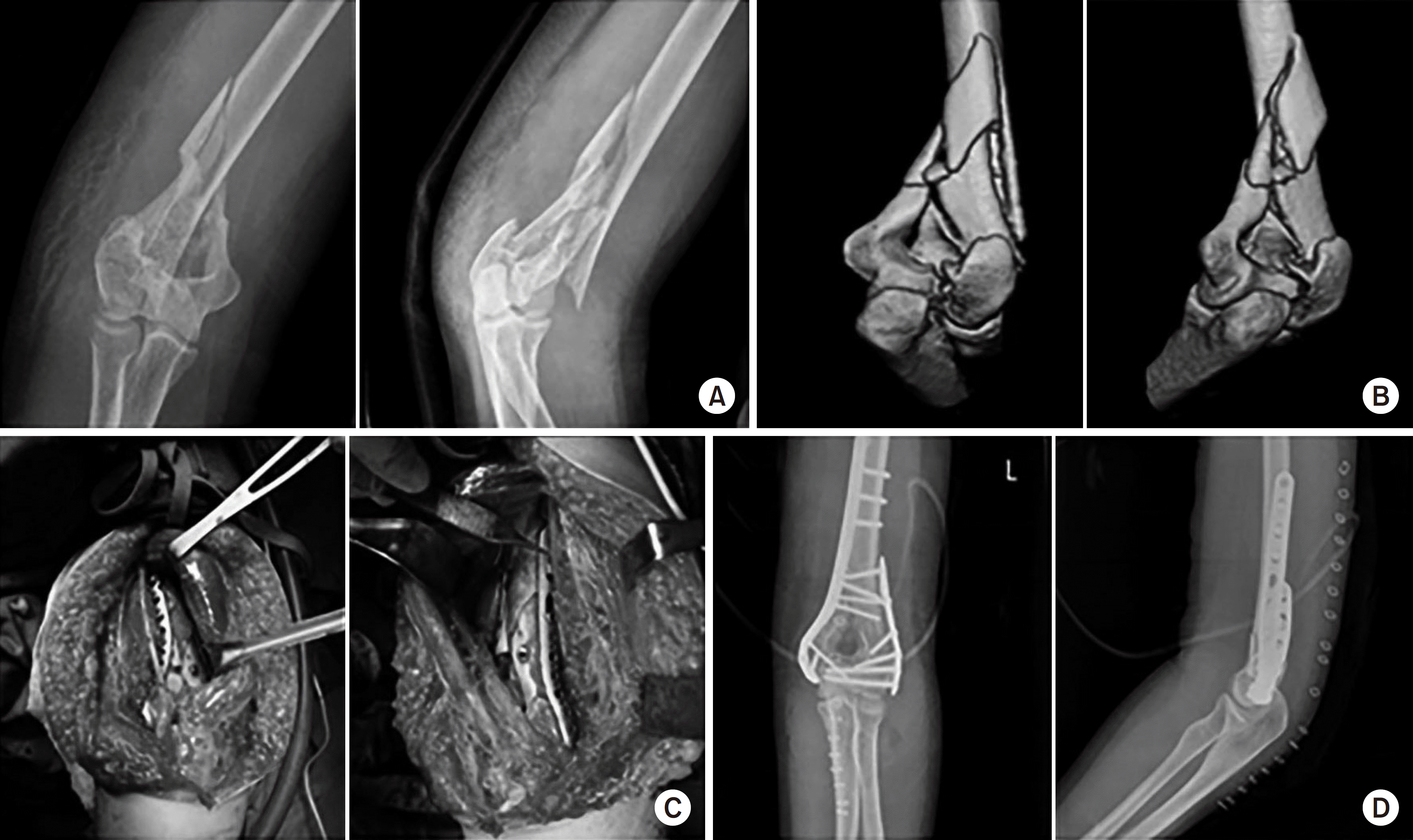
 | Fig. 3.A 39-year-old female fell on the ground and injured her elbow. (A, B) Anteroposterior X-ray and lateral X-ray and computed tomographies of a type C2 IntraArticular fracture of the distal humerus. (C) Intraoperative photos of the paratricipital approach. (D) Postoperative anteroposterior and lateral X-rays of the patient fixed with a plate and screws and maintained anatomical reduction. The patient had an excellent result. |
Table 1.
Demographics and Clinical Results of Patients
| Case No. | Age (yr) | Sex | Vector | Plating type | Complication | ROM (°) | Flex. (°) | Ext. (°) | MEPS |
|---|---|---|---|---|---|---|---|---|---|
| 1 | 60 | F | TA | Orthogonal | 140 | 140 | 0 | Excellent | |
| 2 | 63 | F | SD | Orthogonal | 135 | 140 | 5 | Good (mild pain) | |
| 3 | 56 | F | SD | Orthogonal | UN (resolved after | 135 | 140 | 5 | Excellent |
| metal removal) | |||||||||
| 4 | 39 | F | FD | Parallel | 140 | 145 | 5 | Excellent | |
| 5 | 58 | F | SD | Parallel | 120 | 120 | 0 | Excellent | |
| 6 | 54 | F | SD | Parallel | 115 | 135 | 20 | Good (mild pain) | |
| 7 | 49 | M | SD | Parallel | 130 | 145 | 15 | Excellent | |
| 8 | 79 | F | SD | Parallel | UN (improved spontaneously) | 130 | 140 | 10 | Excellent |
| 9 | 79 | F | SD | Parallel | 140 | 140 | 0 | Excellent | |
| 10 | 88 | F | SD | Parallel | 135 | 135 | 0 | Excellent | |
| 11 | 50 | F | SD | Parallel | 145 | 145 | 0 | Excellent | |
| 12 | 86 | M | TA | Parallel | UN (improved spontaneously) | 140 | 140 | 0 | Excellent |
| Mean | 63.4 | 133.8±8.8∗ | 138.8±6.8∗ | 5.0±6.7∗ |
 XML Download