

 PDF
PDF ePub
ePub Citation
 Print
Print


Abstract
Purpose
The study examined the fusion site and characteristics of the subtalar arthrodesis after intraarticular calcaneal fractures using computed tomography.
Materials and Methods
The clinical results and computed tomographic analysis of the fusion site were reviewed in 18 patients who were followed-up for a minimum of six months after undergoing subtalar arthrodesis due to traumatic arthritis caused by an intraarticular calcaneal fracture from December 2012 to April 2017.
Results
An evaluation of clinical results after subtalar arthrodesis revealed statistically significant improvements. In all cases, arthritis was found in the injured articular surface, which was displaced superolaterally from the initial primary fracture line of the calcaneus. Six months after arthrodesis, the subtalar fusion rate was 80.0% (16/20). Of these, 14 cases had a cannulated screw inserted in the uninjured site that is medial to the primary fracture line. Joint fusion was observed on the uninjured articular surface in 17 cases (85.0%).
Conclusion
Joint fusion was initially achieved at the uninjured posterior facet after subtalar arthrodesis due to traumatic arthritis caused by a displaced intraarticular calcaneal fracture. This suggests that meticulous surgical techniques and cannulated screw positioning at the uninjured site will promote joint fusion.
References
1. Easley ME, Trnka HJ, Schon LC, Myerson MS. Isolated subtalar arthrodesis. J Bone Joint Surg Am. 82:613–624. 2000.

2. Ferrao PN, Saragas NP, Strydom A. Isolated subtalar arthrodesis. JBJS Essent Surg Tech. 6:e12. 2016.
3. Tuijthof GJ, Beimers L, Kerkhoffs GM, Dankelman J, Dijk CN. Overview of subtalar arthrodesis techniques: options, pitfalls and solutions. Foot Ankle Surg. 16:107–116. 2010.
4. Bèzes H, Massart P, Delvaux D, Fourquet JP, Tazi F. The operative treatment of intraarticular calcaneal fractures. Indications, technique, and results in 257 cases. Clin Orthop Relat Res. 290:55–59. 1993.
5. Myerson MS. Primary subtalar arthrodesis for the treatment of comminuted fractures of the calcaneus. Orthop Clin North Am. 26:215–227. 1995.
6. Vulcano E, Ellington JK, Myerson MS. The spectrum of indications for subtalar joint arthrodesis. Foot Ankle Clin. 20:293–310. 2015.
7. Jones CP, Coughlin MJ, Shurnas PS. Prospective CT scan evaluation of hindfoot nonunions treated with revision surgery and low-intensity ultrasound stimulation. Foot Ankle Int. 27:229235. 2006.
8. Coughlin MJ, Grimes JS, Traughber PD, Jones CP. Comparison of radiographs and CT scans in the prospective evaluation of the fusion of hindfoot arthrodesis. Foot Ankle Int. 27:780–787. 2006.
9. Sanders R, Vaupel ZM, Erdogan M, Downes K. Operative treatment of displaced intraarticular calcaneal fractures: longterm (10–20 years) results in 108 fractures using a prognostic CT classification. J Orthop Trauma. 28:551–563. 2014.
10. Cohen MM, Vela ND, Levine JE, Barnoy EA. Validating a new computed tomography atlas for grading ankle osteoarthritis. J Foot Ankle Surg. 54:207–213. 2015.
11. Dorsey ML, Liu PT, Roberts CC, Kile TA. Correlation of arthrodesis stability with degree of joint fusion on MDCT. AJR Am J Roentgenol. 192:496–499. 2009.
12. Kitaoka HB, Alexander IJ, Adelaar RS, Nunley JA, Myerson MS, Sanders M. Clinical rating systems for the ankle-hindfoot, midfoot, hallux, and lesser toes. Foot Ankle Int. 15:349–353. 1994.
13. DiDomenico LA, Butto DN. Subtalar joint arthrodesis for elective and posttraumatic foot and ankle deformities. Clin Podiatr Med Surg. 34:327–338. 2017.
14. Davies MB, Rosenfeld PF, Stavrou P, Saxby TS. A comprehensive review of subtalar arthrodesis. Foot Ankle Int. 28:295–297. 2007.
15. Chahal J, Stephen DJ, Bulmer B, Daniels T, Kreder HJ. Factors associated with outcome after subtalar arthrodesis. J Orthop Trauma. 20:555–561. 2006.
16. Kitaoka HB, Patzer GL. Subtalar arthrodesis for posterior tibial tendon dysfunction and pes planus. Clin Orthop Relat Res. 345:187–194. 1997.
17. Vilá y Rico J, Jiménez Díaz V, Bravo Giménez B, Mellado Romero MÁ, Ojeda Thies C. Results of arthroscopic subtalar arthrodesis for adult-acquired flatfoot deformity vs posttraumatic arthritis. Foot Ankle Int. 37:198–204. 2016.
18. Scranton PE Jr. Results of arthrodesis of the tarsus: talocalcaneal, midtarsal, and subtalar joints. Foot Ankle. 12:156–164. 1991.
19. Ishikawa SN, Murphy GA, Richardson EG. The effect of cigarette smoking on hindfoot fusions. Foot Ankle Int. 23:996–998. 2002.
Fig. 1.
Case of a 52-year-old male's radiologic image series undergoing subtalar arthrodesis. (A) The initial simple foot lateral X-ray shows an intraarticular calcaneal fracture. (B) The initial coronal view of the subtalar joint posterior facet in computed tomography (CT) image shows an intraarticular calcaneal fracture. The black line indicates the uninjured region and the white line indicates the injured region. (C) Simple lateral foot X-ray was performed after surgery. (D) The CT coronal view shows arthritic changes in the subtalar joint. (E) Simple lateral foot X-ray was performed after subtalar arthrodesis. The CT sagittal view shows partial union of the subtalar joint after subtalar arthrodesis (F), CT coronal view shows partial union of the subtalar joint after subtalar arthrodesis (G).
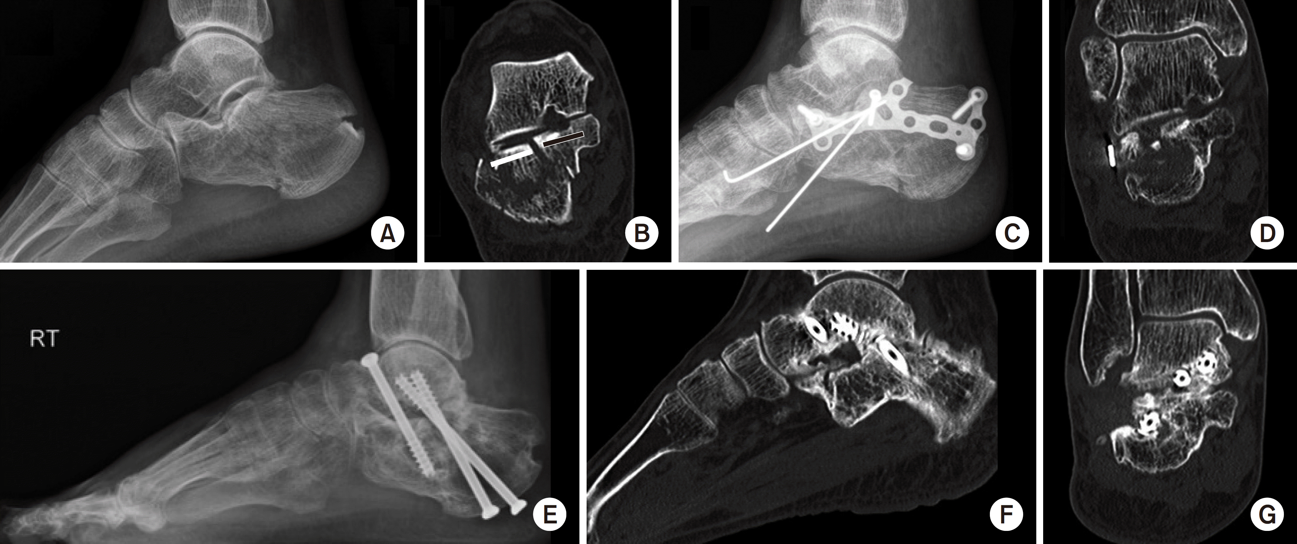
Fig. 2.
The fusion ratio was calculated by dividing the total sum of fused segment length, shown as the short dashed arrow, by the total sum of posterior subtalar joint length, shown as the long solid arrow.
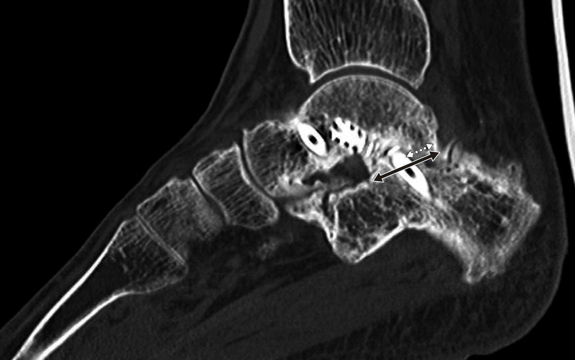
Table 1.
Computed Tomography Ankle OA Grading Scale
Table 2.
Summary of the Demographic Data, Radiologic and Clinical Results
Sandes classification of calcaneus fracture. †0, none; 1, autologous bone; 2, allogenous bone. BG: bone graft, Re-Op: re-operation, F/U: follow-up, PreOp: preoperative, VAS: visual analogue scale, AOFAS: American Orthopaedic Foot and Ankle Society, M: male, F: female, R: right, L: left, FH: fall from height, TA: traffic accident
 XML Download