

 PDF
PDF ePub
ePub Citation
 Print
Print


Abstract
Purpose
The purpose of this study was to investigate the radiologic and serologic factors related to postoperative union using intramedullary (IM) internal fixation in atypical femoral fractures (AFF), which are closely related to bisphosphonates (BPs) for osteoporosis.
Materials and Methods
From February 2008 to December 2016, 65 patients (71 cases) who had undergone IM nail fixation after diagnosis of AFF were enrolled in this study. Patients were divided into group A, who experienced union within 6 months and group B, who did not experience union within 6 months. They were evaluated for duration of BPs use, radiologic factors and serological factors.
Results
The mean duration of BPs use was 6.17 years in group A and 8.24 years in group B (p=0.039). In the subtrochanteric area, there were 14 cases (27.5%) in group A and 14 cases (70.0%) in group B. In the femoral shaft, there were 37 cases (72.5%) in group A and 6 cases (30.0%) in group B (p=0.001). On the preoperative, the flexion in the coronal plane was 5.9o (2.1o-9.2o) in group A and 8.0o (3.1o-12.1o) in group B (p=0.041). On the postoperative, conversion to valgus was 15 cases (29.4%), 8 cases (40.0%); conversion to neutral was 34 cases (66.7%) and 8 cases (40.0%); conversion to varus was 2 cases (3.9%) and 4 cases (20.0%), each (p=0.037). The fracture site gap was 1.5 mm (0–2.9 mm) on the front side and 1.2 mm (0–2.2 mm) on lateral side and 2.2 mm (0.9–4.7 mm) and 1.9 mm (0.5–3.5 mm), each (p=0.042, p=0.049). Among serological factors, there was no significant difference between the two groups.
Conclusion
Factors adversely affecting the union should be recognized before surgery, such as longterm BPs use or a severe degree of bending of the femur in the coronal plane. During surgery, proper reduction and spacing of the fracture site on the coronal plane should allow adequate reduction of the anterior and posterior surfaces. Obtaining anatomic reduction would be most beneficial for union, but if that is not possible, obtaining congenital valgus rather than varus on the coronal plane may be helpful for union.
References
1. Johnell O, Kanis JA. An estimate of the worldwide prevalence and disability associated with osteoporotic fractures. Osteoporos Int. 17:1726–1733. 2006.

2. Schousboe JT, Taylor BC, Fink HA, et al. Cost-effectiveness of bone densitometry followed by treatment of osteoporosis in older men. JAMA. 298:629–637. 2007.
3. Elliot-Gibson V, Bogoch ER, Jamal SA, Beaton DE. Practice patterns in the diagnosis and treatment of osteoporosis after a fragility fracture: a systematic review. Osteoporos Int. 15:767778. 2004.
4. Colón-Emeric CS. Ten vs five years of bisphosphonate treatment for postmenopausal osteoporosis: enough of a good thing. JAMA. 296:2968–2969. 2006.
5. Ha YC, Kim KW, Kim SH, Park YG. Bone biopsy of atypical subtrochanteric fracture in patient with prolonged bisphosphonate therapy: a case report. Korean J Bone Metab. 18:131–135. 2011.
6. Shane E, Burr D, Ebeling PR, et al. Atypical subtrochanteric and diaphyseal femoral fractures: report of a task force of the American Society for Bone and Mineral Research. J Bone Miner Res. 25:2267–2294. 2010.
7. Koh A, Guerado E, Giannoudis PV. Atypical femoral fractures related to bisphosphonate treatment: issues and controversies related to their surgical management. Bone Joint J. 99:295–302. 2017.
8. Shane E, Burr D, Abrahamsen B, et al. Atypical subtrochanteric and diaphyseal femoral fractures: second report of a task force of the American Society for Bone and Mineral Research. J Bone Miner Res. 29:1–23. 2014.
9. Lee YK, Yoon BH, Koo KH. Epidemiology and clinical features of atypical femoral fractures. J Korean Orthop Assoc. 48:175–179. 2013.
10. Schilcher J. High revision rate but good healing capacity of atypical femoral fractures. A comparison with common shaft fractures. Injury. 46:2468–2473. 2015.
11. Prasarn ML, Ahn J, Helfet DL, Lane JM, Lorich DG. Bisphosphonate-associated femur fractures have high complication rates with operative fixation. Clin Orthop Relat Res. 470:2295–2301. 2012.
12. Whelan DB, Bhandari M, McKee MD, et al. Interobserver and intraobserver variation in the assessment of the healing of tibial fractures after intramedullary fixation. J Bone Joint Surg Br. 84:15–18. 2002.
13. Lim HS, Kim CK, Park YS, Moon YW, Lim SJ, Kim SM. Factors associated with increased healing time in complete femoral fractures after longterm bisphosphonate therapy. J Bone Joint Surg Am. 98:1978–1987. 2016.
14. Gómez-Barrena E, Rosset P, Lozano D, Stanovici J, Ermthaller C, Gerbhard F. Bone fracture healing: cell therapy in delayed unions and nonunions. Bone. 70:93–101. 2015.
15. Abrahamsen B, Eiken P, Eastell R, et al. Cumulative alendronate dose and the longterm absolute risk of subtrochanteric and diaphyseal femur fractures: a register-based national cohort analysis. J Clin Endocrinol Metab. 95:5258–5265. 2010.
16. Abrahamsen B, Einhorn TA. Beyond a reasonable doubt? Bisphosphonates and atypical femur fractures. Bone. 50:1196–1200. 2012.
17. Odvina CV, Zerwekh JE, Rao DS, Maalouf N, Gottschalk FA, Pak CY. Severely suppressed bone turnover: a potential complication of alendronate therapy. J Clin Endocrinol Metab. 90:1294–1301. 2005.
18. Neviaser AS, Lane JM, Lenart BA, Edobor-Osula F, Lorich DG. Low-energy femoral shaft fractures associated with alendronate use. J Orthop Trauma. 22:346–350. 2008.
19. Winquist RA, Hansen ST Jr, Clawson DK. Closed intramedullary nailing of femoral fractures. A report of five hundred and twenty cases. J Bone Joint Surg Am. 66:529–539. 1984.
20. Kempf I, Grosse A, Rigaut P. The treatment of noninfected pseudarthrosis of the femur and tibia with locked intramedullary nailing. Clin Orthop Relat Res. 212:142–154. 1986.
21. Webb LX, Winquist RA, Hansen ST. Intramedullary nailing and reaming for delayed union or nonunion of the femoral shaft. A report of 105 consecutive cases. Clin Orthop Relat Res. 212:133–141. 1986.
22. Wu CC, Chen WJ. Treatment of femoral shaft aseptic nonunions: comparison between closed and open bone-grafting techniques. J Trauma. 43:112–116. 1997.
23. Canadian Orthopaedic Trauma Society. Nonunion following intramedullary nailing of the femur with and without reaming. Results of a multicenter randomized clinical trial. J Bone Joint Surg Am. 85:2093–2096. 2003.
24. Wolinsky PR, McCarty E, Shyr Y, Johnson K. Reamed intramedullary nailing of the femur: 551 cases. J Trauma. 46:392–399. 1999.
25. Sims SH. Subtrochanteric femur fractures. Orthop Clin North Am. 33:113–126. viii,. 2002.
26. Vanderschot P, Vanderspeeten K, Verheyen L, Broos P. A review on 161 subtrochanteric fractures: risk factors influencing outcome: age, fracture pattern and fracture level. Unfallchirurg. 98:265–271. 1995.
27. Garland DE, Rieser TV, Singer DI. Treatment of femoral shaft fractures associated with acute spinal cord injuries. Clin Orthop Relat Res. 197:191–195. 1985.
28. Teo BJ, Koh JS, Goh SK, Png MA, Chua DT, Howe TS. Postoperative outcomes of atypical femoral subtrochanteric fracture in patients on bisphosphonate therapy. Bone Joint J. 96:658–664. 2014.
29. Fogagnolo F, Kfuri M Jr, Paccola CA. Intramedullary fixation of pertrochanteric hip fractures with the short AO-ASIF proximal femoral nail. Arch Orthop Trauma Surg. 124:31–37. 2004.
30. Kwon BT, Kwon SH. Prediction of type of proximal femur fracture by analysis of serum makers. Osteoporosis. 13:31–35. 2015.
31. Lee HS, Lee CS, Jang JS, Lee JD, Um SM. Changes of serum alkaline phosphatase and osteocalcin during fracture healing. J Korean Orthop Assoc. 37:411–415. 2002.
32. Leung KS, Fung KP, Sher AH, Li CK, Lee KM. Plasma bone-specific alkaline phosphatase as an indicator of osteoblastic activity. J Bone Joint Surg Br. 75:288–292. 1993.
Fig. 1.
(A) Coronal view of the angle formed by two straight lines that pass the center of the proximal and distal parts, parallel to each other (letter ‘A’). (B) Lateral view of the same two lines (letter ‘B’).
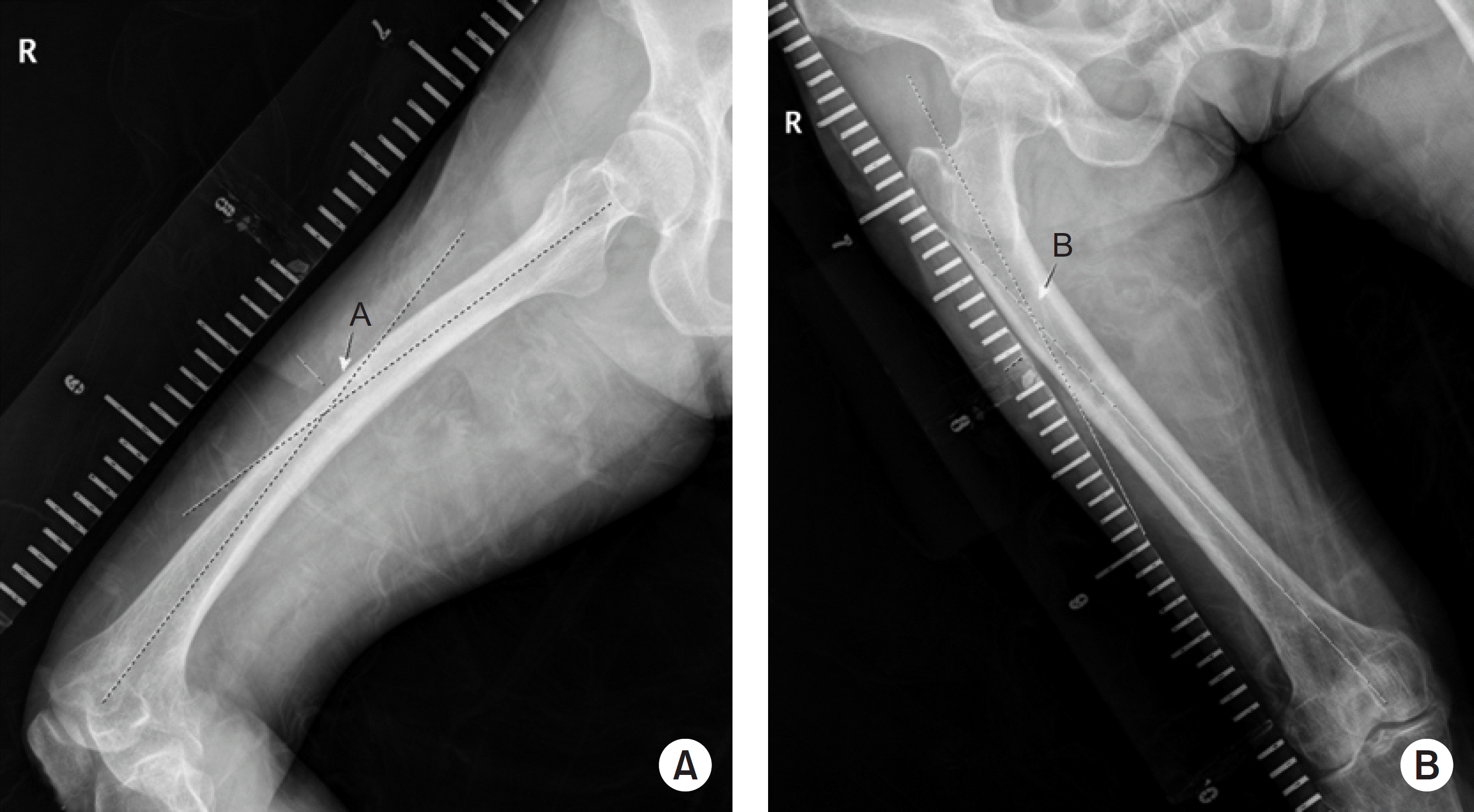
Fig. 2.
In the coronal plane, the angle that the proximal and distal fragments make on the same line as the fracture line based on the unaffected side (letter ‘C’), The angle that the distal and proximal fragments make on the basis of the fracture site of the affected side (letter ‘D’). If C-D is 0: neutral, (–): valgus, (+): varus.
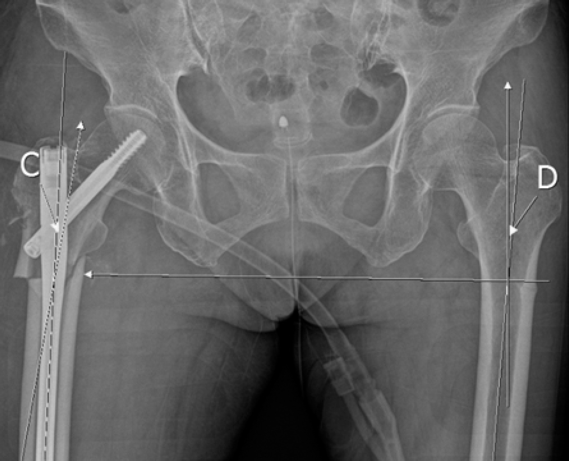
Fig. 3.
(A) The angle that the proximal and distal fragments make based on the fracture site in the coronal plane (letter ‘E’). (B) The value of the unaffected site on the basis of the affected site (letter ‘F’). If E-F is 0: neutral, (+): flexion, (–): extension.
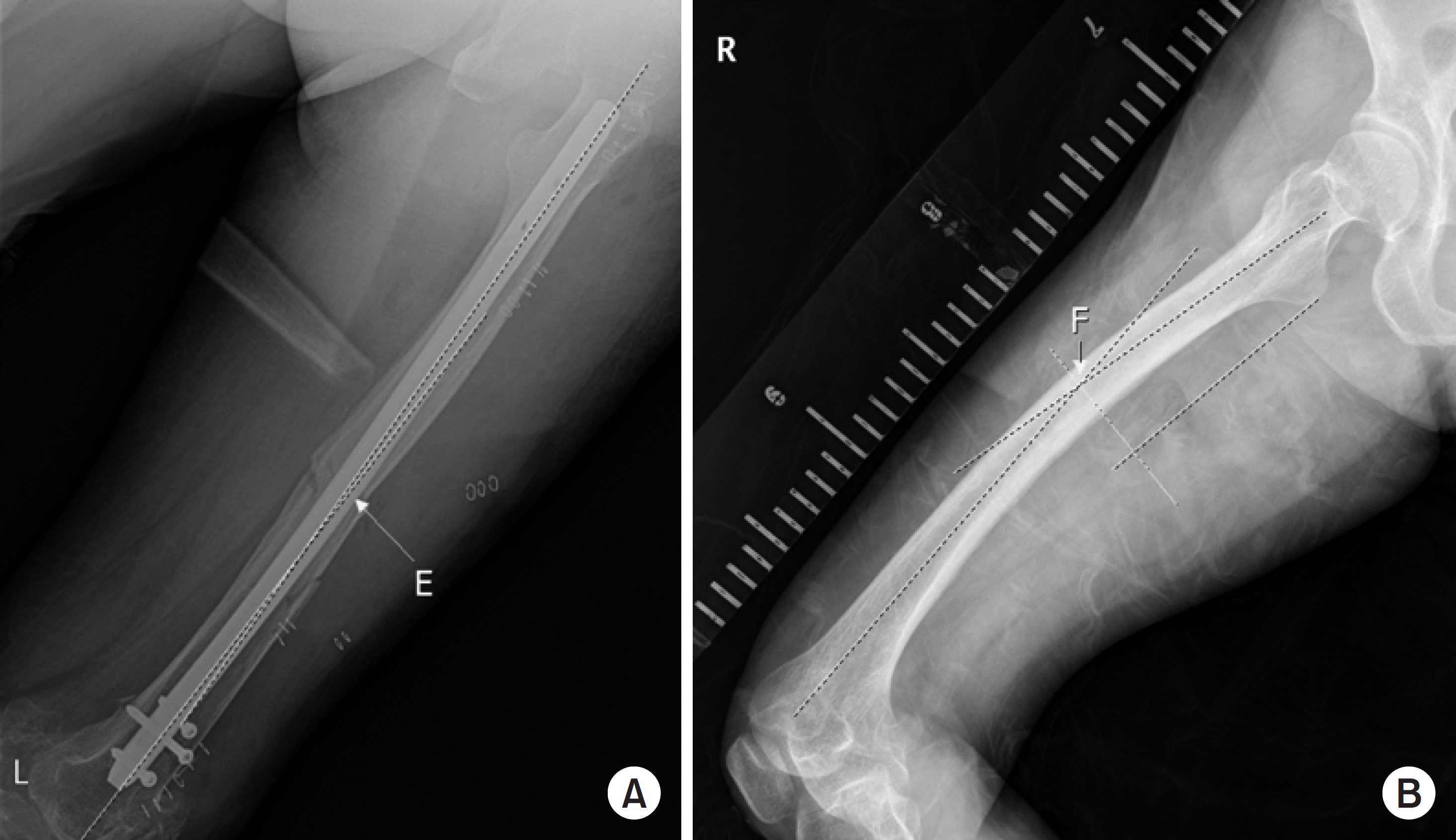
Fig. 4.
(A) Incidence of delayed union according to the correction angle in the coronal plane. (B) Incidence of delayed union according to the sum of the remaining anterior, posterior, medial and lateral gap sizes obtained from anteroposterior and lateral radiographs. Group A experienced union within 6 months of surgery. Group B did not experience union within 6 months.
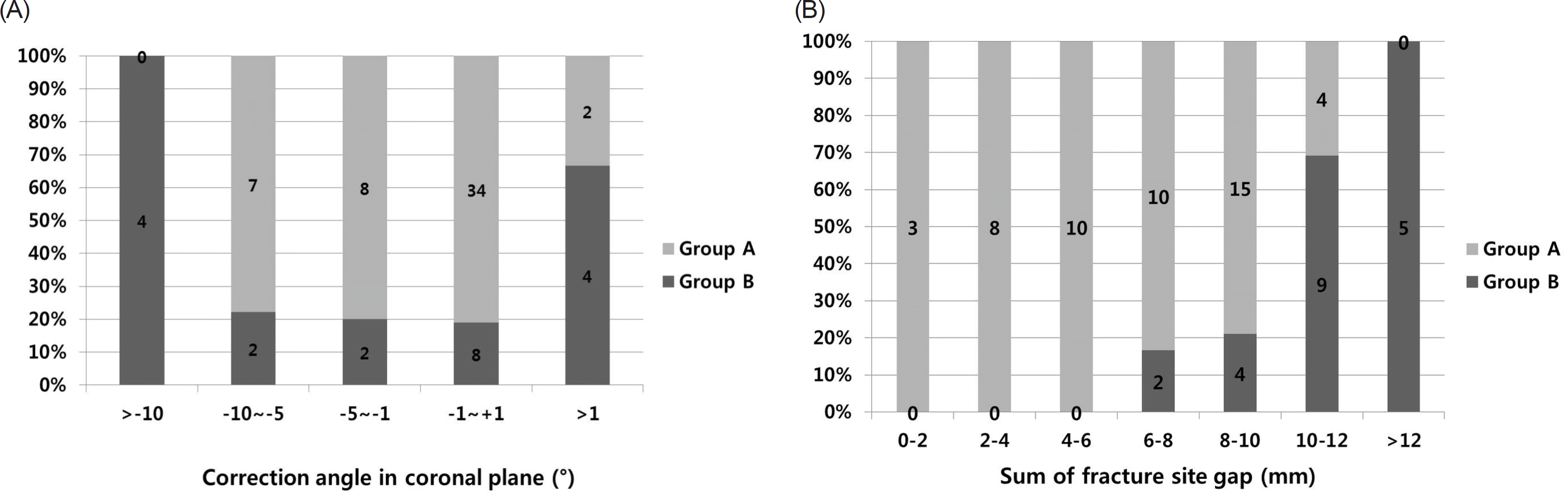
Table 1.
Baseline Patients Characteristics
Values are presented as mean±standard deviation, number only, or number (%). Group A experienced union within 6 months of surgery. Group B did not experience union within 6 months. BMI: body mass index, BMD: bone mineral density, HTN: hypertension, DM: diabetes mellitus, RA: rheumatoid arthritis, Dz.: disease, ASA: American Society of Anesthesiologists.
Table 2.
Fracture Site and Bisphosphonate Administration History
Table 3.
Radiographic Characteristics
Table 4.
Serologic Characteristics
 XML Download