

 PDF
PDF ePub
ePub Citation
 Print
Print


INTRODUCTION
Colorectal cancer (CRC) is the third most common cancer in the world, ranked after lung and breast cancer; and is the second most common cause of cancer death [1]. In the recent decade, incidence of this type of cancer increased rapidly in developing countries especially in regions that significant lifestyle alteration has occurred [2]. Studies have suggested that lifestyle factors play an important role in the etiology of CRC [345]. In this context, the unhealthy diet is responsible for approximately 30%–50% of the CRC incidence [6]. It has been proposed that some dietary factors including‚ high consumption of red meat have not only been associated with an increased risk of CRC, but have also affected the survival of these patients [2345]. Hence, developing strategies to improvement of CRC by altering the diet or specific dietary components has now become a hot topic in research [6].
Recently, studies have focused on the role of immunonutrients on CRC amelioration. These components modulate the suppressed immunologic responses in cancer patients [78]. Among several immunonutrients, glutamine, arginine, and omega-3 fatty acids have the strongest immune-modulatory action [9]. Arginine, one of semi-essential amino acid that used in all cells, by increasing of nitric oxide (NO) concentration can show its beneficial effects in many diseases [10]. NO is a climacteric substance for numerous biological processes such as neurotransmission and macrophage-mediated immunity [11]. Regarding the impact of this amino acid on cancer cells‚ recent evidence showed that arginine has a dual role (both stimulant and inhibitor) in the growth of tumors, where its function is dependent of the stage of the cancer [10]. Several studies indicated that arginine through increasing the NO concentration as well as decreasing the ornithine decarboxylase (ODC) activity, which are necessary for cell growth and differentiation‚ can reduce crypt cell hyper proliferation in CRC patients [121314].
As the studies in this context are inconclusive and the relation between arginine intake and CRC improving has not been systematically reviewed yet, we conducted a systematic review of the available evidence to elucidate the overall effects of arginine in this sense.
MATERIALS AND METHODS
Present systematic review was conducted and reported based on the Preferred Reporting Items for Systematic Reviews and Meta-Analyses guideline [15]. The prespecified study protocol is available from the authors upon request.
A systematic search of electronic databases such as PubMed, Scopus, ISI Web of Science, Cochrane library, and Google Scholar were performed up to March 2019. Our literature search was performed by 2 independent investigators (A.H. and J.K.), without any language or time limitation. Following terms were used in the search of abovementioned databases: (“arginine” OR “L-arginine” OR “D-arginine” OR “oral arginine” OR “dietary arginine” OR “supplement of arginine”) AND (“Colorectal cancer” OR “Colorectal Neoplasms” OR “Colorectal Adenoma”). To identify publications not found from computerized search, the reference lists of all eligible papers were investigated.
Selection of studies
Two reviewers (M.K. and J.K.) independently checked the title and abstract of retrieved articles to find the eligible studies. Where a decision could not be made based on title/abstract, full-text version of pertinent articles was checked to determine the suitable studies. Finally, we included all articles that focused on the effects of oral, supplement and dietary arginine consumption on the CRC. Studies were excluded if they were the clinical trials or were conducted in animals or in vitro models. Moreover, studies reporting the association between serum arginine and CRC without arginine intake or administration of drugs with arginine were excluded. Disagreements between reviewers were resolved by consensus.
Data extraction
Following information was extracted from selected papers using a pre-specified form: first author, published year, location, sex, study design, dose of arginine intake, follow up time, respondent, number of participants, confounding variables considered and main outcome. Data extraction was done independently by 2 authors (M.K. and J.K.) and any different opinions was settled by panel discussion.
Assessment of quality
The quality of the selected articles was evaluated using a validated scale for randomized controlled trials (Jadad scale) [16]. This 5-point quality scale included method of randomization concealment (0–2 points), blinding (0–2 points), and dropout rate (0–1 point). Trials having scores equal or greater than 3 were categorized as high quality and scores of 2 or less as low-quality. As before, data extraction and assessment of quality were done separately by 2 researchers (M.K. and J.K.). There was little disagreement between the 2 assessors over these stages, which were resolved by discussion and consensus.
RESULTS
Overview of studies identified
Detailed process of the study selection was illustrated in Figure 1. Generally, 523 kinds of literature were identified via the initial electronic search; 9 of them [171819202122232425] were selected according to inclusion and exclusion. These articles were from the Asian and European countries. The average of intervention period was 3 days. The number of participants varied from 28 to 120 subjects and the mean age was 60 years. The Jadad scale showed that all of the selected trials had high methodological quality. Table 1 provides a summary of the studies characteristics included in this systematic review.
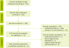
Table 1
Characteristics of included studies
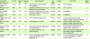
| First author (publication yr) | Country | Sample size | Mean age (yr) | Event followed | RCT design | Duration | Intervention | Jadad scores |
|---|---|---|---|---|---|---|---|---|
| Ma et al. [20] (2007) | China | 60 | 56 | Cell proliferation and ornithine decarboxylase | Parallel | 3 days | 120 mL arginine | 3 |
| Wang et al. [17] (2006) | China | 60 | 53 | Expression of PCNA and surviving, an inhibitor of apoptosis protein | Parallel | 3 days | 120 mL arginine | 3 |
| Hui et al. [19] (2011) | China | 45 | 53 | Expression of inducible nitric oxide synthase, cytotoxicity in tumor cells | Parallel | 3 days | 25 g arginine | 4 |
| Hu et al. [18] (2001) | China | 44 | 59 | Expression of PCNA | Parallel | 3 days | 25 g arginine | 3 |
| Caglayan et al. [21] (2012) | Turkey | 28 | 65 | Tumor infiltrative lymphocytes | Parallel | 7 days | 450 mL of eternal product with Arginine, omega-3 fatty acid and RNA | 3 |
| Finco et al. [22] (2007) | Italy | 28 | 66 | Immunologic cell response | Parallel | 6 days | 750 mL of a diet enriched with arginine, omega-3 fatty acids, and RNA associated with low-fiber foods | 4 |
| Braga et al. [23] (2002) | Italy | 200 | 65 | Immune response, gut oxygenation, and microperfusion | Parallel | 5 days | 750 mL of formula enriched with arginine and n-3 fatty acids | 4 |
| Horie et al. [24] (2006) | Japan | 67 | 69 | SSI, deep incisional SSI, and organ/space SSI | Parallel | 5 days | 750 mL of enteral diet supplemented with arginine, dietary nucleotides, and ω-3 fatty acids | 4 |
| Matsuda et al. [25] (2006) | Japan | 36 | 64 | CD4+ T cells producing intracellular cytokines (interferon-γ and interleukin-4) | Parallel | 5 days | 750 mL of supplemented liquid diet enriched with arginine, ω-3 fatty acids, and RNA | 3 |
RCT, randomized controlled trial; SSI, surgical site infection; PCNA, proliferating cell nuclear antigen.
![]()
Arginine supplement act as a tumor improving
In a study was done by the Wang et al. [17], 60 patients with CRC and 60 patients with colorectal adenoma (CRA) were enrolled and randomly divided into intervention groups that was given 120 mL arginine per day for 3 days and control group that received 120 mL glucose. The expression of proliferating cell nuclear antigen (PCNA) and surviving, an inhibitor of apoptosis protein, was examined immunohistochemically. Although no significant changes were observed in PCNA labeling index for para adenoma after arginine and glucose intervention, but survivin labeling index were significantly lower after taking arginine in colorectal adenocarcinoma and adenoma compare with control subgroups [17]. Hu et al. [18] evaluated changes of tumor PCNA among 44 patients with CRC who were received 25 g arginine hydrochloride for 3 days. Moreover, the changes of cell phase were determined with flow cytometry. The results showed that short period of arginine treatment in pharmaceutical dosage could inhibit PCNA expression from 55.3% to 43.9% in these patients. According to a similar study, expression of Ki67 protein as a cellular marker for proliferation decreased in both groups of colorectal carcinoma and adenoma after short-term application of arginine. In addition, the expression of inducible NO synthase (iNOS) and the levels of the serum NO that induces cytostasis and cytotoxicity in tumor cells, significantly rose in both groups [19].
Ma et al. [20] followed 60 CRC and 60 patients with CRA patients. They were divided into 4 groups, 30 patients in each. In the test groups, subjects received 120 mL arginine for 3 days. The expression of the PCNA, survivin, and NO synthase (NOS) was evaluated immunohistochemically, and ODC activity was assessed spectrophotometrically. Griess assay was used to detect NO in serum. In CRA patients treated with arginine, the PCNA, survivin labeling indexes and ODC activity of the tumor and para tumor mucosa were significantly lower than corresponding pretreatment values. Moreover, iNOS expression in the tumor significantly increased after arginine treatment. In the patients with CRC, serum NO levels were significantly higher than the patients with CRA. Altogether, arginine can restrain crypt cell hyper proliferation, the expression of survivin and block the formation and development of colorectal tumors. This effect of arginine, might be related to the increased serum NO concentration and decreased ODC activity.
Arginine supplementation acts as an immunonutrition
Caglayan et al. [21] followed 28 CRC patients who were given immunonutrition contained arginine, omega-3 fatty acid and RNA added enteral product, standard enteral, total parental nutrition, or normal nutrition (normal feeding planned by a dietitian) regimens. All regimens have the same calorie-nitrogen content that were given during preoperative period for 7-day. Subjects have an endoscopic biopsy before and after these regimens, and the lymphocyte population infiltrating mucosal were estimated in parts of the resected tumor tissue. The tissue specimens were examined and Immunohistochemical analysis was performed by staining with antihuman CD4(+), CD8(+), CD16(+), and CD56(+) antibodies. After nutrition, CD8(+), CD4(+) within the tumor cells were increased after nutrition in given immunonutrition group and CD56(+) cell infiltration in given immunonutrition group was considerably higher than in normal group. Finco et al. [22] followed 28 patients with neoplasms and diverticular disease that were susceptible of laparoscopic surgery. The patients in intervention group were given a diet enriched with arginine, omega-3 fatty acids, and RNA associated with low-fiber foods before and after operation. In control group, preoperative feeding was low-fiber diet and during postoperative period, oral nutrition was restored. Clinical trends and adverse reactions to early nutrition were monitored. After operation, the nutritional (albumin, prealbumin), immunological (lymphocyte subpopulations, immunoglobulins), and biohumoral elements were estimated. There was a significant increase in CD4 lymphocytes in group 1 before surgery as compared with baseline parameters. These results showed, perioperative immunonutrition including arginine, could be safe and useful in increasing the perioperative immunologic cell response and might be improve the preparation and relaxation of the intestinal loops. According to another study, it was show that preoperative intervention contained enteral diet supplemented with immunonutrition such as arginine for 5 days is effective for preventing surgical site infection (SSI) in patients with CRC without malnutrition. Moreover, the frequency of superficial incisional SSI, deep incisional SSI and organ/space SSI in the immunonutrition in compare with control group was significantly lower [24]. Braga et al. [23] followed 200 patients with colorectal neoplasm. They showed that the subjects who received immunonutrients only before operation or prolonged after surgery, had a significantly better immune response, gut oxygenation, and microperfusion than the other subjects. According to Matsuda et al. [25] study, 36 patients with CRC was followed. The control group was received formula enriched with arginine, omega-3 fatty acids and RNA. The results reported that preoperative immunonutrition corrects impaired T-helper (Th)1/Th2 balance in both cancer-bearing state and the postoperative period. We should consider that in all immunonutrition studies, arginine has given with another immunonutrient, so it is not clear that these effects were related to only about arginine.
DISCUSSION
To our knowledge, this was the first systematic review to collect recent evidence on effects of arginine and CRC. Out of 9 studies selected for this review 4 studies [17181920] reported an inverse association between arginine and crypt cell hyper proliferation and 5 studies [2122232425] showed that arginine by strengthen the immune system response could prevent the CRC recurrence after resection of tumor.
A protective effect of arginine consumption against cancer, especially CRC, is biologically plausible. Arginine and its metabolites have been shown to be safe and effective in inhabitation of carcinogenesis both in vitro bioassays and in vivo animal models [10]. This effect can be caused by the ability of arginine to restrain crypt cell hyper proliferation [20]. Colonic crypt cell hyper proliferation has been suggested to play a significant role in the multistep processes involved in the formation and development of CRC [26]. Increased PCNA expression‚ a DNA clamp that is essential for the replication process and as a cellular proliferative and functions index, was seen among patients with CRC [262728293031]. Also, it can be used to predict the degree of malignancy and the prognosis of these patients. As a result‚ any factor such as arginine that can inhibit this nuclear antigen can lead to improve the tumors prognosis. It also suppress hyper proliferation and carcinogenesis process of colorectal tumor cells [20].
According to previous reports‚ the progression of the cell cycle and control of apoptosis (programmed cell death) are intimately linked with carcinogenesis. Survivin, a new inhibitor of apoptosis protein, is expressed in the G2-M phase of the cell cycle in a cycle-regulated manner [32]. Over expression of survivin in cancer may overcome the apoptotic checkpoint that cause transformed cells progression through mitosis which is closely related to colorectal tumorigenesis. However, l-arginine treatment can markedly inhibit survivin overexpression [3334].
The role of NO in tumor cell apoptosis and survival depends on the cell type, the concentration of NO and the duration of cellular exposure to NO. High NO concentration induces cytostasis in tumor cells [353637]; however, a low level of NO plays an active role in the carcinogenesis [3839]. Increased extracellular concentrations of arginine can increaseNO production in endothelial cells and activated macrophages [40], via promoting the translation of NOS2 mRNA in cytokine-stimulated astrocytes and tetrahydrobiopterin, an essential cofactor for the biosynthesis of NO [4142].On the other hand, NO causes cytostasis by inhibiting ODC activity [43]. ODC is the initial limiting enzyme involved in the biosynthesis of polyamines compounds associated with rapid cell proliferation in both normal and neoplastic cells [44454647]. Aberrant regulation of this enzyme results in high basal levels of polyamines in epithelial tumors that cause development and progression of these cell to a malignant phenotype [4849].
After arginine treatment, serum NO level may considerably increase, which may be related to increase iNOS expression and decrease ODC activity. Through this way arginine can inhibit crypt cell hyper proliferation and have a protective role against tumorogenesis [1420].
Arginine, by promoting immune status, may prevent the recurrence of CRC after surgical resection [50]. In experimental models and clinical setting, arginine administration improved bacterial clearance and T-cell function. The arginine-NO pathway has been proposed to be the main defense mechanism for killing intracellular organisms and macrophage toxicity for target cell. Two enzymes‚ Arginase and NOS are crucial components of lymphocyte suppression pathway, and the metabolic products of these enzymes are important moderators of T-cell function [51]. Moreover, it is a major nitrogen source for NO synthesis in macrophages, lymphocytes, polymorph nuclear leukocytes, hepatocytes, vascular endothelial cells. NO prevents tumor cell evasion from the capillary bed into the parenchyma [50]. It emphasized that a good oxygenation is a critical event for the healing of the intestinal anastomosis performed during colorectal surgery. Therefore, NO synthesis from arginine may be responsible for the vasodilator tone and for the increased splanchnic micro perfusion [52].
Since Th cells regulate production of various cytokines and function of immune cells, arginine is able to enhance immune response resulting from the proliferation of Th cells, reinforce the competence of natural killer cells, and strengthen the activity of interleukin-2 and its receptor [53].
Several limitations of this systematic review should be considered. Firstly, the bias and cofounders possibility cannot be excluded. Most of the studies controlled for confounders such as sex, age, and Dukes stage but behaviors factors such as smoking and Arginine interactions with food and drugs are not considered, which might have partially influenced the results. Secondly, most of the selected studies were conducted in Asian countries, so the results might not be well applicable to other populations. Also, several studies have used a mixture of arginine and other immunonutrients. It remains unclear which nutrients exactly caused the immune-enhancing effect. Further, the reviewed studies did not assess adverse events of arginine intake. Despite the short intervention period of the reviewed studies, this study is the first systematic review on the effect of arginine on improving CRC.
CONCLUSION
In summary, arginine supplementation during the initiation stage of carcinogenesis decreased tumor production and crypt cell hyper proliferation, but during the promotion stage stimulated tumor growth. However‚ to achieve more accurate results further rigorous studies in different countries are needed.
 XML Download