

 PDF
PDF ePub
ePub Citation
 Print
Print


Sung-Hee Kim , Jin-Sung Park
, Jin-Sung Park
초록
The patients with myotonic dystrophy (MD) show ocular motor abnormalities including stra-bismus, vergence deficits, and inaccurate or slow saccades. Two theories have been proposed to explain the oculomotor deficits in MD. The central theory attributes the defects of eye movements of MD to the involvement of the central nervous system while the muscular theory attributes to dystrophic changes of the extraocular muscles. A 58-year-old woman with MD showed selective slowing of horizontal saccades and reduced peak velocities for both horizontal canals in head impulse tests, while smooth-pursuit eye movements and vertical head impulse responses were normal. This case suggests that the extraocular muscles-as a final common pathway of the voluntary saccade and reflexive vestibular eye movements-may better explain the defective rapid eye movements observed in MD.
REFERENCES
1.Mahadevan M., Tsilfidis C., Sabourin L., Shutler G., Amemiya C., Jan-sen G, et al. Myotonic dystrophy mutation: an unstable CTG repeat in the 3' untranslated region of the gene. Science. 1992. 255:1253–1255.

2.Osanai R., Kinoshita M., Hirose K. Saccadic slowing in myotonic dystrophy and CTG repeat expansion. J Neurol. 1998. 245:674–680.
3.Di Costanzo A., Toriello A., Mottola A., Di lorio G., Bonavita V., Tedeschi G. Relative sparing of extraocular muscles in myotonic dystrophy: an electrooculographic study. Acta Neurol Scand. 1997. 95:158–163.
4.Emre M., Henn V. Central eye movement disorder in a case of myotonic dystrophy. Neuro-Ophthalmology. 1985. 5:21–25.
5.Hennerici M., Fromm C. Isolated complete gaze palsy: an unusual ocular movement deficit probably due to a bilateral parapon-tine reticular formation (PPRF) lesion. Neuro-Ophthalmology. 1981. 1:165–173.
6.Johnston JL., Sharpe JA. Sparing of the vestibulo-ocular reflex with lesions of the paramedian pontine reticular formation. Neurology. 1989. 39:876.
7.Kuwabara T., Lessell S. Electron microscopic study of extraocular muscles in myotonic dystrophy. Am J Ophthalmol. 1976. 82:303–309.
8.Lessell S., Coppeto J., Samet S. Ophthalmoplegia in myotonic dystrophy. Am J Ophthalmol. 1971. 71:1231–1235.
9.Hess A., Pilar G. Slow fibres in the extraocular muscles of the cat. J Physiol. 1963. 169:780–798.
10.Anastasopoulos D., Kimmig H., Mergner T., Psilas K. Abnormalities of ocular motility in myotonic dystrophy. Brain. 1996. 119(Pt 6):1923–1932.
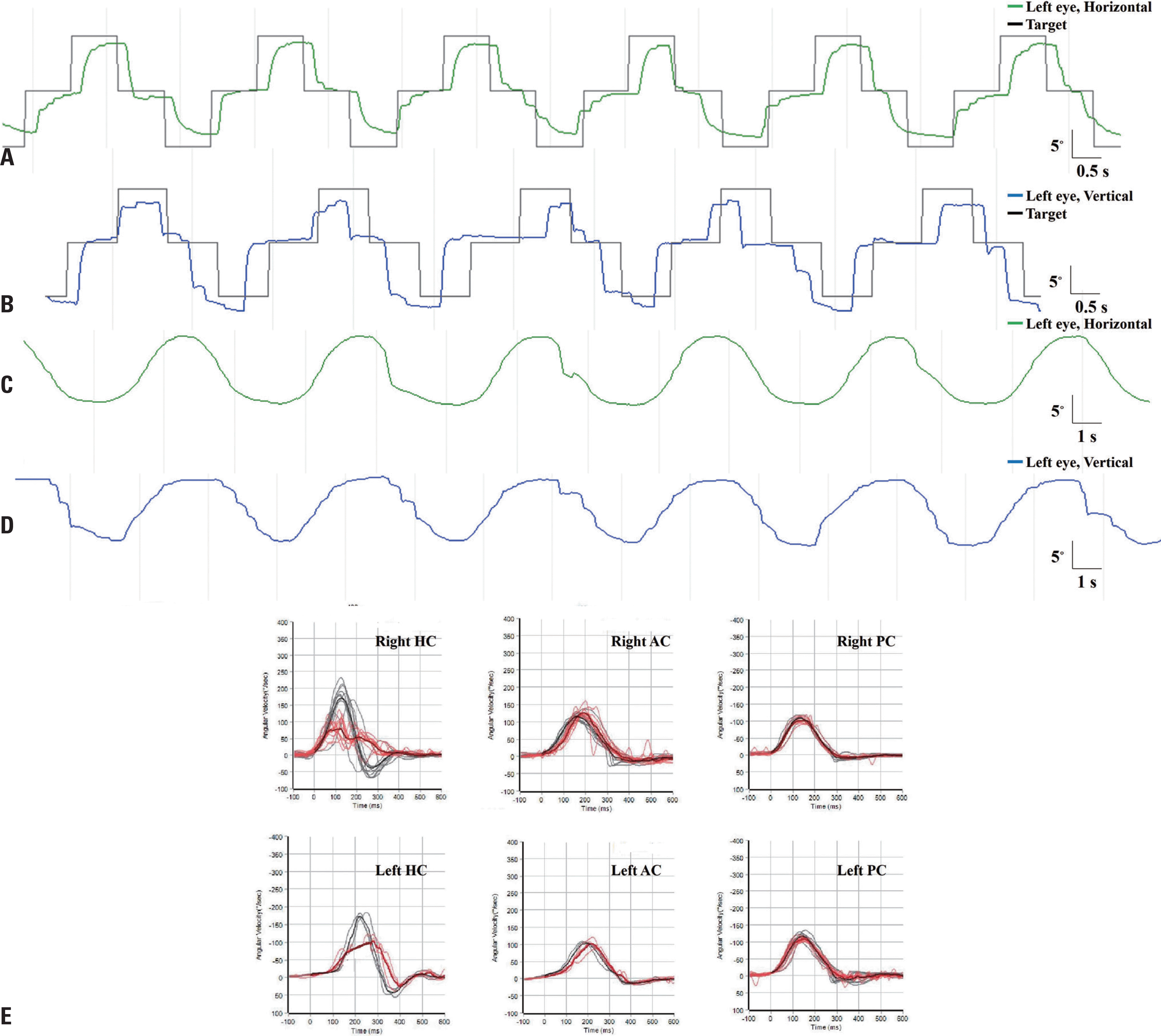
Fig. 1.
A 58-year-old woman with myotonic dystrophy showed slowing and hypometria of horizontal saccades (A). The amplitudes and velocities of the vertical saccades were intact, although anticipatory eye movements were frequently observed (B). Smooth pursuit was normal in both horizontal directions (C) and in the upward direction (D). Video head impulse tests revealed decreased peak velocities for bilateral horizontal canals without catch-up saccades, but pre-served responses for the vertical canals (E). Upward deflection indicated rightward eye motion in the horizontal plane and upward eye motion in the vertical plane. S, seconds; HC, horizontal canal; AC, anterior canal; PC, posterior canal.
 XML Download