

 PDF
PDF Citation
 Print
Print


INTRODUCTION
Most of temporal arteritis is known as giant cell arteritis (GCA), which is a granulomatous vasculitis that occurs in older adults, mainly those over 50 years old [1]. Although it is rare, young patients under 40 years of age could also develop vasculitis in the temporal artery, and this requires some differential diagnosis [2]. The patient may have systemic vasculitis involving temporal artery, or the elderly type arteritis such as GCA, though it is rarely seen in the young, or juvenile temporal arteritis (JTA), which is a eosinophilic arteritis that occurs only locally in the temporal artery [23].
Since the first report of JTA in 1975 [4], a total of 22 cases have been reported so far [234567891011121314151617181920], but its etiology, pathogenesis, and long-term prognosis are still unknown. Although JTA is a localized disease, it is often accompanied by peripheral blood eosinophilia, so some authors have suggested the concept of JTA with eosinophilia [81213]. However, its association with pathogenesis, clinical features, and prognosis of JTA remains unclear. Herein, we report the case of a 24-year-old man with concurrent JTA and hypereosinophilic syndrome (HES), and present a review of the literature on JTA, specifically focusing on the association of combined peripheral blood eosinophilia and the course of JTA.
CASE REPORT
A 24-year-old Korean man presented with a painful nodular lesion on his left temple that worsened from a month ago. He had visited a primary care center, where blood tests revealed eosinophilia, and then referred to our hospital for further evaluation. He had a history of chronic rhinosinusitis, and had been using a fluticasone/salmeterol inhaler for 6 months as empirical treatment for cough and dyspnea. He denied taking any herbal medicines, raw foods, or other drugs that could increase eosinophil count.
A physical examination revealed that tender nodular lesions on both temples (the left side was worse) and the palpable lymph node (LN) on the right inguinal area. Laboratory findings included total leukocyte, eosinophil, and hemoglobin level of 20,900/mm3, 8250/μL, and 17.4 g/dL, respectively. The following tests were within normal ranges: serum electrolytes, creatinine, alanine aminotransferase, aspartate aminotransferase, prothrombin time, activated partial thromboplastin time, and C-reactive protein. The erythrocyte sedimentation rate (ESR) was slightly elevated (26 mm/hr; normal, 0–20 mm/hr). Total IgE (PRIST, Shin Jin Medics Inc., Ilsan, Korea) was 103 IU/mL (normal, <100 IU/mL), the level of eosinophil cationic protein was markedly elevated as > 200 μg/L (normal, <18 μg/L), and vitamin B12 was not increased (388 pg/mL; normal, 197–771 pg/mL). Antineutrophil cytoplasmic antibodies against proteinase 3 or myeloperoxidase and antinuclear antibodies were negative. Serologic test for common parasites (Clonorchis sinensis, Paragonimus westermani, Cysticercus, Sparganum, and Toxocara canis), hepatitis B, C, and human immunodeficiency virus were negative as well.
In the tests performed to diagnose asthma, the results showed 1.28 mg/mL for methacholine PC20 and 64 ppb for a fractional exhaled nitric oxide, which were consistent with asthma. Computed tomography (CT) scan for chest and abdomen revealed that several LN enlargements at bilateral axillary areas, wedge-shaped nonenhancing portion at the spleen suggesting splenic infarction (Fig. 1A), and enlarged LN at right inguinal area (Fig. 1B). An echocardiogram showed no remarkable finding. A bone marrow biopsy showed eosinophilic hyperplasia but no evidence of myeloproliferative disease or chromosomal abnormality. The patient tested negative on PCR for BCR/ABL and FISH for FIP1L1/PDGFRα and FIP1L1/PDGFRβ.
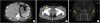 | Fig. 1Computed tomography findings. (A) Wedge-shaped nonenhancing portion (arrow) at the spleen suggesting splenic infarct. (B) Enlarged lymph node (arrowhead) in the right inguinal region. (C) Fusiform dilatation in the both superficial temporal arteries (arrow) with wall thickening. R, right; L, left.
|
CT angiography and 3-dimensional-brain showed fusiform dilatation in the both superficial temporal arteries with wall thickening (Fig. 1C). The patient underwent excisional biopsy of bilateral superficial temporal arteries (Fig. 2A) and the right inguinal LN simultaneously. The histopathological findings of the left superficial temporal artery revealed that marked eosinophilic infiltration in arterial wall and perivascular tissue with eosinophilic abscess, focal destruction of vascular wall, and organizing thrombi, but granuloma formation with multinucleated giant cells was not seen (Fig. 2B, C). The right inguinal LN showed reactive follicles and massive eosinophil infiltration, but no evidence of malignancy.
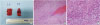 | Fig. 2Biopsy of the superficial temporal artery lesions. (A) The specimens of bilateral superficial temporal arteries after surgical excision. The histopathological findings of the left superficial temporal artery showing marked eosinophilic infiltration in arterial wall and perivascular tissue, but not giant cells or granulomatous lesion (B: H&E, ×100; C: ×400).
|
Based on these results, the diagnosis of concurrent JTA and HES was made. The patient underwent surgical excision of both superficial temporal arteries for his JTA, and administered systemic steroid (intravenous methylprednisolone 90 mg [1 mg/kg] per day) for HES. There was no recurrence of JTA after surgical excision during 2 years of follow-up, and he was well maintained without any symptoms due to eosinophilia with low-dose steroid (prednisone 10 mg/day).
DISCUSSION
If vasculitis of the temporal arteries develops in the young, physicians should consider the following diagnoses: (1) JTA, (2) systemic vasculitis including eosinophilic granulomatosis with polyangiitis (EGPA), thromboangiitis obliterans, and polyarteritis nodosa, involving the temporal artery, and (3) the elderly type vasculitis such as GCA, though it is rarely seen in young adults [2321].
In our case, the pathologic findings of the temporal arteries showed panarteritis with eosinophilic infiltrates but no giant cell or granulomatous lesion was seen, so that we could excluded GCA. Next, we considered the possibility of EGPA. Although he had asthma, chronic rhinosinusitis, and peripheral blood eosinophilia, the pathologic findings of both temporal arteries were not consistent with EGPA, because there was no prominent extravascular eosinophil infiltration, necrotizing vasculitis, or granuloma formation [22]. Therefore, for his temporal arteritis, we concluded that it was consistent with JTA.
However, he had other problems such as several LN enlargement, splenic infarct, and eosinophil count over 8,000/μL. We considered the following diagnosis: JTA with eosinophilia, JTA with Kimura disease, and concurrent JTA and HES. The pathologic finding of excised right inguinal LN showed massive eosinophil infiltration, and this could be due to both Kimura disease and HES [2324]. However, the eosinophil count was extremely high, and there was splenic infarct that suggested thrombosis, which is one of the organ involvements of hypereosinophilia [25], thus we concluded that it was more compatible with HES than Kimura disease. Finally, the diagnosis of concurrent JTA and HES was made.
In 1975, Lie et al. [4] first reported four cases of JTA in the young. These patients complained of painless nodules on their temple, and the pathologic findings revealed nongiant cell inflammation of temporal arteries [4]. From then on until February 2019, according to our literature review through PubMed and Scopus, 18 more cases have been reported [23567891011121314151617181920]. Including the present case, a total of 23 cases of JTA were reported and their mean age was 27 years (range, 7 to 44 years) and there was the male predominance (18 men and 5 women). Only seven cases out of 23 cases involved both side of temples. Clinical manifestation of JTA is as following: age younger than 40 to 50 years, a palpable nodule or vessel engorgement in the temple (painful or painless), and a localized disease so that there is no systemic involvement of vasculitis and usually shows normal ESR [234]. Peripheral blood eosinophilia could be present [2].
The histopathologic findings include intima hyperplasia and disruption of internal elastic lamina, which could also present in GCA, and lymphoeosinophilic infiltrate and its perivascular extension, and endothelial hyperplasia, which usually present in JTA, not in GCA [21114]. The most important difference from GCA is that multinucleated giant cells and granulomatous infiltrates are not observed in JTA [1014].
JTA is a localized disease so that excision is curative and steroid treatment is usually not required, and after excision, recurrence is known to be rare [2]. Of the 22 cases identified through literature search, the excision of affected temporal artery was done in 19 cases [2345679101112141516171819], and only one case reported that the symptoms persisted after resection [14]. Two of the 18 cases that resolved after excision recurred later on the opposite side [1218]. Of the 3 cases without excision, 2 were treated with steroid because of combined disease such as Kimura disease [20] or HES [12], and 1 was treated with tocopherol nicotinate [8], and these patients reported no recurrence.
Of the total 23 cases including our case, we found 10 cases of JTA combined with eosinophilia, and a summary of those cases presented in Table 1. Recurrence after excision only treatment was more frequent in the cases of JTA with eosinophilia than without eosinophilia (2 of 6 cases vs. 0 of 12cases). That is, prognosis of JTA may be worse when present with eosinophilia. In addition, one of the recurred cases reported that eosinophil count was elevated at recurrence [18]. Although only a small number of cases were available for the trend of eosinophil count after excision [8131819], eosinophilia may persist even if temporal arteritis improves without systemic steroids, and may lead to recurrence because documented recurrences occurred only in the patients with JTA with eosinophilia who treated locally. According to very recent case series (n = 12) from France, 41.7% (5 of 12) had peripheral eosinophilia, and this figure was similar to our estimate of 43.4% (10 of 23). However, the relation between the presence of eosinophilia and the recurrence of JTA was not presented in that study [26].
Table 1
Summary of the cases of juvenile temporal arteritis with blood eosinophilia reported in the literature
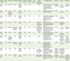
| Year/case No./Ref. | Age/sex | Ethnicity | Comorbidity | Symptom, sites | Systemic symptoms | Eosinophil (/mm3) | ESR (mm/hr) | Organ involvement of eosinophilia | Pathologic finding | Treatment | Course | Eosinophilia |
|---|---|---|---|---|---|---|---|---|---|---|---|---|
| 1996/1 [8] | 39/M | Japanese | Asthma | Painless nodule, bilateral | None | 2,660 | 6 | None | Obliteration of the vessel lumen. Disruption of IEL. Panarteritis with eosinophilic infiltration. No GC. | Tocopherol nicotinate | No recurrence for 2 years | Remained (15%–30%) |
| 2004/2 [3] | 31/M | N/A (published by Greek authors) | None | Painless nodule, left | None | 562 | 3 | None | Arteritis with eosinophilic infiltration. No GC. | Excision | No recurrence | N/A |
| 2005/3 [11] | 23/M | Japanese | None | Painless nodule, left | None | 2,755 | 3 | None | Marked intimal thickening with fibrosis. Eosinophilic infiltrates. Focal disruptions of the IEL. No GC. | Excision | No recurrence for 8 months | N/A |
| 2006/4 [12] | 25/M | Japanese | Asthma | Mild HA, left | Malaise | 1,331 | 3 | Initially none. Skin rash occurred with relapse. | Organizing thrombus. Destruction of the arterial wall. Marked eosinophil infiltrates. | Excision | Recurred on opposite side (right) | N/A |
| 2009/5 [13] | 28/M | Japanese | Previously diagnosed with HES | Painless nodule, right | None | 690 | N/A | Pruritic plaques. Mononeuritis multiplex on both legs. | Obliteration of the vessel lumen. Disruption of IEL. Arteritis with eosinophilic infiltration. No GC. | Steroid | No recurrence | Resolved (90/mm3) |
| 2011/6 [16] | 24/F | Korean | None | Painless nodule, left | None | 16% | 24 | None | Intimal thickening. Focal disruptions of the IEL, EEL. Many eosinophils. No GC. | Excision | No recurrence for 2 months | N/A |
| 2016/7 [18] | 39/M | N/A (published by Italian authors) | None | Painless nodule, right | None | 1,200 | 5 | None | Nongranulomatous panarteritis with eosinophilic infiltrate. Disrupted IEL. No GC. | Excision | Recurred on the right. Second excision and steroid. | Elevated when recurred. Resolved after steroid. |
| 2017/8 [19] | 39/M | Japanese | Systemic sclerosis | Painless nodule, left | None | 2,520 | 10 | None | Nongranulomatous panarteritis with lymphoeosinophilic infiltrates. No GC. Disruption of the IEL. | Excision | No recurrence | Remained |
| 2018/9 [20] | 37/M | Korean | None | Painless nodule, bilateral | None | 15.4% | N/A | LN enlargement, which revealed as KD. | The hyperplastic endothelium. Disruption of the IEL. Eosinophilic infiltrates on the vessel wall. No GC. | Excision only left. Steroid followed by AZA. | The right side resolved after 5 months. No recurrence for 7 years | N/A |
| This case/10 | 24/M | Korean | Asthma, CRS | HA, bilateral | LUQ pain | 8,250 | 26 | Splenic infarct, LN enlargement | Marked eosinophilic infiltration in arterial wall and perivascular tissue. No GC. | Excision. Steroid due to HES. | No recurrence after 2 years. | Low dose steroids were required for eosinophil control. |
ESR, erythrocyte sedimentation rate; N/A, not available; GC, giant cell; IEL, internal elastic lamina; EEL, external elastic lamina; HA, headache; LN, lymph node; KD, Kimura disease; AZA, azathioprine; CRS, chronic rhinosinusitis; LUQ, left upper quadrant; HES, hypereosinophilic syndrome.
![]()
In conclusion, when the young patient present with nodular lesion at temple, JTA is one of the differential diagnosis, and excisional biopsy is essential. JTA is a localized disease, and usually excision is enough for the symptom relief. However, if eosinophilia is present, physicians should be aware of the chance of its recurrence and systemic involvement of eosinophilic disease.
 XML Download