

 PDF
PDF ePub
ePub Citation
 Print
Print


INTRODUCTION
Sentinel lymph node biopsy (SLNB) is a standard procedure for axillary staging in clinically node-negative breast cancer patients. Several clinical trials have documented that there are no statistical differences in survival or regional lymph node (LN) recurrence between patients undergoing SLNB and those undergoing axillary LN dissection (ALND) [1234]. Additionally, these studies provide evidence that SLNB offers accurate information about axillary nodal status, and it is less invasive. The results of the International Breast Cancer Study Group 23-01 study indicated that further axillary surgery was not necessary when only micrometastasis was seen in the sentinel lymph node (SLN) [5]. Furthermore, after the American College of Surgeons Oncology Group Z0011 study, the criteria for performing ALND were relaxed for patients who had one or two metastatic SLNs and were scheduled for breast conserving surgery, adjuvant chemotherapy, and adjuvant radiation therapy [6]. The use of SLNB in patients with breast cancer can improve their quality of life by reducing postoperative side effects. Therefore, SLNB is extensively used and studied.
Frozen section examination is a method for the intraoperative evaluation of SLNs. This modality enables single-stage surgery for immediate ALND when metastases are found in the SLNs and may help avoid additional axillary surgery [7]. ALND is still recommended for patients with positive SLNs who do not fulfill the Z0011 criteria. Additionally, frozen section examination provides immediate axillary nodal status for patients for whom imaging findings and preoperative pathologic results are discordant. Frozen section examinations can be performed for patients who desire immediate reconstruction following total mastectomy to avoid a second surgery. Thus, this modality continues to be a valuable assessment technique to facilitate decisions regarding additional ALND. However, frozen section examination is a limited diagnostic procedure and is less accurate than permanent section examination [8]. Reported false-negative rates of intraoperative frozen section examination for breast cancer range from 9%–59% [79]. False-negative results for SLNs are the most common form of inaccurate result, primarily due to the size of the metastatic foci, but the accuracy of frozen section examination can be influenced by many factors during intraoperative evaluation [1011121314]. Therefore, to avoid either confusion in interpreting frozen section examination results or disastrous harm to patients, frozen section examination results should be studied in light of the potential for false-negative results for SLNs.
Most previous studies that reported false-negative rates for frozen section examination used a logistic regression model to identify relationships between the response and predictive variables by generalization of the clinicopathological findings associated with metastatic SLNs. Additionally, previous studies neglected the fact that retrieved SLNs were correlated because they belonged to a single patient. A single patient could have one false-negative result or multiple false-negative results, and these findings should be viewed differently. The results of frozen section examination from a single patient would be more closely correlated than those from multiple patients; therefore, ignoring these correlations could result in bias [15]. It is important to adopt appropriate regression models that can analyze correlated data. Such regression models include mixed effect regression and generalized estimation equation (GEE) [16]. The use of mixed effect regression is suitable for continuous outcome variables, whereas GEE may be used for variables assessing other types of outcomes [17]. Therefore, we used GEE to evaluate factors related to false-negative results of SLNB in cases where several SLNs were present in the same patient and to observe variables that might have a possible, yet hitherto unknown, correlation.
This study aimed to evaluate the diagnostic performance of frozen section examination for SLN by analyzing the clinicopathological characteristics and treatment-related variables in patients with primary invasive breast cancer.
METHODS
Patient selection
Patients with primary breast cancer who underwent SLNB between January 2007 and December 2016 were retrospectively selected from the medical database of the Yonsei University Severance Hospital (Seoul, Korea). Among 5,897 patients, patients receiving neoadjuvant chemotherapy (n = 612), patients with carcinoma in situ (n = 943), patients with axillary LN metastasis confirmed by preoperative fine needle aspiration, patients who received neoadjuvant chemotherapy (n = 118), and patients with missing data (n = 5) were excluded. A total of 4,219 patients with primary invasive breast cancer who underwent breast conserving surgeries or mastectomies with SLNB with or without ALND were included in this study. The medical database contained the clinicopathological characteristics of all patients, including their chief complaints, physical examinations, preoperative imaging evaluation findings (mammography, ultrasonography, magnetic resonance imaging, and positron emission tomography-computed tomography), treatment methods, and pathologic reports. This study was conducted in accordance with the Declaration of Helsinki, and was approved by the Institutional Review Board of the Severance Hospital, Yonsei University Health System (2017-1804-001).
Evaluation of SLNs and ALND policy
SLNs were detected using a technetium-99m sulfur colloid diluted in normal saline. SLNs with counts greater than 10% of the most radioactive node were identified as “hot” nodes. Clinically suspicious palpable nodes in the surgical field were also resected as SLNs. The hot nodes detected by SLN mapping were labeled as radioactive SLNs in descending order of radioactive intensity, and suspicious cold nodes detected by palpation alone were labeled as clinical SLNs. The specimens were delivered to the Department of Pathology for generating frozen sections. The technicians removed the adipose tissue and sliced along the long axis of the LN. Two frozen section levels were examined by one breast-specialized pathologist and one trainee. The remaining unfrozen tissues were fixed in formalin and embedded in paraffin. Serial sections were stained with hematoxylin and eosin (H&E) and used for routine pathological examination as permanent sections. Immunohistochemistry (IHC) analysis of cytokeratin was used only if cells suspected to be metastases were seen on H&E-stained sections. LNs were considered metastatic if the metastatic foci were greater than 0.2 mm, as described in the American Joint Committee on Cancer (AJCC) staging manual, 8th edition [18]. Isolated tumor cells, defined as tumor foci measuring up to 0.2 mm, were considered nonmetastases. Metastases were classified as micrometastases or macrometastases, based on a threshold size of 2 mm for the metastatic deposits. ALND was performed based on the surgeon's preference. In this retrospective study period, many surgeons applied ALND when the SLN was positive, even after publication of the Z0011 trial.
Histopathologic characteristics
Postoperative pathologic stage was classified based on the AJCC staging, 8th edition [18]. Histologic grade was assessed using the modified Bloom-Richardson grading system. The tubule formation, nuclear pleomorphism, and mitotic counts were each assigned scores ranging from 1 to 3 points. The scores for these 3 criteria were added to yield an overall final score [19]. Tumors were considered positive for the estrogen receptor (ER) and the progesterone receptor (PR) with ≥1% nuclear-stained cells [20]. Human epidermal growth factor receptor 2 (HER2) status was scored from 0 to 3+, according to the American Society of Clinical Oncology/College of American Pathologists guidelines [21]. In cases with a HER2 status of 2+, in situ hybridization (ISH) was performed, and HER2 amplification was defined as a HER2 gene/chromosome 17 copy number ratio of >2.0. The HER2 status was considered positive in cases with IHC scores of 3+ and in those that showed gene amplification by ISH. On the basis of the ER, PR, and HER2 findings, the molecular subtypes were categorized into 4 subgroups as follows: luminal A: ER- or PR-positive, or both, and HER2-negative; luminal B: ER- or PR-positive, or both, and HER2-positive or more than 14% Ki-67; HER2-enriched: ER- and PR-negative and HER2-positive; and triple-negative breast cancer (TNBC): ER- PR-, and HER2-negative.
Data and statistical analysis
The variables for suspicious findings in the axillary LNs (ALNs) included the presence of a palpable mass in the axillary area on physical examination and suspicious findings in the axillary area during preoperative imaging studies. Most patients who had suspicious preoperative findings in the ALNs underwent ALN biopsy by fine needle aspiration. Our cohort included only patients with negative results after fine needle aspiration. During SLNB, dissection was performed in the fat tissue to minimize damage to the SLNs. In some cases, only lymphatic channels that showed radioisotope uptake were resected with bulky fat tissue, and they were sent to the pathology department. In some specimens, LNs were not reported in frozen section examination results. Additionally, the technician may have improperly classified specimens containing 2 or more SLNs while generating the frozen sections. Therefore, in some cases, the number of retrieved LNs reported in the frozen section examination were different from those reported in the permanent section examination. Occasionally, the frozen section examination results indicated the presence of atypical cells. The findings of these atypical cells were equivocal and they were difficult to designate as non-metastatic LNs or metastatic LNs. Atypical cells on frozen section examination results were considered non-metastatic LNs.
False-negative results were defined as SLNs in which micro- and/or macrometastasis were not found in the frozen section examination, but which subsequently showed metastasis on permanent section examination (i.e., only cases in which metastasis was found in the deeper, permanent section but not in the frozen section). Sensitivity was calculated by dividing the true-positive (TP) SLN results by the TP plus false-negative results. The negative predictive value was calculated by dividing the true-negative (TN) results by the TN plus false-negative results. Accuracy was defined as the proportion of patients with TP or TN results among patients with successful SLNBs. We divided the patients into 2 groups according to the accuracy of their frozen section examination, namely, the false-negative group and the accurate frozen section examination group. Differences between these groups were evaluated using the chi-square test or Fisher exact test. While performing the SLNBs, several LNs from the same patient were resected. The clinical information on the SLNs was based on repeatedly measured data. Therefore, the GEE model was used to explore the parameters associated with the false-negative rates based on each SLN. All statistical tests were 2-sided, and P < 0.05 was considered significant. SAS ver. 9.4 (SAS Institute, Cary, NC, USA) was used for the analysis.
RESULTS
Diagnostic performance of frozen section examination
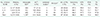
A total of 12,422 SLN biopsies were performed in 4,219 patients. A total of 1,397 SLNs from 908 patients were confirmed to be metastatic based on the pathologic reports of permanent section examinations. By comparing the results of frozen section examination with those of permanent section examination, it was observed that there were 11,025 TN, 1,207 TP, and 190 false-negative results. The diagnostic performance of frozen section examination for SLNB is summarized in Table 1. A total of 190 diagnoses of SLNs in 173 patients were changed from non-metastatic to metastatic based on the permanent pathologic results. One hundred fifty-nine patients had only one false-negative result; the remaining 14 had 2 or more false-negative results. No patients underwent ALND without ALN metastasis, owing to the high specificity of frozen section examination. Because of the false-negative results of frozen section examination, 130 patients underwent secondary ALND.
Characteristics of patients
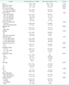
The mean age of the patients at the time of surgery was 51.8 years (standard deviation, 10.83). The mean number of retrieved SLNs was 2.73 (standard deviation, 1.52). Clinicopathological characteristics of the patients are shown in Table 2. The number of retrieved SLNs, number of positive SLNs, suspicious findings in ALNs, number of neoplastic foci, operation type, histology, lymphovascular invasions, TNM stage, ER positivity, and HER2 amplification were significantly different between the 2 groups. Additionally, there were more patients with high TNM stage in the false-negative group than in the accurate frozen section examination group.
Prediction of false-negative results of frozen section examinations
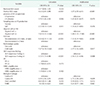
Several variables were significantly associated with falsenegative results on univariate analysis (Table 3). Treatmentrelated variables, including number of metastatic SLNs, missed targeting by the surgeon during SLNB, and missed sampling by the technician during the generation of frozen sections, as well as clinicopathological factors, including suspicious findings in the ALNs, size of SLN metastases, atypical cells on frozen section examination, radioisotope uptake, luminal B subtype, and TNBC subtype, were associated with the false-negative rate. Although most variables were associated with a higher false-negative rate, TNBC subtype was less likely to be associated with false-negative results (odds ratio, 0.3; P = 0.001). The number of positive SLNs, presence of atypical cells on frozen section examination, SLN size, radioisotope uptake, suspicious findings in the ALNs, and luminal B subtype were associated with false-negative results on multivariate analysis. The presence of suspicious findings in the ALNs increased the incidence of false-negative results by 2.36 times on multivariate analysis.
Characteristics and performance by subgroup
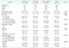
We performed subgroup analysis of several specific parameters associated with the diagnostic performance of frozen section examination. When analyzing by molecular subtype (Table 4), the frozen section examination of SLNs was most accurate in the TNBC subtype and least accurate in the luminal B subtype. The mean number of SLNs resected during intraoperative assessment was similar between subtypes. However, the mean positive LN counts were lowest in the TNBC subtype. The incidence of preoperative suspicious ALN findings was higher in the TNBC subtype than in the luminal B subtype. In contrast, the proportion of patients with pathological stage greater than N1 was higher in the luminal B subtype than in the TNBC subtype. We also performed subgroup analysis in 29 patients with atypical cells in the frozen section examination (Table 5). Invasive lobular carcinomas (ILCs) were present in a significantly larger proportion of patients with atypical cells than in those without. Additionally, the size of the primary tumor was larger in patients with atypical cells than in those without.
DISCUSSION
This study determined the diagnostic performance of intraoperative frozen section examination for SLNs and the factors associated with false-negative results. It is important to evaluate ALN status in patients with breast cancer to determine the disease stage, aid decisions for additional treatment after surgery, and predict prognosis. Intraoperative frozen sections have been used in several institutions because they confer the benefit of single-stage surgery for patients who require ALND. However, frozen section examination for ALN assessment may yield false-negative results. Therefore, it is necessary to understand the factors associated with false-negative results during frozen section examination.
In some studies, only one representative SLN was selected and analyzed. We were able to analyze the repeatedly measured data using a GEE model. In addition to the clinicopathological characteristics, our data also included surgical variables. Therefore, we could accurately analyze the false-negative result of the frozen section examination of each SLN. However, there are some limitations to our study. This was a retrospective study; therefore, a selection bias may have been present.
The false-negative rate increased with the number of resected SLNs. On logistic regression, the number of retrieved LNs did not influence the false-negative rate. However, as SLNBs are repeated, the probability of false-negative results in each assessment may cumulatively increase. In some cases, atypical cells were observed in frozen sections. This may have been related to inflammation of the LNs or structural changes that occurred during frozen section generation [22]. Atypical cells may also be seen in ILCs [23], as nodal involvement is a single-cell infiltration of the LN parenchyma, and it mimics a sinus histiocyte in ILCs [2223]. The proportion of patients with ILC was high among patients with atypical cells in the frozen section examination. This may be due to the metastatic pattern of ILC. Lymphatic metastasis may modify or block the lymphatic channel [24]. In such cases, there may be a shift in the axillary mapping results. Therefore, in addition to the LNs detected in the mapping method, hard or large LNs may be metastatic. These cold LNs need to be evaluated for metastasis.
The comparison of the diagnostic performance of frozen section examination of SLNs at our institution with those at other institutions suggests that the procedure is feasible [8142526]. Proper interpretation of the results of intraoperative frozen section examination is important because this determines the decision of additional surgeries, especially for patients who require immediate reconstruction or do not meet the Z0011 criteria. However, frozen sections are morphologically inferior to permanent sections [13]. Moreover, frozen sections are difficult to interpret because the applicability of IHC for frozen sections is limited. Furthermore, frozen section generation can result in permanent tissue loss. In our study, patients with confirmed pathologic N2 and N3 stages, for whom ALND is mandatory under the Z0011 trial, accounted for less than 2% (n = 71) of the patient population. Furthermore, the accuracy of frozen section examination is mainly influenced by the size of the metastatic node, and secondary further axillary surgery should be omitted when only micrometastatic SLNs are present with false-negative results due to metastatic nodal size. Therefore, the false-negative results due to metastatic node size do not change our practice. Our data indicate that intraoperative frozen section examination of the SLNs should not be routinely performed in all early breast cancer patients. Further in-depth studies to identify patients who will benefit from intraoperative frozen section should be performed.
 XML Download