

 PDF
PDF Citation
 Print
Print


INTRODUCTION
The function of the popliteal tendon (PopT) in horses remains undescribed. In humans, it is considered to act as a stabilizer of the posterior-lateral region of the knee; the more the knee is flexed, the stronger the stabilization action. The PopT prevents the tibia from posterior displacement by internally rotating it during flexion, thus preventing it from gliding forward. This action prevents meniscal impingement between the tibia and femur [1] thus protecting not only the meniscus but also the joint cartilage [2]. Its action is closely related to those of the lateral collateral ligament (LCL) and the popliteo-fibular ligament, which means that if one of these structures is affected, it is likely that the others are also affected [3]. In humans, the PopT constitutes the main proximal attachment of the popliteus muscle onto the femur; its insertion seats below and slightly proximal to the proximal insertion of the LCL, over the lateral epicondyle region. The tendon lies between the joint capsule and the synovial membrane [2]. In horses, the PopT is intra-articular and divides the caudal pouch of the lateral femorotibial joint into proximal and distal compartments within its synovial diverticulum. The PopT travels below the LCL, between the lateral meniscus and the joint capsule [4].
Several small accessory attachments of the PopT have been described in humans; some of which are very common, e.g., over the fibula or meniscus or at the joint capsule [5]. The PopT itself has been classified into four different types, according to the number of parts. Type I is composed of only one part and occurs in 13.7% of human knees. Type II has two separate parts and is present in 34.2% of knees. Type III is composed of three separate parts and occurs in 23.3% of knees. Type IV also has three parts, but the parts are stacked together rather than separate, and this type is present in 28.8% of human knees [6]. It is important to recognize these anatomical variations, as they could be confused with lesions in diagnostic image examination or during arthroscopic examination [7]. Insertional variations of the equine PopT have not yet been described.
The objectives of this study were to obtain a detail anatomical description of the equine popliteal structure and, secondly, to identify the possible anatomical variations of the PopT.
MATERIALS AND METHODS
Thirty equine cadavers were used for the anatomical study. All horses were euthanized for reasons unrelated to this study. Written approval for necropsy was obtained from the animals' owners. Horse ages ranged from one to 16 years old. Twelve mares, 15 geldings, and 3 stallions were included. The examined breeds were Spanish (n = 8), French Saddle (n = 15), Arabian (n = 4), Friesian (n = 1), and cross breed (n = 2). The insertion of the PopT was examined on both hind limbs of each horse, and tendon characteristics and the site of the insertion were recorded.
RESULTS
The popliteus muscle inserted distally onto the caudal surface of the tibia in all horses. The myotendinous structure was easily identified. Interestingly, the PopT had an attachment over the lateral meniscus in all examined limbs (Fig. 1). Three different tendon types were identified in the 60 hind limbs examined. The types were classified according to the number of components forming the tendon (Fig. 2). When the tendon included only one part, it was classified as type I, and this was identified in 40% of the limbs. When the tendon was composed of two parts, it was classified as type II; this variant was identified in 26.6% of the limbs. Type III, with three parts forming the tendon, was observed in 33.4% of the limbs. Insertional variations were not linked to breed or sex, as variation was observed within a breed, and there were different tendon types among the female, male, and castrated horses. Importantly, there was no variation within the same animal. Furthermore, the site of insertion of the tendon also varied. The insertions were located either cranial, underneath, or caudal to the proximal insertion of the LCL (Fig. 3). The most common insertion area was in the cranial location, identified in 66.7% of the limbs, while 8.3% were inserted underneath and 25% were inserted caudal to the LCL.
Fig. 1
Meniscal attachment of the popliteal tendon. After the removal of superficial structures, transection of the peroneus tertius tendon (black arrows), and transection of the lateral collateral ligament (yellow star), the popliteal tendon (red arrows) is pulled apart while a finger lifts the attachment (green arrow) that this tendon has over the lateral meniscus (L). Cranial is to the left, proximal to the top.
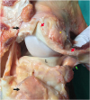
Fig. 2
Popliteal tendon types. The 3 different types of popliteal tendon are schematized in (A). Type I is shown in (B), type II in (C), and type III in (D). Cranial is to the left, proximal to the top. Black arrows indicate the septa demarking the different components of popliteal tendon types II and III.
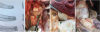
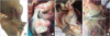
Fig. 3
Proximal insertion sites of the popliteal tendon. (A) A cadaveric model illustrates the three different locations where the insertion of the popliteal tendon was observed (red lower-case letters). Cranial is to the left, proximal to the top. Tibia (T), lateral condyle (F). With regard to the LCL, (B) illustrates a cranial insertion, (C) an underneath insertion location, and (D) a caudal insertion. Yellow stars indicate the LCL, and black stars indicate the insertion site of the popliteal tendon.
LCL, lateral collateral ligament.
DISCUSSION
Similar to findings in humans, this study shows, for the first time, that there are individual variations in equine PopT. Proximal PopT insertional variations are common in humans, and it has been reported that these variations could be mistaken for lesions during diagnostic imaging examinations [7]. In contrast to that in human anatomy, the equine PopT had a constant attachment to the meniscus (Fig. 1). When the joint is in motion, these attachments may prevent the tendon from displacing abaxially and, as reported in humans, help protect the lateral meniscus [1]. Three different tendon types were identified in the examined horses (Fig. 2) with the types classified according to the number of components forming the tendon. The anatomy of the horse was slightly different from that observed in humans, as the different components could be differentiated but were always stacked together. In that respect, the equine PopT resembles the human type IV PopT. In human type II and III the different parts of the tendon are completely separate [6], and this feature was not observed in any of the 30 horses examined. Additionally, we identified three different areas for the proximal insertion of the PopT (Fig. 3) as well as three different tendon types; these variations are important considerations during imaging examination of the region.
In conclusions, this study demonstrates, for the first time, that the equine popliteal tendon has a constant attachment to the lateral meniscus, that it has at least three conformational variants, and that the insertion site can also vary in relation to the proximal insertion of the LCL. Interestingly, variation was observed from horse to horse, but not within the same animal. Anatomical variation in this structure should be considered when examining the equine stifle.
 XML Download