

 PDF
PDF ePub
ePub Citation
 Print
Print


Jeong-Hoon Kim , Jong-Eun Kim, Young-Bum Park, Keun-Woo Lee
, Jong-Eun Kim, Young-Bum Park, Keun-Woo Lee
Abstract
After the teeth were extracted, maxillary and mandibular alveolar ridges show the opposite resorption pattern and as a result, the mandibular arch is enlarged than maxillary arch relatively. In this situation, we should evaluate both alveolar ridge relationship and arrange the artificial teeth properly for stability of removable prosthesis. This case is a 77 years old male patient who wishes to make removable prosthesis and has atrophic alveolar ridge. By use of model scanner and CAD software, the angle between interalveolar crest line and occlusal plane was easily measured. Depending on the measurement, the artificial teeth are arranged in unilateral cross bite and after completion, patient was satisfied with the denture which showed proper stability, retention, support.
Figures and Tables
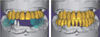
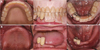 | Fig. 1Intraoral examination. (A) with existing prosthesis: Denture teeth wear and denture base resin discoloration, (B) without denture: Alveolar ridge atrophy.
|
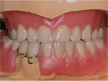
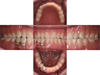
 | Fig. 3(A) Final impression taking (Maxilla: Polyvinyl siloxane, Mandible: Polyether) for maxillary complete denture and mandibular surveyed crown, working cast and occlusion rim fabrication, (B) Jaw relation record, (C) Working cast mounting.
|
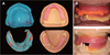
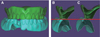
 | Fig. 4Working casts scanning. (A) Maxillary and mandibular working cast, (B) Mandibular occlusal rim scan data, (C) Separation of occlusion rim scan data.
|
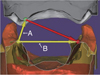 | Fig. 5Measuring the angle between interalveolar crest line (yellow line : A) and occlusal plane (yellow line : B): Occlusal rim scan data.
|

References
1. Atwood DA, Coy WA. Clinical, cephalometric, and densitometric study of reduction of residual ridges. J Prosthet Dent. 1971; 26:280–295.

2. Sanghvi SJ, Bhatt NA, Bhargava K. An evaluation of cross-bite ridge relationships. A study of articulated jaw records of 150 edentulous patients. J Prosthet Dent. 1981; 45:24–29.
3. Weinberg , Lawrence A. Tooth position in relation to the denture base foundation. J Prosthet Dent. 1958; 8:398–405.
4. Bilhan H, Geckili O, Ergin S, Erdogan O, Ates G. Evaluation of satisfaction and complications in patients with existing complete dentures. J Oral Sci. 2013; 55:29–37.
5. Kawahata N, Kamada Y, Ohtsuka A, Kamashita Y, Nishi Y, Hamano T, Nagaoka E. A visual method for analysing buccolingual position of artificial posterior teeth. Part 1: use of the ridge crest. J Oral Rehabil. 1998; 25:914–920.
6. Palla S. Occlusal considerations in complete dentures. Science and practice of occlusion. Chicago: Quintessence;1997. p. 457–467.
7. Baba NZ. Materials and processes for CAD/CAM complete denture fabrication. Curr Oral Health Rep. 2016; 3:203–208.
8. Steinmassl O, Offermanns V, Stöckl W, Dumfahrt H, Grunert I, Steinmassl PA. In vitro analysis of the fracture resistance of CAD/CAM denture base resins. Materials (Basel). 2018; 11:E401.
9. Steinmassl O, Dumfahrt H, Grunert I, Steinmassl PA. CAD/CAM produces dentures with improved fit. Clin Oral Investig. 2018; 22:2829–2835.
10. Goodacre BJ, Goodacre CJ, Baba NZ, Kattadiyil MT. Comparison of denture tooth movement between CAD-CAM and conventional fabrication techniques. J Prosthet Dent. 2018; 119:108–115.
 XML Download