Journal List > J Cardiovasc Imaging > v.27(3) > 1130003

Kim, Koh, Lee, and Chung: Three-dimensional EchoNavigator System Guided Transcatheter Closure of Paravalvular Leakage
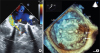
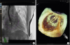
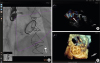
A 75-year-old female who visited the Outpatient Department presented with dyspnea (NYHA III) and syncope. She underwent mitral valve replacement (MVR) in 1988 and 2000. The laboratory data showed hemolytic anemia (hemoglobin 6.4 g/dL, reticulocyte count 7.72%, total bilirubin 1.76 mg/dL) and she needed red blood cell (RBC) transfusion every 3-4 weeks. There was severe eccentric paravalvular leakage (PVL) on color Doppler transesophageal echocardiography (TEE) (Figure 1A). The patient refused tri-do valve surgery and the cardiac surgeon expressed that MVR might not reduce PVL due to fibrotic changes to the mitral annulus after re-do MVR. We decided to do transcatheter implantation of a vascular plug into the slit between the prosthetic valve and mitral annulus. The EchoNavigator system (Phillips Healthcare, Best, The Netherlands) was used to find the slit and wiring (Figure 2, Movie 1). Two Amplatzer Vascular Plugs™ (8 mm and 10 mm) were deployed (Figure 3). After the procedure, hemolysis was improved and no further RBC transfusions were required.
Three-dimensional (3D) TEE-guided transcatheter closure of PVL is an effective treatment modality for PVL after surgical valvular replacement.1) In 2017, there were eight cases of PVL following transcatheter valve replacement in Belgium and Poland. In one case, 3D-echocardiography was fused with fluoroscopy images in real time using Echonavigator.2) This is the first Korean case in which the Echo-navigator was used to close PVL. EchoNavigator offers real-time fusion of live fluoroscopic and echocardiographic images for intuitive guidance during structural heart disease procedures.

Figures and Tables

Figure 1

Pre-procedural transesophageal echocardiography (TEE). (A) Two-dimensional TEE color Doppler image demonstrated severe paravalvular leakage (yellow arrow). (B) Three-dimensional TEE image demonstrated dark holes between the prosthetic mitral valve and mitral annulus (white arrows), which was localized at the medial side of the anterior mitral annulus, near to the aortic valve. AV: aortic valve, LA: left atrium, LAA: left atrial appendage, LV: left ventricle.

jcvi-27-227-g001
Figure 2

Fusion image from the EchoNavigator System (Phillips Healthcare, Best, The Netherlands) during the procedure. Green dot, demonstrated on fluoroscopic view (A), was marked from the real-time 3-dimenstion transesophageal echocardiography image (B). The guide wire was targeted to the green dot on fluoroscopic view and was successfully passed through the slit.

jcvi-27-227-g002
Figure 3

Fusion image from EchoNavigator System (Phillips Healthcare, Best, The Netherlands) after deployment. (A) Final positions of two Amplatzer™ vascular plugs are shown by fluoroscopy. (B) Color Doppler image demonstrated markedly reduced paravalvular leakage (white arrow) compared to Figure 1A. (C) Three-dimensional transesophageal echocardiography demonstrated final position of the vascular plugs (white circle).

jcvi-27-227-g003

Notes

Conflict of Interest The authors have no financial conflicts of interest.

References

1. Cruz-Gonzalez I, Rama-Merchan JC, Rodríguez-Collado J, et al. Transcatheter closure of paravalvular leaks: state of the art. Neth Heart J. 2017; 25:116–124.
crossref
2. Hascoet S, Smolka G, Bagate F, et al. Multimodality imaging guidance for percutaneous paravalvular leak closure: Insights from the multi-centre FFPP register. Arch Cardiovasc Dis. 2018; 111:421–431.
crossref

SUPPLEMENTARY MATERIAL

Movie 1

Fusion image from the EchoNavigator System (Phillips Healthcare, Best, The Netherlands) during the procedure. Green dot, demonstrated on fluoroscopic view, was marked from the real-time 3-dimenstion transesophageal echocardiography image. The guide wire was targeted to the green dot on the fluoroscopic view and was successfully passed through the slit.
TOOLS
ORCID iDs

Byung Ho Kim
https://orcid.org/0000-0001-7325-3939

Yoon-Seok Koh
https://orcid.org/0000-0002-0688-3858

Kwan-Yong Lee
https://orcid.org/0000-0002-0480-1046

Woo-Baek Chung
https://orcid.org/0000-0002-6933-2957

Similar articles