



 PDF
PDF ePub
ePub Citation
 Print
Print


So-Yeun Kim1, 2 , Eun-Young Kwon1, Kyoung-Hwa Jung1, Hye-Mi Jeon1, Young-Jae Baek1, Mi-Jung Yun2, Jung-Bo Huh2
, Eun-Young Kwon1, Kyoung-Hwa Jung1, Hye-Mi Jeon1, Young-Jae Baek1, Mi-Jung Yun2, Jung-Bo Huh2
Abstract
There are several unfavorable conditions regarding alveolar bone condition that may compromise the denture patient's satisfaction. Chewing efficiency may not be satisfactory when alveolar bone is deficient, and the denture stability could hardly be achieved when alveolar bone shape is irregular. Implant overdenture can be useful to provide satisfactory denture experience compared to conventional denture. The attachment for implant overdenture can be classified into bar attachment and solitary attachment. When the positions of the implants are in the mandibular anterior region, bar attachment may be favorable to obtain a rigid support of the entire denture. When implants are distributed both on anterior and posterior region, a solitary attachment could be considered for ease of removal and maintenance. This report presents implant overdenture cases with the patients that had unilateral mandibular alveolar bone atrophy conditions. Different abutments were chosen based on the individual patient's mandibular alveolar bone condition and the treatments were successful in terms of patient satisfaction.
Figures and Tables
Fig. 1
Pre-operative panoramic radiograph of the 1st case showing severe bone loss on the right side of the mandible.
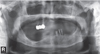
Fig. 2
(A) Duplicated denture template for diagnosis containing six radiopaque materials, (B) Panoramic section of dental computed tomography, (C) Coronal sections at four sites, (D) Indications on the model of selected placement position, (E) Surgical guide with holes on three selected sites.
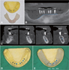
Fig. 3
(A) Maxillary wax denture and mandibular tooth arrangement, (B) Evaluation of diagnostic wax dentures, (C) Mounted cast with maxillary wax denture showing mandibular alveolar bone resorption, (D) Space evaluation for bar design using putty index, (E) Fabricated milled bar pattern, (F) Casted milled bar with locator attachment.
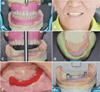
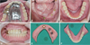
Fig. 4
(A, D, E) Definitive prosthesis, (B) Extraoral photograph after placement of prosthesis, (C) Intraoral view of milled bar.
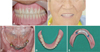

Fig. 6
Pre-operative panoramic radiograph of the 2nd case showing severe mandibular bone loss, especially on the right side.
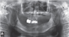
Fig. 7
Diagnostic arrangement of mandibular tooth on the computer aided design (CAD). (A, B) Occlusal, (C) Right, (D) Frontal and (E) Left view.
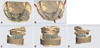
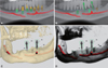
Fig. 8
Planning of implant placement on the implant planning software. (A, B) Panoramic section. Three sites were selected to avoid inferior alveolar nerve canal, (C) CT stereoscopic view showing implant fixture exposure on alveolar bone, (D) Stereoscopic view with reduced concentration.

References
1. van der Bilt A, Olthoff LW, van der Glas HW, van der Weelen K, Bosman F. A mathematical description of the comminution of food during mastication in man. Arch Oral Biol. 1987; 32:579–586.

2. Wismeijer D, Van Waas MA, Vermeeren JI, Mulder J, Kalk W. Patient satisfaction with implant-supported mandibular overdentures. A comparison of three treatment strategies with ITI-dental implants. Int J Oral Maxillofac Surg. 1997; 26:263–267.
3. Naert I, Quirynen M, Theuniers G, van Steenberghe D. Prosthetic aspects of osseointegrated fixtures supporting overdentures. A 4-year report. J Prosthet Dent. 1991; 65:671–680.
4. Trakas T, Michalakis K, Kang K, Hirayama H. Attachment systems for implant retained overdentures: a literature review. Implant Dent. 2006; 15:24–34.
5. Gotfredsen K, Holm B. Implant-supported mandibular overdentures retained with ball or bar attachments: a randomized prospective 5-year study. Int J Prosthodont. 2000; 13:125–130.
6. Abi Nader S, de Souza RF, Fortin D, De Koninck L, Fromentin O, Albuquerque RF Junior. Effect of simulated masticatory loading on the retention of stud attachments for implant overdentures. J Oral Rehabil. 2011; 38:157–164.
7. Burns DR. Mandibular implant overdenture treatment: consensus and controversy. J Prosthodont. 2000; 9:37–46.
8. Närhi TO, Geertman ME, Hevinga M, Abdo H, Kalk W. Changes in the edentulous maxilla in persons wearing implant-retained mandibular overdentures. J Prosthet Dent. 2000; 84:43–49.
9. Lee JY, Yoon JY, Oh NS. The use of surgical guide stent for implant placement. J Korean Acad Prosthodont. 2014; 52:366–375.
10. English CE. Bar patterns in implant prosthodontics. Implant Dent. 1994; 3:217–229.
11. Takeshita S, Kanazawa M, Minakuchi S. Stress analysis of mandibular two-implant overdenture with different attachment systems. Dent Mater J. 2011; 30:928–934.
12. Bergendal T, Engquist B. Implant-supported overdentures: a longitudinal prospective study. Int J Oral Maxillofac Implants. 1998; 13:253–262.
13. Geckili O, Bilhan H, Bilgin T. Locator® attachments as an alternative to ball attachments in 2-implant retained mandibular overdentures. J Can Dent Assoc. 2007; 73:691–694.
14. Min BK, Shin EJ, Vang MS, Yang HS, Park SW, Yun KD, Lim HP. Overdentures of the patient with Parkinson's disease: A case report. J Korean Acad Prosthodont. 2015; 53:352–358.
 XML Download