

 PDF
PDF ePub
ePub Citation
 Print
Print


Kwang Man Park , Richard Leesungbok, Suk Won Lee
, Richard Leesungbok, Suk Won Lee
Abstract
Orthognathic surgery of skeletal Class III malocclusion improves oral function and facial appearance. The greater amount of skeletal discrepancy, the greater amount of teeth movement required for decompensation, and this often causes pathological changes in periodontal tissue especially in lower anterior dentition. We made a Top-Down treatment plan with personalized analysis using Face Hunter, Plane System and ARCUS Digma II, in order to resolve severe mobility and cross-bite of lower anterior teeth for 49-year-old female patient who had undergone orthognathic surgery 20 years ago due to skeletal Class III malocclusion and mandibular prognathism. Lower anterior teeth were extracted and alveoloplasty was done. After healing of the wound, immediate loading was conducted immediately after implant placement. Final restorations were fabricated Zirconia using CAD/CAM, and inserted intraorally screw-retained type. During 6-month follow-up, no abnormal episodes of restorations were observed, and obtained satisfactorily both of functional and esthetic outcomes.
Figures and Tables
Fig. 1
Initial intraoral view. (A) Occlusal view of upper dentition, (B) Occlusal view of lower dentition, (C) Right lateral view, (D) Left lateral view, (E) Frontal view of habitual bite, (F) Frontal view of CR bite, (G) Lateral view of habitual bite, (H) Lateral view of CR bite.
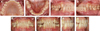
Fig. 2
Willis method. Measurements on the facial profile view of preoperative stage (A≒B), (A) Distance from the center of the pupil of the eye to rima oris, (B) Distance from bony ledge under the nose to the bottom of the mandible.
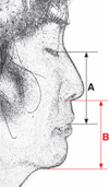
Fig. 3
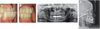
Radiographic records. (A) Panoramic view before extraction of hopeless teeth (06-30-2016), (B) Panoramic view after placement of implant on #45 (06-05-2018), (C) Periapical view after placement of implant on #45 (06-05-2018), (D) After orthognathic surgery on 01-05-2010, (E) Before implant treatment on 06-22-2018.
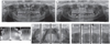
Fig. 4
Extraction of the hopeless teeth. (A) Alveoloplasty after extraction of the lower anterior hopeless teeth on #44, #43, #42, #41, #31, #32, #33, and #34, (B) Delivery of removable temporary denture after suturing.
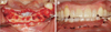
Fig. 5
Diagnostic wax-up on the mounted models. (A) Diagnostic wax-up on the lower anterior teeth, (B, C) Surgical template and immediate provisional restoration.

Fig. 6
The 1st implant surgery. (A) Implant placed on #33, #34, #43 and tooth extraction of #35, (B) Insertion of immediate provisional restoration on the implants.
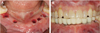
Fig. 7
The 2nd implant surgery. (A, B) Implant remove due to failure on #33, and newly placed on #32,#35, (C, D) Final impression taking, (E) Adjusted immediate provisional restoration.
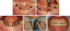
Fig. 8
Facial analysis and design with digital workflow. (A – C) CAD by Face Hunter & Plane System, (D) Arcus digma II data.
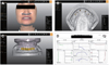
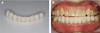
Fig. 9
Delivery of the polymer trial restoration. (A) The 1st CAD/CAM for fabrication of polymer trial restoration, (B) Frontal view after insertion.

References
1. Artun J, Krogstad O. Periodontal status of mandibular incisors following excessive proclination. A study in adults with surgically treated mandibular prognathism. Am J Orthod Dentofacial Orthop. 1987; 91:225–232.

2. Sperry TP. The limitations of orthodontic treatment. Angle Orthod. 1993; 63:155–158.
3. Steiner GG, Pearson JK, Ainamo J. Changes of the marginal periodontium as a result of labial tooth movement in monkeys. J Periodontol. 1981; 52:314–320.
4. Ahn HS, Kim SS, Son WS. A study on the morphological changes of lower incisor and symphysis during surgical-orthodontic treatment in skeletal class III malocclusion. Korean J Orthod. 2002; 32:361–373.
5. Kim Y, Park JU, Kook YA. Alveolar bone loss around incisors in surgical skeletal Class III patients. Angle Orthod. 2009; 79:676–682.
6. Hwang HS, Kim JC, Kim JM. The effect of dental protrusion on the width of attached gingiva. Korean J Orthod. 1998; 28:135–142.
7. Lee JY, Yu HS, Ryu YK. A study on skeletal relapse patterns following orthognathic surgery of Class III patients: comparison between SSRO and IVRO. Korean J Orthod. 1998; 28:461–477.
8. Huang YL, Pogrel MA, Kaban LB. Diagnosis and management of condylar resorption. J Oral Maxillofac Surg. 1997; 55:114–119.
9. Hwang SJ, Haers PE, Seifert B, Sailer HF. Non-surgical risk factors for condylar resorption after orthognathic surgery. J Craniomaxillofac Surg. 2004; 32:103–111.
10. Hwang SJ, Haers PE, Zimmermann A, Oechslin C, Seifert B, Sailer HF. Surgical risk factors for condylar resorption after orthognathic surgery. Oral Surg Oral Med Oral Pathol Oral Radiol Endod. 2000; 89:542–552.
11. Wolford LM. Idiopathic condylar resorption of the temporomandibular joint in teenage girls (cheerleaders syndrome). Proc (Bayl Univ Med Cent). 2001; 14:246–252.
12. Hoppenreijs TJ, Freihofer HP, Stoelinga PJ, Tuinzing DB, van't Hof MA. Condylar remodelling and resorption after Le Fort I and bimaxillary osteotomies in patients with anterior open bite. A clinical and radiological study. Int J Oral Maxillofac Surg. 1998; 27:81–91.
13. Cutbirth M, Van Sickels JE, Thrash WJ. Condylar resorption after bicortical screw fixation of mandibular advancement. J Oral Maxillofac Surg. 1998; 56:178–182.
14. Weber HP, Morton D, Gallucci GO, Roccuzzo M, Cordaro L, Grutter L. Consensus statements and recommended clinical procedures regarding loading protocols. Int J Oral Maxillofac Implants. 2009; 24:180–183.
15. Gallucci GO, Benic GI, Eckert SE, Papaspyridakos P, Schimmel M, Schrott A, Weber HP. Consensus statements and clinical recommendations for implant loading protocols. Int J Oral Maxillofac Implants. 2014; 29:287–290.
16. Leesungbok R. Lee's Top-down implant dentistry. Seoul: Myungmun Publishing;2004.
17. Park JH, Jeong CM, Jeon YC, Lim JS. A study on the occlusal plane and the vertical dimension in Korean adults with natural dentition. J Korean Acad Prosthodont. 2005; 43:41–51.
18. Carlsson GE, Ingervall B, Kocak G. Effect of increasing vertical dimension on the masticatory system in subjects with natural teeth. J Prosthet Dent. 1979; 41:284–289.
19. Malchiodi L, Cucchi A, Ghensi P, Nocini PF. Evaluation of the esthetic results of 64 nonfunctional immediately loaded postextraction implants in the maxilla: correlation between interproximal alveolar crest and soft tissues at 3 years of follow-up. Clin Implant Dent Relat Res. 2013; 15:130–142.
 XML Download