

 PDF
PDF ePub
ePub Citation
 Print
Print


Introduction
Proper implant position is fundamental to achieve esthetic and functional implant-supported prostheses.1 A meticulous preoperative diagnostic and implant planning is mandatory to obtain predictable results. The most conventional way of placing an implant was to create a radiographic guide templates and then converting it to a surgical guide template after cone-beam computed tomography (CBCT) is taken.2 This manual fabrication of guide templates involves possibly inaccurate laboratory procedure, and would lead to the difficulty in placing the implant fixture as planned.3 In recent years, the matching of 3-dimensional (3D) cone-beam computed tomography (CBCT) dataset in digital imaging and communications in medicine (DICOM) format and 3D Mesh dataset in STL format has begun and allowed the introduction of digital surgical guide. Those images are used for planning the placement of implants considering bone, mucosa, and teeth.4 Using digital surgical guides, drilling and placing implants at a predetermined position became more predictable, which decreases errors in the implant placement compared to the traditional method. In addition, the advantages of this digital guide system are minimal invasiveness, accurate implant placement, less post-surgical discomfort and reduced time required for surgery.567
Image surface mapping enables the matching images between CBCT and mesh using at least 3 congruent points.8 This image registration protocol is applicable when enough number of teeth is remained and distributed in oral cavity. This condition is important in order to create a virtual oral cavity same to the corresponding patient by the image superimposition.910 In patients with extensive tooth loss, the use of additional markers has been suggested to improve the accuracy of image registration.111213 Recently, a new protocol using a micro-screw was introduced for enhancing the accuracy of computer-guided implant surgery in a fully edentulous jaw.14 The effects of micro-screw use was shown in terms of image fusion and guide positioning in a clinical report.15 However, the effect of microscrew on the operator performance factors of image registration process in unknown. The purpose of this study was to evaluate whether the use of micro-screw influences the operators' working time and convenience on the image registration process for computer-guided implant surgery. The null hypothesis was that there is no difference between conventional and micro-screw assisted image registration methods in the time and convenience.
Materials and methods
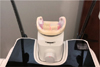
A mandibular dental model was prepared in the form of Kennedy Class I edentulism. Two micro-screws were placed on the each side of retromolar area using an electric torque driver (MEG-TORQ, MegaGen, Daegu, Korea), and scan caps were connected to the micro-screws. Afterwards, the surface of the mandibular model was digitized by using an intraoral scanner (CS 3600, Carestream, Rochester, NY, USA), and the scan image was saved in standard tessellation language (STL) format. Then, radiographic images were taken by using a CBCT scanner (PaXFlex3D, Vatech, Seoul, Korea) with a field of view of 120 × 85 mm, voxel size of 0.2 mm, exposure conditions of 90 kVp, 10 mA, and a 24-s pulsed scan (Fig. 1). Data were saved in the DICOM format.
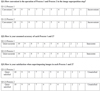
The STL and DICOM data were imported into a dental software program (R2GATE v1.1.1, MegaGen, Daegu, Korea) for computer-aided implant surgery planning and guide fabrication. Using volume rendering, the DICOM data were reformatted as a 3D reconstruction image with which the surface scan data were merged. The image registration was conducted manually by designating 3 congruent points from the two images. The registration process was carried out two times per each operator with different matching conditions (Fig. 2). In the first matching, matching points were limited to the remaining teeth. In the second matching, the scan cap images were included as matching areas, as well as the tooth image. Twelve dental graduates who were blinded to the purpose of study and had same the working year in the dental filed, participated in this study. While performing the registration process, working time was recorded for each condition. After finishing the image registration, operators' convenience and satisfaction on each matching condition was investigated using a questionnaire (Fig. 3).
All data were reported as the mean ± standard deviation. The Mann-Whitney U test were used to compare the amounts of performance time, convenience, satisfaction between the 2 groups. Statistical analyses were performed using SPSS 25.0 for Windows (SPSS Inc., Chicago, IL, USA). A P value < .05 was considered significant.
Results
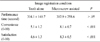
The results of this study is shown in Table 1. The performance time was not statistically different between image registration conditions (P > .05). As for operator convenience and satisfaction evaluations, micro-screw assisted image registration condition showed higher values than the condition teeth alone used (P < .001). In the analyses among factors, performance time and operator satisfaction did not show tendency. In the other hand, operator convenience and satisfaction showed positive correlation (Fig. 4).
Discussion
The premise of this article is that the presence of micro-screw in the oral cavity can increase the time efficiency and convenience on the image matching of radiographic and digital scan data in computer-guided implant surgery as an additional fiducial marker. The finding of the study demonstrated that the micro-screw use did not reduce the performance time, but increased convenience of the process. Thus, the null hypothesis, there is no difference between conventional and micro-screw assisted image registration methods in the convenience and time, was partially rejected.
In the past several years, with the introduction of CBCT and 3D printing into the field of implant dentistry, computer-aided design/computer-aided manufacturing (CAD/CAM) technology brought a great evolution of new treatment concepts to dental implant treatment.16 CBCT and 3D implant planning software provides clinicians not only 3D information of patient's anatomic structures, but also data regarding the patient's final prosthesis. These digital data can be combined with the CAD/CAM technology and further lead to a digital workflow ending with the production of stereolithographic template via a prototyping system.1718
Nowadays, two types of techniques are available in transferring the planned implant position information to the clinical situation,: dynamic and static.19 Dynamic guided implant surgery uses the navigation technique with a software provides a real-time feedback which allows to adjust the implant position and any modification if necessary. However, this dynamic system is not frequently used mostly due to the high costs of the equipment requested and complicated software. The static or application of surgical templates is less flexible in regard to changing the surgical plan as the information is only transmitted through the template. Nonetheless, this static guided implant surgery has been preferred due to no requirement a sophisticated process to carry out, insuring the predictability, a reduced invasiveness of surgical procedures,20 a shorten healing period, and less postoperative discomfort.21
Participants in this study were dental graduates with no exposure to conventional way of image registration or advanced technique of merging 3D images from CBCT and surface scan. This homogeneous group allowed investigating the efficiency of these matching images techniques in an objective and non-biased manner. This is of particular importance because results from a different study population including experienced clinicians or professional designers could be confounding factors to interpret. Whereas, this investigation addressed only the efficiency and operator's preference on both merging 3D data set techniques. Several other aspects need further investigations. This study yielded initial evidence that the effects of use of micro-screws on the operator performance factors.
Even there was not a significant difference of time performnance with assistance of micro-screws compairing to conventional technique but greater number of participants agreed that they preffered using micro-screws in matiching image due to their assumptiom of accuracy for final merged image which had been observed directly during the processes. The level of difficulty judged by participants was significantly lower with assistance of micro-screws. These subjective difference in the process of merging images also affected participants' perception of effectiveness.
As for the aspects of accuracy, several attempts have been performed to facilitate the image superimposition in digital guide fabrication. Widmann et al.13 proved the use of implants with ball attachments to increase the number of reference points for image matching in edentulous jaws and concluded that a fixed reference improves registration procedure. Oh et al.12 suggested the insertion of additional resin markers on the palatal gingiva, increasing the reference points and improving the registration of CBCT and digital surface scan data.
The first limitation of this study is that the number of participants is small. The second restraint is that this study was performed with a dental model as a clinical scenario that excluded the effect of actual treatment conditions. Patient consent for placement of micro-screws associated with treatment is also of implant concern. Although results of this study set the first brick to build statement that the insertion of a micro-screw in the posterior area of an edentulous ridge changed the conditions for guided implant surgery, more large scaled clinical studies are need to confirm the effects of improving satisfaction and convenience in use of micro-screw.
Conclusion
Within the limits of the study, it can be concluded that a higher satisfaction of operators may be achieved if surgical templates are designed by superimposing a surface scan with CBCT with assistance of micro-screws as one of fiducial markers in order to transform the virtual plan into reality. Further clinical studies are needed to support the finding of this study.


 XML Download