

 PDF
PDF ePub
ePub Citation
 Print
Print


INTRODUCTION
Hepatocellular carcinoma (HCC) is the sixth most common malignancy worldwide, especially in several regions of Africa and Asia,1 and the burden of this devastating cancer is expected to further increase in coming years. The incidence of HCC is complex and multifactorial,23 and its molecule mechanism has not been fully elucidated. Better understanding of the mechanisms underlying HCC progression is crucial for improving the diagnosis and successful treatment of HCC.
Evidence from experimental and clinical studies increasingly pointed that hypoxia plays a fundamental role in solid tumor development,4 and that it appears to be strongly associated with tumor propagation, malignant progression, or resistance to therapy.45 Although hypoxia plays an important role in the metastasis of HCC, its underlying mechanism still remains largely unclear.
Long noncoding RNA (lncRNA) is a class of non-coding RNA with a length greater than 200 nucleotides (nt). LncRNAs have been implicated in a large range of biological procession, such as epigenetic regulation, transcriptional regulation, and posttranscriptional regulation.6 LncRNAs were involved in hypoxia-promoted tumor progression. For instance, lncRNA BC005927 could be induced by hypoxia in gastric cancer (GC) cells and mediates hypoxia-induced GC cell metastasis, and increased BC005927 expression was correlated with a higher tumor-node-metastasis stage.7 Also, lncRNA-BX111 promoted metastasis and progression of pancreatic cancer through regulating zinc finger E-box binding homeobox transcription factor 1 (ZEB1) under hypoxia, and overexpression of lncRNA-BX111 dramatically enhanced proliferation and invasion of pancreatic cancer cells.8 LncRNA metastasis-associated lung adenocarcinoma transcript 1 (MALAT1) has been reported to be abnormally expressed in various types of cancers. Previous studies have shown that MALAT1 plays a pivotal role in the differentiation, proliferation, apoptosis, and migration of many tumor cells.9101112 Also, the role of MALAT1 in HCC has been reported as well. Hou, et al.13 found that MALAT1 promoted the migration and invasion of HCC by upregulating silent information regulator 1 via microRNA (miR)-204 sponging. However, whether MALAT1 would influence HCC cells under hypoxic conditions remains unknown.
In this study, we first revealed that MALAT1 was involved in HCC cell proliferation, survival, migration, and invasion through miR-200a sponge activity cells under hypoxia, revealing a new mechanism of MALAT1 involved in hypoxic HCC progression.
MATERIALS AND METHODS
Cell culture and oxygen conditions
HCC cell lines Huh7, SNU-423, PLC, and Hep3B were obtained from Shanghai Institute of Biochemistry and Cell Biology, Chinese Academy of Sciences (Shanghai, China). All cells were cultured in Dulbecco's modified Eagle's medium (DMEM, Thermo Fisher Scientific, Waltham, MA, USA) supplemented with 10% of fetal bovine serum (FBS, Hyclone, Logan, UT, USA) and 1% of penicillin/streptomycin stock solution (Sigma, St. Louis, MO, USA). Hypoxic conditions were maintained in a humidified variable aerobic workstation at 37℃ (N-control). To induce hypoxia, oxygen concentrations were reduced from 20% to 1% (H-control), while carbon dioxide (CO2) remained at 5%.
Reagents and transfection
MALAT1 expression plasmid (MALAT1), pcDNA3.1 vector (vector), small interfering RNA (si-RNA) against MALAT1 (si-MALAT1), si-RNA negative control (si-NC), miR-200a mimic (miR-200a), mimic negative control (miR-NC), miR-200a inhibitor (anti-miR-200a), and inhibitor negative control (anti-miR-NC) were synthesized by GENEWIZ Co. Ltd. (Suzhou, China). Hep3B cells (70% confluence in 6-well plates) were transfected with above-mentioned plasmids or oligos using Lipofectamine 3000 (Thermo Fisher Scientific).
Quantitative reverse transcription PCR
Total RNA was extracted from cells using Trizol reagent (Thermo Fisher Scientific). MicroRNAs (miRNAs) were isolated using miRNeasy mini kits (Qiagen, Hilden, Germany) reversely transcribed into complementary DNA (cDNA) using TaqMan® MicroRNA Reverse Transcription kit (Biosystems, Forster City, CA, USA). To quantify mRNAs, reverse transcription was performed using Prime Script™ RT reagent kit (Takara, Shiga, Japan). Quantitative PCR was performed using TaqMan® Universal PCR Master Mix II (Biosystems), qPCR primers were as follows: U6-R, 5′-CTCGCTTCGGCAGCAGCACA-3′; U6-F, 5′-AACGCTTCACGAATTT-GCGT-3′; MALAT1-F, 5′-ATGCGA GTTGTTCTCCGTCT-3′; MALAT1-R, 5′-TATCTGCGGTTTCCTCAAGC-3′; miR-200a-F, 5′-CACCGCCTCCCATTGTC-3′; miR-200a-R, 5′-CACAGGAAGTCAGTTCAGACC-3′. Relative expression levels of miRNA or lncRNA (normalized to U6 small nuclear RNA) were analyzed by 2−ΔΔCt method.
MTT assay
Hep3B cells were seeded into 96-well plates (2×10³ cells/well), and were transfected with miR-200a, miR-NC, anti-miR-200a, si-MALAT1, si-NC, or si-MALAT1+anti-miR-200a. At 24 h after transfection, cells were challenged with hypoxia for 24 hours, after which 20 µL of MTT (5 mg/mL, Sigma) was added into each well and incubated for another 4 h at 37℃. The mixed medium was then discarded, and 150 µL dimethyl sulfoxide (DMSO, Sigma) was added to dissolve the precipitates. Cell proliferation was evaluated by measuring the absorbance at 570 nm using a microplate reader (Molecular Devices, Sunnyvale, CA, USA).
Transwell invasion and migration assay
We seeded 1×10⁵ of Hep3B cells, which were suspended in 500 µL serum-deprived culture medium, into the upper compartment of Transwell apparatus (Corning Inc., Corning, NY, USA). For cell invasion assay, the membranes of upper compartments were matrigel pre-coated, and un-coated ones were used for cell migration assay. Cells were cultured for 24 h, and cells migrated to the underside of upper compartment membrane in response to culture medium supplemented with 5% FBS in lower compartment were fixed with methanol and stained with crystal violet. The number of migrated cells were counted in five randomly picked view under microscope (Leica, Wetzlar, Germany).
Cell apoptosis assay
Cell apoptosis was analyzed using FITC Annexin V Apoptosis Detection Kit (BD Biosciences, Franklin Lakes, NJ, USA). Hep3B cells were collected and digested with trypsin, washed with pre-cooled PBS, and resuspended in 200 µL binding buffer. Cells were then labeled with 10 µL Annexin V-FITC and 5 µL propidium iodide (PI) in dark for 15 min at room temperature. Cell apoptotic rate was detected with FACS Calibur flow cytometer and Cell Quest software (BD Biosciences).
Luciferase reporter assay
A wild-type (WT) fragment of MALAT1 containing putative miR-200a binding site and its mutated (MUT) seed sequence were purchased from Shanghai Bioengineering Co., Ltd. (Guangzhou, China), and were inserted downstream of a luciferase reporter gene on pmirGLO dual-luciferase miRNA target expression vectors (Promega, Madison, WI, USA), named as MALAT1-WT and MALAT1-MUT, respectively. Hep3B cells were cotransfected with MALAT1-WT or MALAT1-MUT and miR-200a or miR-NC using Lipofectamine 3000 (Thermo Fisher Scientific). Cell were harvested at 48 h after transfection. Dual-luciferase reporter assay system (Promega) was used to detect luciferase activity according to the manufacturer's instructions.
Statistical analysis
All data were expressed as mean±SD from three separate experiments. All statistical analyses were performed by SPSS 20.0 statistical software (IBM Corp., Armonk, NY, USA). Student's t-test or ANOVA was performed for significance test. A p value less than 0.05 was considered significant.
RESULTS
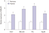
MALAT1 was upregulated in HCC cells by hypoxia
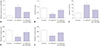
To investigate MALAT1 expression levels in HCC cells in response to hypoxia, Huh7, SNU-423, PLC, and Hep3B cells were incubated under hypoxic (1% O2) or normoxic (20% O2) condition. MALAT1 expression levels in these cells were detected by qRT-PCR. We found that MALAT1 expression was increased in all HCC cells after being exposed to hypoxic condition (Fig. 1). Hep3B cell line was used for further analysis.
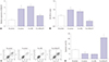
Knockdown of MALAT1 suppressed growth and induced apoptosis of Hep3B cells after hypoxia challenge
Based on the results mentioned above, the effects of MALAT1 knockdown on proliferation and apoptosis of Hep3B cells under hypoxic condition were further explored. First, Hep3B cells were transfected with MALAT1 siRNA or negative control siRNA. Hep3B cells after transfection were then cultured under hypoxic condition for 24 hours, followed by re-oxygenation for 48 hours. qRT-PCR assay showed that the relative expression of MALAT1 was significantly decreased in si-MALAT1-transfected Hep3B cells compared with negative control siRNA transfected ones (Fig. 2A). MTT assay showed that the proliferation of Hep3B cells was significantly increased by hypoxia challenge, which was significantly attenuated by MALAT1 knockdown (Fig. 2B). Cell apoptosis assay revealed that hypoxia challenge reduced Hep3B cell apoptosis in vitro, which was significantly attenuated by MALAT1depletion (Fig. 2C and D).
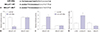
Knockdown of MALAT1 suppressed migration and invasion of Hep3B cells promoted by hypoxia
To investigate the role of MALAT1 in HCC cell migration and invasion under hypoxia, Transwell assay was performed to analyze the migration and invasion of Hep3B cells with MALAT1 depletion under hypoxia. We found that hypoxia improved the migration and invasion abilities of Hep3B cells, which were significantly inhibited by MALAT1 silencing (Fig. 3).
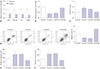
MALAT1 interacted with miR-200a in hypoxic Hep3B cells
Starbase v2.0 (http://starbase.sysu.edu.cn/starbase2/) suggested that miR-200a has binding sites with MALAT1. Luciferase reporter plasmids containing WT or MUT MALAT1 binding sites of miR-200a were established (Fig. 4A). Luciferase reporter assay indicated that miR-200a mimic significantly decreased the luciferase activity of MALAT1-WT group, and miR-200a inhibitor significantly enhanced the luciferase activity of MALAT1-WT group. However, miR-200a mimic or inhibitor showed no significant impact on the luciferase activity in MALAT1-MUT group, respectively (Fig. 4B and C). To further investigate the effect of MALAT1 on miR-200a expression, Hep3B cells were transfected with si-MALAT1, si-NC, pcDNA MALAT1, or pcDNA3.1 vector. We found that miR-200a was significantly upregulated in si-MALAT1-transfected Hep3B cells, while downregulated in pcDNA MALAT1-transfected cells (Fig. 4D).
Upregulation of miR-200a inhibited the proliferation, invasion, migration, and induced apoptosis in Hep3B cells under hypoxia
To explore miR-200a expression in response to hypoxia, Huh7, SNU-423, PLC, and Hep3B cells were incubated 48 h in hypoxic or normoxic conditions. Then, qRT-PCR was performed to detect the expression of miR-200a. We found that miR-200a expression levels were significantly decreased in these HCC cell lines after hypoxia challenge (Fig. 5A). To further investigate the possible roles of miR-200a in Hep3B cells proliferation, apoptosis, migration, and invasion under hypoxia, miR-200a mimic or mimic control was transfected into Hep3B cells. Expression of miR-200a in Hep3B cells was significantly reduced after hypoxia challenge, which was significantly improved by miR-200a mimic transfection (Fig. 5B). MTT and flow cytometry results demonstrated that miR-200a mimic transfection significantly attenuated the effect on Hep3B cell proliferation and anti-apoptosis by hypoxia challenge (Fig. 5C and D). Transwell assay results further showed that miR-200a mimic transfection significantly inhibited cell migration and invasion ability of Hep3B cells promoted by hypoxia (Fig. 5E and F).
Downregulation of miR-200a partially reversed the effect of MALAT1 knockdown on hypoxia-challenged Hep3B cells
To further confirm whether MALAT1 regulates proliferation, apoptosis, migration, and invasion of hypoxia-challenged Hep3B cells by sponging miR-200a, Hep3B cells were co-transfected with si-MALAT1 and miR-200a inhibitor. Knockdown of MALAT1 significantly inhibited the proliferation, migration, and invasion, while inducing apoptosis of hypoxia-challenged Hep3B cells. All of these effects were significantly attenuated by miR-200a inhibitor transfection (Fig. 6).
DISCUSSION
Liver cancer is the second leading cause of cancer-related deaths worldwide,14 and HCC accounts for approximately 90% of all cases of primary liver cancer.15 Increasing evidence suggested noncoding RNAs, including lncRNAs and miRNAs, as important regulator in the development of HCC with potential diagnostic and therapeutic values.16 MALAT1 has been reported to exert oncogenic roles in multiple cancers, including HCC.1718 This study aimed to investigate the role and mechanism of MALAT1 in HCC cells under hypoxia. We performed qRT-PCR to monitor the expression levels of MALAT1 and miR-200a in HCC cell lines (Huh7, SNU-423, PLC, and Hep3B) under hypoxia or normoxia. Our data suggested that MALAT1 was upregulated, while miR-200a was downregulated in HCC cell lines (Huh7, SNU-423, PLC, and Hep3B) by hypoxia challenge. MALAT1 knockdown inhibited the proliferation, migration, and invasion, and induced apoptosis in hypoxia-challenged Hep3B cells, while miR-200a mimic transfection had the same effects. Silencing of miR-200a significantly attenuated the effect of MALAT1 knockdown on hypoxia-challenged Hep3B cells. Notably, we confirmed that miR-200a interacted with MALAT1 in HCC cells. Our results suggest that MALAT1 may contribute to the development of HCC promoted by hypoxia by regulating miR-200a.
MALAT1 is a well-known lncRNA associated with cancer.19 Researchers have revealed that MALAT1 can be an oncogenic factor in HCC.18 For instance, Malakar, et al.20 showed that lncRNA MALAT1 was upregulated in HCC, but also in liver tumors from a mouse model of hepatic carcinogenesis. In addition, MALAT1 could act as a proto-oncogene through Wnt pathway activation and induction of oncogenic splicing factor serine/arginine-rich splicing factor 1. Chen, et al.21 demonstrated that MALAT1 was upregulated in HCC tissues and cell lines, and that MALAT1 might serve as a prognostic indicator for HCC patients. Furthermore, MALAT1 regulated ZEB1 expression by sponging miR-143-3p and promoted HCC progression. These data suggested that MALAT1 may exhibit vital roles in the development and progression of HCC. Hypoxia is a common feature of many solid tumors, including HCC, which can participate in tumor progression. However, the effects of MALAT1 on hypoxia-treated HCC cells have not been examined. In this study, we first found that MALAT1 was increased by hypoxia challenge in HCC cells in vitro, especially in Hep3B cell line. Furthermore, the promotion of cell proliferation, migration, and invasion, as well as inhibition of apoptosis of Hep3B cells induced by hypoxia were attenuated by MALAT1 knockdown. These results implicated that MALAT1 may be involved in hypoxia-promoted HCC cell malignancy and tumor progression.
LncRNAs can regulate gene expression by “sponging” miRNAs.2223 MiRNAs are short noncoding RNA molecules of 17–22 nt in length, and are involved in the oncogenesis and progression of multiple cancers.24 Previous studies have manifested that miRNAs were abnormally expressed in HCC and participated in fundamental biological processes including cell proliferation, differentiation, and apoptosis.2526
Many researchers have reported that miR-200a inhibited HCC cell proliferation,27282930 and that it plays an important role in protecting cardiomyocyte survival under hypoxic conditions. Li, et al.31 pointed that miR-200a level was decreased in H/R-cardiac microvascular endothelial cells (CMECs), and thymosin beta 4 attenuated hypoxia-reoxygenation induced CMECs injury by miR-200a-Nrf2 signaling. Sun, et al.32 manifested that miR-200a was significantly downregulated in ischemic myocardial tissues and hypoxic cardiomyocytes, and suppression of Keap1 by miR-200a exerted cardioprotective effect against hypoxia-induced oxidative stress and cell apoptosis. The role of miR-200a in hypoxic HCC cells remains unknown. In the present study, we found that miR-200a was markedly downregulated in HCC cells by hypoxia, suggesting that miR-200a might be involved in the cancer progression of hypoxic HCC. Starbase v2.0 suggested that MALAT1 and miR-200a has binding sites, which was further authenticated by luciferase reporter assay. We disclosed that miR-200a interacted with MALAT1 and MALAT1 negatively regulated miR-200a expression in Hep3B cells. In addition, upregulation of miR-200a inhibited proliferation, migration, and invasion, and induced apoptosis in hypoxia-challenged Hep3B cells, indicating that miR-200a may participate in hypoxia-challenged HCC progression. Moreover, MALAT1 knockdown had the same effect on Hep3B cells compared with miR-200a mimic transfection. In addition, miR-200a depletion attenuated the effect of MALAT1 knockdown on Hep3B cell proliferation, migration, invasion and apoptosis. These results disclosed that MALAT1 was involved in HCC cell malignancy promoted by hypoxia by sponging miR-200a.
Taken together, we first demonstrated that MALAT1 participated in hypoxia-challenged Hep3B cells proliferation, apoptosis, migration, and invasion in vitro by sponging miR-200a, providing novel insight into the vital role of lncRNA-miRNA functional network in cancer development. MALAT1 might serve as a possible therapeutic target for HCC management. The potential molecular mechanism which was involved in the regulation of MALAT1 and miR-200a, as well as the role of MALAT1 in other cell lines (Huh7 and SNU-423), need to be further investigated.

 XML Download