

 PDF
PDF ePub
ePub Citation
 Print
Print


INTRODUCTION
During orthodontic treatment, fixed appliances such as brackets and ligatures promote plaque accumulation and complicate teeth cleaning. The resulting biofilm can produce acids that can cause demineralization and the formation of visible so-called white-spot lesions.1 These lesions are often irreversible, and the ongoing demineralization process can subsequently lead to the development of more advanced carious lesions that require invasive treatment. Thus, early detection and assessment of initial carious lesions, as well as preventive interventions, are crucial to stop lesion progression. In addition to regular follow-up, dietary recommendations, and repeated oral hygiene instructions, the use of caries-preventing agents can reduce the demineralization risk and promote the remineralization process of teeth.2
Clinical trials have shown the anti-caries efficacy of fluoride-releasing products.134 Fluoride can be supplied locally in the form of mouth-rinsing solutions, gels, varnishes, sealants, and fluoride-releasing materials.5 The use of a varnish is especially advantageous in patients with low compliance because it adheres to the tooth surface for a long duration and is independent of patient cooperation. Fluoride-releasing varnish can be used in the bracket adhesive technique to prevent demineralization of the teeth.6 Similarly, the use of light-curing sealants with a high filler content can prevent the formation of white-spot lesions due to their increased resistance to abrasion.67 Meanwhile, modern dental care products contain different antimicrobial agents for biofilm control, such as chlorhexidine, enzymes, essential oils, and phenol derivatives.
Due to the multitude of available anti-caries agents, the question arises as to which application form ensures effective protection against initial carious lesions during orthodontic treatment. To test the efficacy of different caries-preventing agents, standardized specimens and reliable diagnostic tools are desirable. The current study was therefore set up to evaluate the efficacy of two widely used fluoride-releasing sealants and a chlorhexidine/ thymol-containing varnish for the prevention of initial carious lesions in a microbial caries model in vitro by using quantitative light-induced fluorescence (QLF). We tested the hypotheses that the application of these agents leads to lower demineralization effects and that there are no differences in effectiveness among the tested products.
MATERIALS AND METHODS
Preparation of enamel blocks
Sixty-five intact, non-carious, unrestored human molars were selected out of a pool of collected teeth in accordance with a protocol approved by the Ethics Committee of the University Göttingen, Germany (No. 16/6/09). From these 65 human molars, standardized enamel blocks with a diameter of 5 mm were produced (Band System 300/310; EXAKT Advanced Technologies GmbH, Norderstedt, Germany). The surfaces of the enamel blocks were polished (Roto Pol-35; Struers GmbH, Willich, Germany) in order to obtain plano-parallel surfaces and ensure equal roughness of all specimens. Previously marked slots were drilled into the specimens in order to perform assessments at the same position during the investigation.
Demineralization solution
For the demineralization process, Streptococcus mutans (Clarke 1924, DSM 20523; Leibniz Institute DSMZ, Braunschweig, Germany) was used. To prepare the inocula, S. mutans was grown on blood agar plates (COS; bioMérieux SA, Marcy l'Etoile, France) for 48 hours. Ten colonies of S. mutans were inserted into 500 mL of glucose-bouillon (Merck KGaA, Darmstadt, Germany; composition in 10 L of distilled water: 50 g NaCl, 100 g peptone from meat pancreatically digested granulated, 100 g granulated meat extract dry, 100 g D(+)-glucose monohydrate and 6 mL NaOH) and incubated at 36.6℃ for 23 hours under microaerophilic conditions (5% oxygen, 10% carbon dioxide, and 85% nitrogen). Contamination of cultures was verified by the Gram method.
Remineralization solution (artificial saliva)
A remineralization solution with the following composition was prepared for the experiments (materials were obtained from the pharmacies of Georg-August-University, Göttingen, Germany): 1.505 g sorbitol, 0.06 g KCl, 0.0425 g NaCl, 0.0025 g MgCl2•6 H2O, 0.0075 g CaCl2•2 H2O, 0.125 g Na2HPO4•12 H2O, 0.25 g carboxymethyl cellulose sodium, and 50 g purified water.
Test material
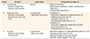
The specimens were randomly allocated to five groups. In three groups (n = 15), different caries-protective agents were applied according to the manufacturer's instructions: group A, chlorhexidine/thymol-containing varnish (Cervitec Plus®; Ivoclar Vivadent AG, Schaan, Liechtenstein); group B, fluoride-releasing chemically cured sealant (Maximum Cure®; Reliance Orthodontic Products, Inc., Itasca, IL, USA); and group C, fluoride-releasing light-cured sealant (Pro Seal®; Reliance Orthodontic Products, Inc.). For group A, a single dose of Cervitec Plus® was applied thinly on the enamel surfaces of specimens using a micro-brush (extra fine; Kerr GmbH, Biberach, Germany). Subsequently, the varnish was dried. For groups B and C, the enamel surfaces of the specimens were etched for 30 seconds with 37% phosphoric acid gel (Ivoclar Vivadent AG) prior to baseline varnish application, rinsed with water for 60 seconds, and dried thoroughly in oil-free air. For group B, both components of Maximum Cure® were mixed in a dappen-dish and applied in a thin uniform layer to the etched surfaces of specimens using a micro-brush. For group C, three drops of Pro Seal® were dispensed onto a mixing pad and a thin, uniform layer was applied on the etched enamel surfaces with a bristle brush. The enamel surfaces were stroked with the same brush to ensure a thin layer and good coverage. Subsequently, the layers were light-cured for 20 seconds (Ortholux™ XT Curing Light; 3M Unitek, Landsberg am Lech, Germany).
Table 1 shows the compositions of these agents. Group D (n = 15) served as a positive control (specimens only underwent the re- and demineralization cycles without application of any product) and group E (n = 5) served as a negative control (specimens were not subjected to the re- and demineralization cycles and only treated with artificial saliva).
Demineralization- and remineralization cycle
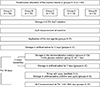
After the application of the test products (groups A–C) and rinsing, the specimens of groups A–D were stored in the demineralization solution and thereafter in artificial saliva for one hour each. These processes were repeated three times per day. Until the next cycle on the following day, all specimens (groups A–E) were stored in artificial saliva (about 15 hours). This procedure was continued for 30 days (Figure 1).
Evaluation of carious lesions
The specimens were imaged using QLF (Inspektor Research Systems BV, Amsterdam, The Netherlands) at baseline and days 7, 14, and 30. Using the QLF software package (version 2.0.0.43; Inspektor Research Systems BV), the average fluorescence loss (ΔF, %) and surface size of the lesions (mm2) were measured. The data were statistically analyzed using Wilcoxon–Mann–Whitney test (α = 0.05).
Statistical analysis
Statistical analysis was performed using the programs SAS (version 9.2; SAS Institute GmbH, Heidelberg, Germany) and Statistica (version 9; StatSoft [Europe] GmbH, Hamburg, Germany). The influences of test products and time on the measurements were investigated separately according to ΔF and size of lesion using two-way (non-parametric) ANOVA. In the case of a significant effect, pair comparisons were performed using the Wilcoxon-Mann–Whitney test. The level of significance was determined by α = 5%.
RESULTS
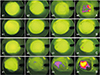
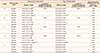
Average ΔF and surface size of the lesion in the specimens are presented in Table 2. Figure 2 shows the QLF images of groups A–D at all measurement points.
The specimens in group A demonstrated a significant increase in ΔF and lesion surface size after 30 days (p = 0.014), no significant difference in comparison with the positive control group (group D; p = 1.000), and a significant difference in comparison with the negative control (group E) after 30 days (p = 0.014).
The specimens in group B showed no changes in both parameters at all measurement points. Although the specimens in group C showed increased ΔF after 14 days, they showed no significant fluorescence change after 30 days (p = 0.392). Groups B and C showed no significant changes in the surface size of lesion compared to the negative control group (group E; p = 1.000) and a significant difference compared to the positive control (group D) after 30 days (p ≤ 0.028).
DISCUSSION
The current study assessed the effectiveness of a chlorhexidine/thymol-containing varnish and two fluoride- releasing sealants. While the two fluoride-releasing sealants (Maximum Cure® and Pro Seal®) showed greater caries-preventing ability, carious lesion formation was observed even with the use of chlorhexidine/thymolcontaining varnish (Cervitec Plus®). Therefore, our hypotheses that the fluoride-releasing sealants could prevent initial carious lesions and that the tested products did not differ in their ability to prevent the formation of carious lesions were rejected.
Fluorides play a central role in caries prevention.4 In orthodontics, in addition to the daily supervised tooth brushing with the application of fluoride, fluoride-releasing bonding materials or fluoride-releasing sealants for brackets and bands are also used for caries prevention.8 These products can continuously release fluoride over a long period and are therefore effective for tooth surfaces.9 Both the fluoride-releasing sealants (Maximum Cure® and Pro Seal®) assessed in this study are used to prevent demineralization of etched areas where orthodontic brackets are affixed and to improve the adhesion of bonding materials.6 Light-cured sealants are believed to be superior to chemically cured sealants due to their higher degree of polymerization, which can yield a more complete/stable coverage of the enamel surface.7 In the current study, Maximum Cure® and Pro Seal® showed no significant differences in ΔF and surface size of the lesions after 30 days. Previous studies have also shown that both products influence the extent and progression of demineralization effectively.671011 No significant differences were observed in the effectiveness of chemically cured and light-cured sealants after 30 days. Demito et al.12 demonstrated a reduction in demineralization depth of up to 38% after application of fluoride-releasing varnish compared to a reference group without fluoridation. The current study showed that the unprotected enamel surfaces that were exposed to demineralization- and remineralization cycles tend to develop erosive/white-spot lesions after 14 days.13
Chlorhexidine-containing products are used with the aim of reducing the demineralization risk by influencing the bacterial metabolism and by reducing the amount of S. mutans,14 and thymol was used as a purified active compound in characterizing different microorganisms' susceptibilities. Thymol has been reported to be one of the most active antimicrobials among the constituents of essential oils.15 Although several studies have demonstrated that supplemental application of the chlorhexidine/thymol-containing Cervitec Plus® has a tendency to inhibit demineralization, other studies have found no evidence of caries prevention.16171819 The current study also showed no anti-cariogenic effect of Cervitec Plus®. On the 14th day, the specimens with Cervitec Plus® showed a reduction in fluorescence, and on the 30th day, there was no significant difference between the group A and the group D. In contrast to the two fluoride-based agents investigated, Cervitec Plus® is applied to the cleaned tooth surface without any prior enamel etching process. Therefore, there may be less adhesion between the varnish and the tooth surface than between the sealant and the tooth surface. As a result, the varnish may have chipped off and the resulting discontinuities may have led to a reduced protective effect. Another possible explanation for the lower anti-cariogenic effect of Cervitec Plus® compared to the sealers is that S. mutans tends to recolonize over long-term application of chlorhexidine.20 Zaura-Arite and ten Cate21 also showed that a fluoride-releasing sealant has a greater demineralization-inhibiting effect than Cervitec Plus®. In contrast, Petersson et al.22 found in a comparative study of Cervitec and the fluoride-releasing Fluor Protector that both products were similarly effective in controlling caries incidence. The combined use of chlorhexidine along with fluoridation could help reduce caries risk.2324 Nevertheless, due to the lack of evidence for chlorhexidinecontaining products, fluoride-releasing products have often been considered the means of choice for preventing initial carious lesions.42025
Compared to most other studies that used a chemicalbased model of artificial caries, the current study used S. mutans in a microbe-based model. In comparison with natural carious lesions, artificial carious lesions allow production of a standardized specimen of any caries stage according to the need. While microbe-based caries models more closely resemble the intraoral situation and their caries development process is very similar to natural carious lesions, the existing models are costlier and require more time than chemical-based models. However, chemical-based models have disadvantages such as surface softening, implementation without intraoral conditions, and less realistic time periods of deand remineralization.26 This model used in the current study allowed us to 1) easily produce carious lesions under biological conditions with a high level of control, 2) show different levels of anti-cariogenic effects of different products, and 3) show the chronological progress of the anti-cariogenic effects.
QLF is considered to be a validated caries diagnostic tool, especially for detection of initial caries.27 Transverse microradiography is known as the gold standard method for determination of mineral loss, but it is destructive and invasive. In contrast, QLF provides non-invasive multiple measurements and the evaluated images can be archived, enabling longitudinal monitoring of caries development or progression.28 Previous studies presented a high correlation between ΔF and mineral loss and confirmed QLF as a suitable diagnostic tool.282930 Ando et al.29 reported that there is a non-linear correlation or even a non-correlation between ΔF and the size of the lesions. In contrast, an imperfect linear correlation between both parameters was observed in the current study regardless of groups and times of measurement.
The limitations of the current study are related to its experimental setup. No histological analysis of carious lesions was conducted after the QLF assessment to validate lesion formation. This analysis could have been performed after 30 days, e.g., using scanning electron microscopy or transverse microradiography. Additionally, the current study was performed in vitro, in which the entirety of clinical conditions and physiological processes could not be fully reproduced. Furthermore, this study lacks a simulation of orthodontic treatment procedures such as fixing of the brackets on the specimens, and it compared test products with different types of application (varnish and sealant), which may impair comparability. These shortcomings should be considered in follow-up studies.
 XML Download