

 PDF
PDF ePub
ePub Citation
 Print
Print


Abstract
Purpose
We report a case of acute visual loss with ophthalmoplegia after prone position spinal surgery who had blood supply dependence on collateral circulation due to occlusion of the Internal carotid artery.
Case summary
A 74-year-old man was referred to the department of ophthalmology for acute visual loss and ophthalmoplegia after lumbar spine surgery performed in prone position. On the initial visit, his right visual acuity was 0.8 and the left visual acuity was negative light perception. Intraocular pressure was normal. There was a relative afferent pupillary defect and ophthalmoplegia of all directions in the left eye. Because of the ptosis of the upper eyelid in the left eye, it was impossible to tune the eye voluntarily. The cherry red spot and pale retina were observed on the fundus examination. On brain magnetic resonance imaging angiography, we found complete obstruction of the left internal carotid artery. He had intravenous injection of 1 g methylprednisolone for 3 days, and discharged with per oral medicine. After 1 month of treatment, the ophthalmoplegia was slightly improved, but visual acuity was not recovered.
Conclusions
In this case, unlike previous reports, acute visual loss and ophthalmoplegia occurred after spinal surgery the patient who had collateral circulation for ocular blood supply because of complete obstruction of the left internal carotid artery. This report highlights the importance of being aware of the anatomical variant in possible complications of external ocular compression after non-ocular surgery.
Figures and Tables
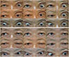 | Figure 1Photographs in the nine cardinal directions. (A) At his initial visit after spinal surgery, limitation of abduction, supraduction and infraduction are shown in the left eye. Because of the ptosis of the upper eyelid, it is impossible to tune the eye voluntarily. The left eye pupil is dilated at 4.5 mm. (B) After 1 month, the external ophthalmoplegia was improved.
|
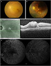 | Figure 2Posterior segment of the left eye. (A) Fundus photographs of patient on initial visit, (B) and last follow up at 7 months later. The cherry red spot on fovea with pale retina was showed, it developed chorioretinal atrophy several months later. (C) The reflectivity and thickness of inner retina was increased on optical coherence tomography, (D) arm to retinal time and arterio-venous transit time delay with delayed filling (28 seconds after injection) was detected on fluorescein angiography.
|
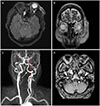 | Figure 3Brain magnetic resonance Image images of the patient. (A) On a fluid attenuated inversion recovery image, left optic nerve exhibits marked enhancement (arrow), (B) extraocular muscles show diffuse swelling (arrows). (C) Reconstructed magnetic resonance angiography image reveals complete occlusion in the left internal carotid artery (ICA) (red arrowheads) from the bifurcation site of left common carotid artery (short arrow), while intact right ICA (arrowheads). The left vertebral artery (long arrow) is enlarged, directly originated from left subclavian artery. (D) Complete occlusion in the left ICA (arrowhead) is visible on the axial view, by contrast to well visible right ICA (arrow).
|
References
1. Halfon MJ, Bonardo P, Valiensi S, et al. Central retinal artery occlusion and ophthalmoplegia following spinal surgery. Br J Ophthalmol. 2004; 88:1350–1352.

2. Stevens WR, Glazer PA, Kelley SD, et al. Ophthalmic complications after spinal surgery. Spine (Phila Pa 1976). 1997; 22:1319–1324.
3. Asok T, Aziz S, Faisal HA, et al. Central retinal artery occlusion and ophthalmoplegia following spinal surgery in the prone position. Med J Malyasia. 2009; 64:323–324.
4. Merchut MP, Gupta SR, Naheedy MH. The relation of retinal artery occlusion and carotid artery stenosis. Stroke. 1988; 19:1239–1242.
5. Shen Y, Drum M, Roth S. The prevalence of perioperative visual loss in the United States: a 10-year study from 1996 to 2005 of spinal, orthopedic, cardiac, and general surgery. Anesth Analg. 2009; 109:1534–1545.
6. Ahn EK, Min KT, Kim JR. Central retinal artery occlusion following general anesthesia. Korean J Anesthesiol. 1992; 25:777–779.
7. Kim C, Kim JH. A case of unilateral blindness occurring during general anesthesia for neurosurgical operation. Korean J Anesthesiol. 1989; 22:770–771.
8. Jin KH, Sohn WS, Kwark HW. Ischemic optic neuropathy following general anesthesia with prone position. J Korean Ophthalmol Soc. 1997; 38:2241–2246.
9. Kim JH, Ha H, Heo H, Lee SH. Visual loss with external ophthalmoplegia after shoulder operation. Clin Neuroophthalmol. 2018; 8:13–17.
10. Kumar N, Jivan S, Topping N, Morrell AJ. Blindness and rectus muscle damage following spinal surgery. Am J Ophthalmol. 2004; 138:889–891.
11. Myers MA, Hamilton SR, Bogosian AJ, et al. Visual loss as a complication of spine surgery. A review of 37 cases. Spine (Phila Pa 1976). 1997; 22:1325–1329.
12. The Korean Society of Neuro-Ophthalmology Society. Anatomy of neuro-ophthalmology. The Korean Society of Neuro-Ophthalmology Society. Neuro-ophthalmology. 3rd ed. Seoul: Ilchokak;2017. v. 1. chap. 1.
13. West J, Askin G, Clarke M, Vernon SA. Loss of vision in one eye following scoliosis surgery. Br J Ophthalmol. 1990; 74:243–244.
14. Hayreh SS, Zimmerman MB. Central retinal artery occlusion: visual outcome. Am J Ophthalmol. 2005; 140:376–391.
15. Uribe AA, Baig MN, Puente EG, et al. Current intraoperative devices to reduce visual loss after spine surgery. Neurosurg Focus. 2012; 33:E14.
 XML Download