

 PDF
PDF ePub
ePub Citation
 Print
Print


Abstract
Purpose
We report a case of utilizing a previous silicone band track in the reoperation of scleral encircling.
Case summary
An 8-year-old male presented with rhegmatogenous retinal detachment in the right eye. Five days after this diagnosis, he received scleral buckling surgery and cryopexy to seal the retinal tear. One month after surgery, a fundus examination showed subretinal fluid at the inferior site of the scleral buckle. He underwent scleral encircling surgery and a cryopexy procedure. The patient has had an uneventful postoperative course, and the retina has remained attached over a follow-up period of 9 months. However, exotropia and hypotropia developed in the right eye. Diagnosis of restrictive strabismus due to tissue adhesion around the silicone band was made. The encircling band was therefore removed and laser photocoagulation was performed 360° around the retina. Twenty-four hours after surgery, a fundus examination showed subretinal fluid. He received 360° scleral encircling surgery not using the 360° conjunctival peritomy. After confirming a previous encircling tract using #0-0 polydioxanone as a guide, #5-0 Nylon was tied to the end of the guide and inserted through the encircling tract with the end sutured with the silicone band. The silicone band was inserted into the encircling tract by pulling the #5-0 Nylon as a guide. Ophthalmoscopy revealed an attached retina with indentation of the scleral buckle at 360°.
Figures and Tables
Figure 1
Fundus photographs and Hess screening test of the patient. (A) Fundus photograph image at the first visit. Retinal tear at 9 o'clock direction and retinal detachment were observed. (B) Postoperative Hess screening test of the patient 9 months after scleral encircling. Exotropia and hypotropia developed in the right eye. (C) Postoperative fundus photograph 1 day after scleral encircling band removal and laser indirect ophthalmoscope photocoagulation showing subretinal fluid, decreased buckle height and laser scar. (D) Postoperative fundus photograph 1 day after re-operation of scleral encircling showing the sufficient buckle height and retinal attachment.
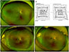
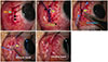
Figure 2
Intraoperative photographs from the surgeon's perspective reveal the operative procedure using the previous encircling tract. (A) Medial conjunctival peritomy (black arrows) and medial rectus muscle dissection (blue arrow) were done. (B) After confirming the previous encircling tract, 0-0 polydioxanone (PDS) (black arrow) was inserted through the encircling tract (yellow arrow). (C) The 5-0 Nylon was tied to the end of 0-0 PDS and using 0-0 PDS as a guide, 5-0 Nylon (blue arrow) was inserted to the encircling tract (yellow arrow). (D) The 5-0 Nylon was sutured to the 40 silicone band and using 5-0 Nylon as a guide (blue arrow), silicone band (white arrow) was inserted to the encircling tract (yellow arrow). (E) Silicone band (white arrows) was placed beneath the four rectus muscles and was fixed by the clip. MR = medial rectus.
Notes
References
1. Shunmugam M, Shah AN, Hysi PG, Williamson TH. The pattern and distribution of retinal breaks in eyes with rhegmatogenous retinal detachment. Am J Ophthalmol. 2014; 157:221–226.e1.

2. Gonin J. The treatment of detached retina by searing the retinal tears. Arch Ophthalmol. 1930; 4:621–625.
3. Schepens CL. Symposium: present status of retinal detachment surgery. Scleral buckling with circling element. Trans Am Acad Ophthalmol Otolaryngol. 1964; 68:959–979.
4. Lincoff HA, Baras I, McLean J. Modifications to the custodis procedure for retinal detachment. Arch Ophthalmol. 1965; 73:160–163.
5. D'Amico DJ. Clinical practice. Primary retinal detachment. N Engl J Med. 2008; 359:2346–2354.
6. Greven CM, Wall AB, Slusher MM. Anatomic and visual results in asymptomatic clinical rhegmatogenous retinal detachment repaired by scleral buckling. Am J Ophthalmol. 1999; 128:618–620.
7. Yao Y, Jiang L, Wang ZJ, Zhang MN. Scleral buckling procedures for longstanding or chronic rhegmatogenous retinal detachment with subretinal proliferation. Ophthalmology. 2006; 113:821–825.
8. Tsui I. Scleral buckle removal: indications and outcomes. Surv Ophthalmol. 2012; 57:253–263.
9. Kreiger AE, Hodgkinson BJ, Frederick AR Jr, Smith TR. The results of retinal detachment surgery. Analysis of 268 operations with a broad scleral buckle. Arch Ophthalmol. 1971; 86:385–394.
10. Kanski JJ, Elkington AR, Davies MS. Diplopia after retinal detachment surgery. Am J Ophthalmol. 1973; 76:38–40.
11. Sewell JJ, Knobloch WH, Eifrig DE. Extraocular muscle imbalance after surgical treatment for retinal detachment. Am J Ophthalmol. 1974; 78:321–323.
12. Wolff SM. Strabismus after retinal detachment surgery. Trans Am Ophthalmol Soc. 1983; 81:182–192.
13. Saleh TA, Gray RH. New retinal detachment following removal of a scleral explant. Eye (Lond). 2003; 17:245–246.
14. Foster RE, Meyers SM. Recurrent retinal detachment more than 1 year after reattachment. Ophthalmology. 2002; 109:1821–1827.
 XML Download