

 PDF
PDF ePub
ePub Citation
 Print
Print


Abstract
Case summary
A 49-year-old female presented with an outpouching punctal mass at the left lower lid that occurred a week prior to her visit. Histopathological examination revealed a cyst filled with keratin arranged in lamina and surrounding the bacterial colony. The epithelial wall was composed of multilaminar, keratinizing squamous epithelium without goblet cells. The features were consistent with a keratinizing cyst. There was no recurrence at 4 months after the excision, and the punctum was patent.
Figures and Tables
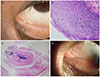
Figure 1
The keratinizing cyst of the left lower lid. (A) A photograph of the left lower eyelid showing a 2 mm, tense, whitish, non-vascular, smooth, cystic, dome-shaped mass at the region of lower punctum. (B) The epithelium was multilayered and showed a regular basaloid germinal layer without any goblet cells or any granular layer. Microphotograph showed keratin fibers on the stratified squamous epithelium (Haemotoxylin and Eosin [H&E] stain, ×400). (C) Microphotograph of the cystic content showing central bacterial colony (arrow) and surrounding keratin fibers arranged in laminar and wavy patterns (H&E stain, ×40). (D) There was no recurrence after 4 months.
References
1. Rumelt S, Pe'er J, Rubin PA. The clinicopathological spectrum of benign peripunctal tumours. Graefes Arch Clin Exp Ophthalmol. 2005; 243:113–119.

2. Ali MJ, Naik MN, Kaliki S. Punctal keratinizing cyst: a clinicopathological correlation of an exceptionally rare lacrimal disorder. Ophthalmic Plast Reconstr Surg. 2015; 31:e66–e68.
3. Kamal S, Ali MJ, Naik MN. Punctal keratinizing cyst: report in a pediatric patient with fourier domain optical coherence tomography features. Ophthalmic Plast Reconstr Surg. 2015; 31:161–163.
4. Yonekawa Y, Jakobiec FA, Zakka FR, Kim N. Keratinizing cyst of the lacrimal punctum. Cornea. 2013; 32:883–885.
5. Suh JY, Jung HM, Ahn HB, Kim MH. Clinical features and treatment of peripunctal tumors. J Korean Ophthalmol Soc. 2012; 53:918–923.
6. Wang L, Bi C, Wang T, et al. A coagulase-negative and non-haemolytic strain of Staphylococcus aureus for investigating the roles of SrtA in a murine model of bloodstream infection. Pathog Dis. 2015; 73:ftv042.
7. Kuniyuki S, Yoshida Y, Maekawa N, Yamanaka K. Bacteriological study of epidermal cysts. Acta Derm Venereol. 2008; 88:23–25.
 XML Download