

 PDF
PDF ePub
ePub Citation
 Print
Print


Abstract
Purpose
To measure and analyze ocular deviations between dominant and non-dominant eyes using video-oculography (VOG) in intermittent exotropia.
Methods
Fourteen subjects who were diagnosed with intermittent exotropia from July 2017 to July 2018 with age of 5 or more, visual acuity of 20/30 or better and corrected visual acuity of 20/25 or more and difference in vision of both eyes of 1 line or less on Snellen optotype were included. The subjects were asked to fixate on a black-on-white optotype at 1 m, which subtended a visual angle of 50 minutes of arc. The video files and data about ocular deviations were obtained using VOG with alternate cover test. We analyzed angles of ocular deviations in dominant and non-dominant eyes.
Results
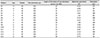
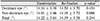
Among the 14 subjects in this study, the mean age were 7.6 ± 1.7 (range 5–9 years). Seven of 14 subjects had the right eye dominance. Six of the 14 subjects were men. There was no significant difference of ocular deviations between the dominant and non-dominant eyes in VOG (p = 0.167). Additionally, there was no significant difference of the values of VOG when one eye was exodeviated or re-fixated (p = 0.244), when both eyes were deviated, and when both eyes were re-fixated (p = 0.195, 0.637).
Conclusions
In this study, there was no significant difference of ocular deviations between the dominant and non-dominant eyes, between when an eye was exodeviated or fixated using VOG. Therefore, it may not be a problem even if alternate prism cover test is performed in any eye in intermittent exotropia of more than 50 prism diopter without amblyopia or refraction abnormality that could affect the uncorrected visual acuity.
Figures and Tables
Figure 1
A video-oculography (VOG) (accuracy: 0.1 degree). (A) Two cameras installed at the top can make measurements of both eye movements (red arrowheads). A tilted semi-transparent glass allows subjects to fixate the target (red arrows). (B) The subjects wearing the VOG goggles were asked to fixate a target point between the two eyes and were instructed to keep the head straight so that the eyes were in primary position during the examination.

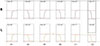
Figure 2
The graphs of video-oculography in the right-dominant eye. (A) is section of cover-uncover test in both eyes, (B) is section of alternative cover test. Initial binocular alignment was verified with both eyes open during the first 5 seconds. Subsequently, each eye was allowed 5 seconds of covered time and 5 seconds of uncovered time, and then the alternate cover test was repeated 3 times, with each eye being covered for 3 seconds.
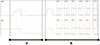
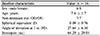
Figure 3
The section of alternative cover test of a patient who has the right-dominant eye. (A) Differences of ocular deviations between the dominant and non-dominant eye at the same section. Six values (A–C) of the non-dominant eye with values of the dominant eye in the same section. We compared LA1 with RA1, LA2 with RA2, LB1 with RB1, LB2 with RB2, LC1 with RC1, and LC2 with RC2. (B) Differences in values when an eye is deviated or re-fixated. In the right eye, RA1 and RA2, RB1 and RB2, RC1 and RC2. In the left eye, LA1 and LA2, LB1 and LB2, LC1 and LC2. (C) Differences in values when the non-dominant eye is deviated and when the dominant eye is deviated. Applied when the non-dominant eye and the dominant eye are re-fixated. Re-fixation: RA2 and LA1, RB2 and LB1, RC2 and LC1, deviation: RA1 and LA2, RB1 and LB2, RC1 and LC2. ‘RA1’ is defined as the values of ‘A1’ section in the right eye.

References
1. Romano R. Worldwide surveys of current management of intermittent exotropia by MD strabologists. Binocul Vis Strabismus Q. 1993; 8:167–176.
2. Ing MR, Pang SW. The racial distribution of strabismus. A statistical study. Hawaii Med J. 1974; 33:22–23.
3. Vail D. Worth and Chavasse's Squint. Am J Ophthalmol. 1960; 50:189.

4. Johns HA, Manny RE, Fern K, Hu YS. The intraexaminer and interexaminer repeatability of the alternate cover test using different prism neutralization endpoints. Optom Vis Sci. 2004; 81:939–946.
5. Holmes JM, Leske DA, Hohberger GG. Defining real change in prism-cover test measurements. Am J Ophthalmol. 2008; 145:381–385.
6. Hatt SR, Mohney BG, Leske DA, Holmes JM. Variability of control in intermittent exotropia. Ophthalmology. 2008; 115:371–376.e2.
7. Yang HK, Hwang JM. The effect of target size and accommodation on the distant angle of deviation in intermittent exotropia. Am J Ophthalmol. 2011; 151:907–913.e1.
8. Pediatric Eye Disease Investigator Group. Inter-observer reliability of the prism and alternate cover test in children with esotropia. Arch Ophthalmol. 2009; 127:59–65.
9. Pediatric Eye Disease Investigator Group. Christiansen SP, Chandler DL, et al. Instability of ocular alignment in childhood esotropia. Ophthalmology. 2008; 115:2266–2274.
10. Choi RY, Kushner BJ. The accuracy of experienced strabismologists using the Hirschberg and Krimsky tests. Ophthalmology. 1998; 105:1301–1306.
11. Yang HK, Han SB, Hwang JM, et al. Assessment of binocular alignment using the three-dimensional Strabismus Photo Analyzer. Br J Ophthalmol. 2012; 96:78–82.
12. van der Geest JN, Frens MA. Recording eye movements with video-oculography and scleral search coils: a direct comparison of two methods. J Neurosci Methods. 2002; 114:185–195.
13. Park N, Park B, Oh M, et al. A quantitative analysis method for comitant exotropia using video-oculography with alternate cover. BMC Ophthalmol. 2018; 18:80.
14. Economides JR, Adams DL, Horton JC. Variability of ocular deviation in strabismus. JAMA Ophthalmol. 2016; 134:63–69.
15. Hrynchak PK, Herriot C, Irving EL. Comparison of alternate cover test reliability at near in nonXMLLink_XYZstrabismus between experienced and novice examiners. Ophthalmic Physiol Opt. 2010; 30:304–309.
16. Von Noorden GK, Campos EC. Binocular vision and ocular motility. Theory and management of strabismus, vol. 6. 1st ed. St. Louis: Mosby;1990. p. 356–376.
17. Burian H. Symposium on horizontal ocular deviations. 1st ed. St. Louis: Mosby;1971. p. 235.
18. Costenbader FD. The physiology and management of divergent strabismus. 1st ed. St. Louis: CV Mosby;1950. p. 349–376.
19. Lee SA, Sunwoo IN, Kim KW. Divergence paralysis due to a small hematoma in the tegmentum of the brainstem. Yonsei Med J. 1987; 28:326–328.
20. Clark R, Demer J, Miller J, Rosenbaum A. Heterotopic rectus extraocular muscle pulleys simulate oblique muscle dysfunction. J AAPOS. 1997; 39.
21. Li Q, Bai J, Zhang J, et al. Assessment of cortical dysfunction in patients with intermittent exotropia: an fMRI study. PLoS One. 2016; 11:e0160806.
22. Ghasia FF, Otero-Millan J, Shaikh AG. Abnormal fixational eye movements in strabismus. Br J Ophthalmol. 2018; 102:253–259.
23. Economides JR, Adams DL, Horton JC. Capturing the moment of fusion loss in intermittent exotropia. Ophthalmology. 2017; 124:496–504.
 XML Download