

 PDF
PDF ePub
ePub Citation
 Print
Print


Abstract
Purpose
We compared and analyzed central corneal thickness (CCT) measurements according to the corneal thickness obtained with noncontact specular microscopy (NCSM) and ultrasound pachymetry (USP).
Methods
CCT was measured in the order of NCSM and USP by a single optometrist in 120 eyes of 120 healthy subjects. The measurements were compared between the devices and the measurement agreements and correlations between the devices were analyzed. To determine if the measurements differed depending on the thickness of the cornea, the patients were divided into three tertile groups from the thinnest patient by CCT measurement using USP, and then the differences in CCT measured by the two devices were analyzed.
Results
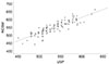
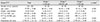
The CCT measurements using NCSM and USP were 548.25 ± 4.64 µm and 533.09 ± 35.96 µm, respectively. NCSM measurements were found to be thicker, showing statistically significant differences between the measurements (p < 0.001). The two examinations showed a high degree of correlation (r = 0.878; p < 0.01). In the three groups, the differences in CCT measurements between NCSM and USP were 12.93 ± 21.88 µm, 16.85 ± 15.89 µm, and 15.70 ± 20.46 µm, respectively, but the differences between the three groups were not statistically significant (p = 0.655).
Figures and Tables
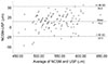 | Figure 1Bland Altman plots between the 2 methods. The middle line is the mean and the lines on the side represent the upper and lower 95% limits of agreement. NCSM = noncontact specular microscopy; USP = ultrasound pachymetry; SD = standard deviation.
|
References
1. De Moraes CG, Juthani VJ, Liebmann JM, et al. Risk factors for visual field progression in treated glaucoma. Arch Ophthalmol. 2011; 129:562–568.

2. Francis BA, Varma R, Chopra V, et al. Intraocular pressure, central corneal thickness, and prevalence of open-angle glaucoma: the Los Angeles Latino Eye Study. Am J Ophthalmol. 2008; 146:741–746.
3. Gordon MO, Beiser JA, Brandt JD, et al. The ocular hypertension treatment study: baseline factors that predict the onset of primary open-angle glaucoma. Arch Ophthalmol. 2002; 120:714–720. discussion 829-30.
4. Herndon LW, Weizer JS, Stinnett SS. Central corneal thickness as a risk factor for advanced glaucoma damage. Arch Ophthalmol. 2004; 122:17–21.
5. Cennamo G, Rosa N, La Rana A, et al. Non-contact tonometry in patients that underwent photorefractive keratectomy. Ophthalmologica. 1997; 211:341–343.
6. Simon G, Small RH, Ren Q, Parel JM. Effect of corneal hydration on Goldmann applanation tonometry and corneal topography. Refract Corneal Surg. 1993; 9:110–117.
7. Tonnu PA, Ho T, Newson T, et al. The influence of central corneal thickness and age on intraocular pressure measured by pneumotonometry, non-contact tonometry, the Tono-Pen XL, and Goldmann applanation tonometry. Br J Ophthalmol. 2005; 89:851–854.
8. Ashwin PT, McDonnell PJ. Collagen cross-linkage: a comprehensive review and directions for future research. Br J Ophthalmol. 2010; 94:965–970.
9. Kim DH, Kim MS, Kim JH. Early corneal-thickness changes after penetrating keratoplasty. J Korean Ophthalmol Soc. 1997; 38:1355–1361.
10. Calvo-Sanz JA, Ruiz-Alcocer J, Sánchez-Tena MA. Accuracy of Cirrus HD-OCT and Topcon SP-3000P for measuring central corneal thickness. J Optom. 2018; 11:192–197.
11. Marsich MW, Bullimore MA. The repeatability of corneal thickness measures. Cornea. 2000; 19:792–795.
12. Miglior S, Albe E, Guareschi M, et al. Intraobserver and interobserver reproducibility in the evaluation of ultrasonic pachymetry measurements of central corneal thickness. Br J Ophthalmol. 2004; 88:174–177.
13. Módis L Jr, Langenbucher A, Seitz B. Scanning-slit and specular microscopic pachymetry in comparison with ultrasonic determination of corneal thickness. Cornea. 2001; 20:711–714.
14. Wheeler NC, Morantes CM, Kristensen RM, et al. Reliability coefficients of three corneal pachymeters. Am J Ophthalmol. 1992; 113:645–651.
15. Solomon OD. Corneal indentation during ultrasonic pachometry. Cornea. 1999; 18:214–215.
16. Villaseñor RA, Santos VR, Cox KC, et al. Comparison of ultrasonic corneal thickness measurements before and during surgery in the prospective evaluation of Radial Keratotomy (PERK) Study. Ophthalmology. 1986; 93:327–330.
17. Choi KS, Nam SM, Lee HK, et al. Comparison of central corneal thickness after the instillation of topical anesthetics: proparacaine versus oxybuprocaine. J Korean Ophthalmol Soc. 2005; 46:757–762.
18. Bland JM, Altman DG. Statistical methods for assessing agreement between two methods of clinical measurement. Lancet. 1986; 1:307–310.
19. Bovelle R, Kaufman SC, Thompson HW, Hamano H. Corneal thickness measurements with the Topcon SP-2000P specular microscope and an ultrasound pachymeter. Arch Ophthalmol. 1999; 117:868–870.
20. Módis L Jr, Langenbucher A, Seitz B. Corneal thickness measurements with contact and noncontact specular microscopic and ultrasonic pachymetry. Am J Ophthalmol. 2001; 132:517–521.
21. Al Farhan HM, Al Otaibi WM, Al Razqan HM, Al Harqan AA. Assessment of central corneal thickness and corneal endothelial morphology using ultrasound pachymetry, non-contact specular microscopy, and Confoscan 4 confocal microscopy. BMC Ophthalmol. 2013; 13:73.
22. Suzuki S, Oshika T, Oki K, et al. Corneal thickness measurements: scanning-slit corneal topography and noncontact specular microscopy versus ultrasonic pachymetry. J Cataract Refract Surg. 2003; 29:1313–1318.
23. Al-Ageel S, Al-Muammar AM. Comparison of central corneal thickness measurements by Pentacam, noncontact specular microscope, and ultrasound pachymetry in normal and post-LASIK eyes. Saudi J Ophthalmol. 2009; 23:181–187.
24. Tai LY, Khaw KW, Ng CM, Subrayan V. Central corneal thickness measurements with different imaging devices and ultrasound pachymetry. Cornea. 2013; 32:766–771.
25. Almubrad TM, Osuagwu UL, Alabbadi I, Ogbuehi KC. Comparison of the precision of the Topcon SP-3000P specular microscope and an ultrasound pachymeter. Clin Ophthalmol. 2011; 5:871–876.
26. González-Pérez J, González-Méijome JM, Rodríguez Ares MT, Parafita MA. Central corneal thickness measured with three optical devices and ultrasound pachometry. Eye Contact Lens. 2011; 37:66–70.
27. Khaja WA, Grover S, Kelmenson AT, et al. Comparison of central corneal thickness: ultrasound pachymetry versus slit-lamp optical coherence tomography, specular microscopy, and Orbscan. Clin Ophthalmol. 2015; 9:1065–1070.
28. Fujioka M, Nakamura M, Tatsumi Y, et al. Comparison of Pentacam Scheimpflug camera with ultrasound pachymetry and noncontact specular microscopy in measuring central corneal thickness. Curr Eye Res. 2007; 32:89–94.
29. Bao F, Wang Q, Cheng S, et al. Comparison and evaluation of central corneal thickness using 2 new noncontact specular microscopes and conventional pachymetry devices. Cornea. 2014; 33:576–581.
30. Módis L Jr, Szalai E, Németh G, Berta A. Evaluation of a recently developed noncontact specular microscope in comparison with conventional pachymetry devices. Eur J Ophthalmol. 2010; 20:831–838.
31. Çevik SG, Duman R, Çevik MT, et al. Comparison of central corneal thickness estimated by an ultrasonic pachymeter and non-contact specular microscopy. Arq Bras Oftalmol. 2016; 79:312–314.
32. Jung YG, Song JS, Kim HM, Jung HR. Comparison of corneal thickness measurements with noncontact specular microscope and ultrasonic pachymeter. J Korean Ophthalmol Soc. 2004; 45:1060–1065.
33. Kim HS, Kim JH, Kim HM, Song JS. Comparison of corneal thickness measured by specular, US pachymetry, and Orbscan in Post-PKP eyes. J Korean Ophthalmol Soc. 2007; 48:245–250.
34. Yang YS, Koh JW. Utility of the Noncontact specular microscopy for measurements of central corneal thickness. J Korean Ophthalmol Soc. 2014; 55:59–65.
35. Lee MJ, Shin YU, Lim HW, et al. Central corneal thickness measured by noncontact specular microscopy, dual rotating scheimpflug camera and ultrasound pachymetry. J Korean Ophthalmol Soc. 2015; 56:1520–1526.
36. Wells M, Wu N, Kokkinakis J, Sutton G. Correlation of central corneal thickness measurements using Topcon TRK-1P, Zeiss Visante AS-OCT and DGH Pachmate 55 handheld ultrasonic pachymeter. Clin Exp Optom. 2013; 96:385–387.
37. Tam ES, Rootman DS. Comparison of central corneal thickness measurements by specular microscopy, ultrasound pachymetry, and ultrasound biomicroscopy. J Cataract Refract Surg. 2003; 29:1179–1184.
38. Nissen J, Hjortdal JO, Ehlers N, et al. A clinical comparison of optical and ultrasonic pachometry. Acta Ophthalmol (Copenh). 1991; 69:659–663.
39. Uçakhan OO, Ozkan M, Kanpolat A. Corneal thickness measurements in normal and keratoconic eyes: pentacam comprehensive eye scanner versus noncontact specular microscopy and ultrasound pachymetry. J Cataract Refract Surg. 2006; 32:970–977.
40. Ing JJ, Ing HH, Nelson LR, et al. Ten-year postoperative results of penetrating keratoplasty. Ophthalmology. 1998; 105:1855–1865.
41. Kohlhaas M, Stahlhut O, Tholuck J, Richard G. Changes in corneal thickness and endothelial cell density after cataract extraction using phacoemulsification. Ophthalmologe. 1997; 94:515–518.
42. Olsen T. Corneal thickness and endothelial damage after intracapsular cataract extraction. Acta Ophthalmol (Copenh). 1980; 58:424–433.
43. Olsen T, Eriksen JS. Corneal thickness and endothelial damage after intraocular lens implantation. Acta Ophthalmol (Copenh). 1980; 58:773–786.
44. Ventura AC, Wälti R, Böhnke M. Corneal thickness and endothelial density before and after cataract surgery. Br J Ophthalmol. 2001; 85:18–20.
45. Cheng H, Bates AK, Wood L, McPherson K. Positive correlation of corneal thickness and endothelial cell loss. Serial measurements after cataract surgery. Arch Ophthalmol. 1988; 106:920–922.
46. Amon M, Menapace R, Radax U, Papapanos P. Endothelial cell density and corneal pachometry after no-stitch, small-incision cataract surgery. Doc Ophthalmol. 1992; 81:301–307.
47. Hashmani N, Hashmani S, Hanfi AN, et al. Effect of age, sex, and refractive errors on central corneal thickness measured by Oculus Pentacam (R). Clin Ophthalmol. 2017; 11:1233–1238.
48. Tayyab A, Masrur A, Afzal F, et al. Central corneal thickness and its relationship to intra-ocular and epidmiological determinants. J Coll Physicians Surg Pak. 2016; 26:494–497.
49. Hashemi H, Yazdani K, Mehravaran S, et al. Corneal thickness in a population-based, cross-sectional study: the Tehran Eye Study. Cornea. 2009; 28:395–400.
50. Mercieca K, Odogu V, Fiebai B, et al. Comparing central corneal thickness in a sub-Saharan cohort to African Americans and Afro-Caribbeans. Cornea. 2007; 26:557–560.
51. Prasad A, Fry K, Hersh PS. Relationship of age and refraction to central corneal thickness. Cornea. 2011; 30:553–555.
52. Channa R, Mir F, Shah MN, et al. Central corneal thickness of Pakistani adults. J Pak Med Assoc. 2009; 59:225–228.
53. Wang Q, Liu W, Wu Y, et al. Central corneal thickness and its relationship to ocular parameters in young adult myopic eyes. Clin Exp Optom. 2017; 100:250–254.
54. Nomura H, Ando F, Niino N, et al. The relationship between age and intraocular pressure in a Japanese population: the influence of central corneal thickness. Curr Eye Res. 2002; 24:81–85.
 XML Download