

 PDF
PDF ePub
ePub Citation
 Print
Print


Abstract
Purpose
We evaluated the usefulness of the Tomey TMS-4N™ computerized videokeratoscopy (Tomey, Nagoya, Japan) for dry eye by analyzing the correlation between the regularity indices of TMS-4N™ and conventional dry eye parameters or dry eye severity.
Methods
In this retrospective study, the medical records of 193 dry eye patients (386 eyes) were analyzed. The regularity indices of TMS-4N™ such as the surface asymmetry index (SAI), surface regularity index (SRI), potential visual acuity (PVA), and irregular astigmatism index (IAI) were compared with conventional dry eye parameters (corrected visual acuity [CVA], ocular surface disease index [OSDI] score, tear film break-up time [TBUT], corneal staining score [CFS], and Schirmer's I test). We also analyzed correlations between the regularity indices of TMS-4N™ and dry eye severity according to the Korean Corneal Disease Study Group.
Results
The regularity indices of TMS-4N™ such as SAI, SRI, PVA, and IAI increased according to the severity of dry eye. The regularity indices correlated significantly and positively with the CVA and CFS, but were significantly and negatively correlated with the TBUT. The OSDI score did not correlate with the regularity indices.
Figures and Tables
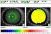 | Figure 1Examination images using Tomey TMS-4N™ (Tomey, Nagoya, Japan). The image is analyzed by a program which identifies the location of 256 circumferential points around each ring's reflection. CYL = simulated keratometric cylinder change; SRI = surface irregularity index; PVA = potential visual acuity; SAI = surface asymmetry index.
|
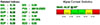 | Figure 2Indices from Klyce Corneal Statistics in Tomey TMS-4N™ (Tomey, Nagoya, Japan). Surface asymmetry index has measured the difference between the keratometry all ring 180 degrees apart across the corneal surface. Surface irregularity index measures the local variations in the corneal center. Potential visual acuity shows the visual acuity calculated by surface regularity index. Irregular astigmatism index measures the average sum of area corrected for the difference between each ring over the entire corneal surface. SAI = surface asymmetry index; SRI = surface irregularity index; CYL = simulated keratometric cylinder change; PVA = potential visual acuity; CVP = coefficient of variation of corneal power; ACP = average corneal power; SDP = standard deviation of corneal power; CEI = corneal eccentricity index; IAI = irregular astigmatism index; AA = analyzed area; EDP = elevation/depression power; EDD = elevation/depression diameter.
|
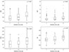 | Figure 3Differences and correlation between dry eye severity and videokeratoscopic regularity indices. Each videokeratoscopic index was positively correlated with severity of dry eye. Data were analyzed using one-way analysis of variance, Jonckheere-Terpstra test. SRI = surface regularity index; SAI = surface asymmetry index; PVA = potential visual acuity; IAI = irregular astigmatism index. *, o defined as a data point that is located outside the fence of the box plot (* = extreme values, o =out values).
|
Table 1
Clinical characteristics of study population
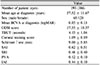
Values are presented as mean ± standard deviation unless otherwise indicated..
BCVA = best corrected visual acuity; logMAR = the logarithm of minimal angle of resolution; OSDI = ocular surface disease index; TBUT = tear break-up time; SAI = surface asymmetry index; SRI = surface regularity index; PVA = potential visual acuity; IAI = irregular astigmatism index.
![]()

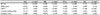
Table 3
Spearman correlation analysis between videokeratoscopic indices with conventional dry eye tests
![]()

Notes
References
1. Craig JP, Nicholas KK, Akpek EK, et al. TFOS DEWS II definition and classification report. Ocul Surf. 2017; 15:276–283.

2. Stapleton F, Alves M, Bunya VY, et al. TFOS DEWS II epidemiology report. Ocul Surf. 2017; 15:334–365.
3. Ahn JM, Lee SH, Rim TH, et al. Prevalence of and risk factors associated with dry eye: the Korea National Health and Nutrition Examination Survey 2010–2011. Am J Ophthalmol. 2014; 158:1205–1214.e7.
4. Roh HC, Lee JK, Kim M, et al. Systemic comorbidities of dry eye syndrome: the Korean National Health and Nutrition Examination Survey V, 2010 to 2012. Cornea. 2016; 35:187–192.
5. Paulsen AJ, Cruickshanks KJ, Fischer ME, et al. Dry eye in the beaver dam offspring study: prevalence, risk factors, and health-related quality of life. Am J Ophthalmol. 2014; 157:799–806.
6. Wolffsohn JS, Arita R, Chalmers R, et al. TFOS DEWS II diagnostic methodology report. Ocul Surf. 2017; 15:539–574.
7. Patel S, Murray D, McKenzie A, et al. Effects of fluorescein on tear breakup time and on tear thinning time. Am J Optom Physiol Opt. 1985; 62:188–190.
8. Mengher LS, Bron AJ, Tonge SR, Gilbert DJ. Effect of fluorescein instillation on the pre-corneal tear film stability. Curr Eye Res. 1985; 4:9–12.
9. Mengher LS, Bron AJ, Tonge SR, Gilbert DJ. A non-invasive instrument for clinical assessment of the pre-corneal tear film stability. Curr Eye Res. 1985; 4:1–7.
10. Nichols KK, Mitchell GL, Zadnik K. The repeatability of clinical measurements of dry eye. Cornea. 2004; 23:272–285.
11. Pinschmidt NW. Evaluation of the Schirmer tear test. South Med J. 1970; 63:1256. passim.
12. Feldman F, Wood MM. Evaluation of the Schirmer tear test. Can J Ophthalmol. 1979; 14:257–259.
13. Wright JC, Meger GE. A review of the Schirmer test for tear production. Arch Ophthalmol. 1962; 67:564–565.
14. Lemp MA. Report of the National Eye Institute/Industry workshop on Clinical Trials in Dry Eyes. CLAO J. 1995; 21:221–232.
15. de Paiva CS, Lindsey JL, Pflugfelder SC. Assessing the severity of keratitis sicca with videokeratoscopic indices. Ophthalmology. 2003; 110:1102–1109.
16. Ozkan Y, Bozkurt B, Gedik S, et al. Corneal topographical study of the effect of lacrimal punctum occlusion on corneal surface regularity in dry eye patients. Eur J Ophthalmol. 2001; 11:116–119.
17. Liu Z, Pflugfelder SC. Corneal surface regularity and the effect of artificial tears in aqueous tear deficiency. Ophthalmology. 1999; 106:939–943.
18. Hyon JY, Kim HM, Lee D, et al. Korean guidelines for the diagnosis and management of dry eye: development and validation of clinical efficacy. Korean J Ophthalmol. 2014; 28:197–206.
19. McGinnigle S, Naroo SA, Eperjesi F. Evaluation of dry eye. Surv Ophthalmol. 2012; 57:293–316.
20. Yoon KC, Im SK, Kim HG, You IC. Usefulness of double vital staining with 1% fluorescein and 1% lissamine green in patients with dry eye syndrome. Cornea. 2011; 30:972–976.
21. Bron AJ, Evans VE, Smith JA. Grading of corneal and conjunctival staining in the context of other dry eye tests. Cornea. 2003; 22:640–650.
22. Lee SJ, Kim HY, Park YM, Lee JS. Comparison of therapeutic effects of 3% diquafosol tetrasodium with aging in dry eye. J Korean Ophthalmol Soc. 2016; 57:734–741.
23. Tsubota K. Tear dynamics and dry eye. Prog Retin Eye Res. 1998; 17:565–596.
24. Chen JJ, Rao K, Pflugfelder SC. Corneal epithelial opacity in dysfunctional tear syndrome. Am J Ophthalmol. 2009; 148:376–382.
25. Choi KW, Moon SW, Joo MJ. Wavefront aberration changes after the instillation of artificial tear in dry eyes. J Korean Ophthalmol Soc. 2006; 47:186–191.
26. Shiotani Y, Maeda N, Inoue T, et al. Comparison of topographic indices that correlate with visual acuity in videokeratography. Ophthalmology. 2000; 107:559–564.
27. Liu Z, Pflugfelder SC. The effects of long-term contact lens wear on corneal thickness, and surface regularity. Ophthalmology. 2000; 107:105–111.
28. Dursun D, Monroy D, Knighton R, et al. The effects of experimental tear film removal on corneal surface regularity and barrier function. Ophthalmology. 2000; 107:1754–1760.
29. Németh J, Erdélyi B, Csákány B. Corneal topography changes after a 15 second pause in blinking. J Cataract Refract Surg. 2001; 27:589–592.
30. Battat L, Macri A, Dursun D, Pflugfelder SC. Effects of laser in situ keratomileusis on tear production, clearance, and the ocular surface. Ophthalmology. 2001; 108:1230–1235.
31. Goto T, Klyce SD, Zheng X, et al. Gender- and age-related differences in corneal topography. Cornea. 2001; 20:270–276.
32. Maeda N, Sato S, Watanabe H, et al. Prediction of letter contrast sensitivity using videokeratographic indices. Am J Ophthalmol. 2000; 129:759–763.
33. Schein OD, Tielsch JM, Munõz B, et al. Relation between signs and symptoms of dry eye in the elderly. A population-based perspective. Ophthalmology. 1997; 104:1395–1401.
34. Nichols KK, Nichols JJ, Mitchell GL. The lack of association between signs and symptoms in patients with dry eye disease. Cornea. 2004; 23:762–770.
 XML Download