

 PDF
PDF ePub
ePub Citation
 Print
Print


Abstract
Methods
Twenty-six patients (49 eyes) with at least 6 months of myopia prior to OK treatment were evaluated. Changes in the spherical equivalent (SE) refractive error and axial length were compared before and after OK use. Changes in the SE and axial length were also compared between two groups according to the myopic progression before baseline: Group 1 with myopic progression < 1 D/year and Group 2 with myopic progression > 1 D/year.
Results
The myopic progression rate decreased from −1.1 to −0.3 D/year after OK treatment (p < 0.001). Greater increases in axial length were observed in patients who were younger and had less myopia at baseline, a higher rate of myopia progression before baseline, and a shorter axial length at baseline (p < 0.001, p < 0.004, p < 0.007, and p < 0.001, respectively). The increase in axial length was significantly greater in the group with greater myopic progression before baseline (0.2 mm/year) than in the group with less myopic progression (0.1 mm/year) (p = 0.001).
Figures and Tables
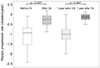 | Figure 1Myopic progression rates of before and after orthokeratology lens (OK) treatment. The mean myopic progression rate from diagnosis to OK (before OK) is −1.1 D/year, which is significantly higher than −0.3 D/year from OK to last follow up (after OK) (*p < 0.001, Wilcoxon Signed-Rank test). The mean myopic progression of 1 year before OK is −1.1 D, which is significantly higher than −0.2 D in 1 year after OK (*p < 0.001, Wilcoxon Signed-Rank test).
|
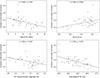 | Figure 2Increase in axial length and age at orthokeratology lens (OK), spherical equivalent refractive error (SE) at OK, SE change (diopter, D)/year and axial length at OK. Significant correlations were found between increase in axial length and age at OK, SE at OK, SE change (D)/year and axial length at OK (Pearson's correlation coefficient, r = −0.482; p < 0.001, r = 0.409; p = 0.004, r = −0.380; p = 0.007, r = −0.554; p < 0.001).
|
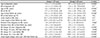
Table 2
Comparison between 2 groups according to the myopic progression before orthokeratology treatment
Values are presented as mean ± standard deviation (range). ‘Group 1’ means myopic progression rate less than 1 D/year. ‘Group 2’ means myopic progression rate more than 1 D/year.
SE = spherical equivalent refractive error; D = diopter; OK = orthokeratology lens.
*Two sample t-test; †Mann Whitney U-test.
![]()
References
1. Fan DS, Lam DS, Lam RF, et al. Prevalence, incidence, and progression of myopia of school children in Hong Kong. Invest Ophthalmol Vis Sci. 2004; 45:1071–1075.

2. Li SM, Li SY, Kang MT, et al. Near work related parameters and myopia in Chinese children: the Anyang childhood eye study. PLoS One. 2015; 10:e0134514.
3. Cho P, Cheung SW, Edwards M. The longitudinal orthokeratology research in children (LORIC) in Hong Kong: a pilot study on refractive changes and myopic control. Curr Eye Res. 2005; 30:71–80.
4. Walline JJ, Jones LA, Sinnott LT. Corneal reshaping and myopia progression. Br J Ophthalmol. 2009; 93:1181–1185.
5. Kakita T, Hiraoka T, Oshika T. Influence of overnight orthokeratology on axial elongation in childhood myopia. Invest Ophthalmol Vis Sci. 2011; 52:2170–2174.
6. Hiraoka T, Kakita T, Okamoto F, et al. Long-term effect of overnight orthokeratology on axial length elongation in childhood myopia: a 5-year follow-up study. Invest Ophthalmol Vis Sci. 2012; 53:3913–3919.
7. Santodomingo-Rubido J, Villa-Collar C, Gilmartin B, Gutiérrez-Ortega R. Factors preventing myopia progression with orthokeratology correction. Optom Vis Sci. 2013; 90:1225–1236.
8. Charm J, Cho P. High myopia-partial reduction ortho-k: a 2-year randomized study. Optom Vis Sci. 2013; 90:530–539.
9. Cho P, Cheung SW. Retardation of myopia in Orthokeratology (ROMIO) study: a 2-year randomized clinical trial. Invest Ophthalmol Vis Sci. 2012; 53:7077–7085.
10. Chen C, Cheung SW, Cho P. Myopia control using toric orthokeratology (TO-SEE study). Invest Ophthalmol Vis Sci. 2013; 54:6510–6517.
11. Saw SM, Shankar A, Tan SB, et al. A cohort study of incident myopia in Singaporean children. Invest Ophthalmol Vis Sci. 2006; 47:1839–1844.
12. Mutti DO, Mitchell GL, Moeschberger ML, et al. Parental myopia, near work, school achievement, and children's refractive error. Invest Ophthalmol Vis Sci. 2002; 43:3633–3640.
13. Hammond CJ, Snieder H, Gilbert CE, Spector TD. Genes and environment in refractive error: the twin eye study. Invest Ophthalmol Vis Sci. 2001; 42:1232–1236.
14. Swarbrick HA, Alharbi A, Watt K, et al. Myopia control during orthokeratology lens wear in children using a novel study design. Ophthalmology. 2015; 122:620–630.
15. Chan KY, Cheung SW, Cho P. Orthokeratology for slowing myopic progression in a pair of identical twins. Cont Lens Anterior Eye. 2014; 37:116–119.
16. Hyman L, Gwiazda J, Hussein M, et al. Relationship of age, sex, and ethnicity with myopia progression and axial elongation in the correction of myopia evaluation trial. Arch Ophthalmol. 2005; 123:977–987.
 XML Download