Journal List > Allergy Asthma Immunol Res > v.11(5) > 1129644

Hur and Broide: Genes and Pathways Regulating Decline in Lung Function and Airway Remodeling in Asthma

Abstract

Asthma is a common disorder of the airways characterized by airway inflammation and by decline in lung function and airway remodeling in a subset of asthmatics. Airway remodeling is characterized by structural changes which include airway smooth muscle hypertrophy/hyperplasia, subepithelial fibrosis due to thickening of the reticular basement membrane, mucus metaplasia of the epithelium, and angiogenesis. Epidemiologic studies suggest that both genetic and environmental factors may contribute to decline in lung function and airway remodeling in a subset of asthmatics. Environmental factors include respiratory viral infection-triggered asthma exacerbations, and tobacco smoke. There is also evidence that several asthma candidate genes may contribute to decline in lung function, including ADAM33, PLAUR, VEGF, IL13, CHI3L1, TSLP, GSDMB, TGFB1, POSTN, ESR1 and ARG2. In addition, mediators or cytokines, including cysteinyl leukotrienes, matrix metallopeptidase-9, interleukin-33 and eosinophil expression of transforming growth factor-β, may contribute to airway remodeling in asthma. Although increased airway smooth muscle is associated with reduced lung function (i.e. forced expiratory volume in 1 second) in asthma, there have been few long-term studies to determine how individual pathologic features of airway remodeling contribute to decline in lung function in asthma. Clinical studies with inhibitors of individual gene products, cytokines or mediators are needed in asthmatic patients to identify their individual role in decline in lung function and/or airway remodeling.

INTRODUCTION

Asthma is a common disorder of the airways characterized by airway inflammation and in a subset of asthmatics by decline in lung function and airway remodeling. The estimated prevalence of asthma in the United States is approximately 8.6% for adults and 8.3% for children in 2016.1 In Korea, the prevalence of asthma is estimated as 3.1% of adults in 2017,2 and 5.3%–9.1% of children (age 6–15 years) in 2010. A subset of asthmatics develops decline in lung function and airway remodeling which this review focuses on. Airway remodeling is characterized by structural changes including airway smooth muscle hypertrophy/hyperplasia, subepithelial fibrosis due to thickening of the reticular basement membrane, mucus metaplasia of the epithelium, and angiogenesis.3 Epidemiologic studies suggest that both genetic and environmental factors may contribute to decline in lung function and airway remodeling in a subset of asthmatics.
Epidemiologic studies of longitudinal lung function over a 15-year period in adult asthmatics compared to non-asthmatics demonstrated that asthmatics had a greater decline in forced expiratory volume in 1 second (FEV1) over time than non-asthmatic controls.4 In that large study of 17,506 subjects, asthmatics had a greater decline in FEV1 (38 mL/year) than non-asthmatics (22 mL/year). In addition, asthmatics who smoked tobacco had a greater decline in lung function than those who did not. Studies have also examined lung function changes in childhood asthmatics. As children have lungs that continue to grow until the age of 18 to 25 years,5 childhood asthmatics with airway remodeling may or may not attain normal lung function by adulthood before they exhibit decline in lung function. For example, longitudinal measurements of lung function in 684 childhood asthmatics followed up for 13 years demonstrated that those with persistent childhood asthma may have either normal lung function growth (25% of childhood asthmatics) or impaired lung function growth outcomes (75% of childhood asthmatics).6 A normal pattern of lung-function growth in childhood is characterized by a normal steep increase during adolescence, a plateau in early adulthood and a gradual decline at old ages. Abnormal trajectories include 1) reduced lung function growth (i.e. do not attain normal adult lung function) (23% of childhood asthmatics), 2) normal lung function growth and an early decline in lung function (26% of childhood asthmatics), and 3) reduced lung function growth and an early decline in lung function (26% of childhood asthmatics).6 At the last lung function test in that 13-year longitudinal study of lung function in childhood asthma, 11% of childhood asthmatics met the global initiative of chronic obstructive lung disease criteria for lung function impairment that was consistent with chronic obstructive pulmonary disease (COPD).6 Such longitudinal epidemiologic studies of lung function in adults and children with asthma demonstrate that the potential for a subset of adult asthmatics to progress with an accelerated lung function decline over time, or for a subset of childhood asthmatics either to not attain normal long function and/or to have an accelerated decline in lung function. As not all asthmatics exhibit these lung function changes, it is likely that genetic and environmental factors contribute to the different patterns of lung function growth and/or decline in each asthmatic subject. In terms of genetic factors for decline in lung function in childhood-onset asthmatics, a recent genome-wide association study (GWAS) study investigated genes associated with decline in lung function in adults with self-reported childhood asthma who were adult tobacco smokers.7 Among 10,199 adult smokers, 730 (7%) reported childhood asthma.7 The subjects were smokers and reported a history of childhood asthma, had reduced lung function as adults, and an increased risk for COPD (odds ratio, 3.42; 95% confidence interval [CI], 2.81–4.18).7 Genetic associations were also found in these subjects who had childhood asthma with known asthma loci, including IL1RL1, IL13, LINC01149 and GSDMB.7 These findings in childhood asthmatics who smoke underscore the interaction of genetic factors in childhood asthma and environmental factors such as tobacco smoke in contributing to decline in lung function and the development of features of early COPD.
In this review, we provide update of advances in the understanding of the genetic and environmental factors contributing to pathways of decline in lung function and airway remodeling in asthma as well as highlight current gaps in our understanding.

GENES REGULATING DECLINE IN LUNG FUNCTION AND/OR AIRWAY REMODELING

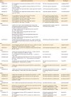
A combination of genetic and environmental factors leads to the development of asthma. Although over 100 genes have been linked to the development of asthma, the most reproduced asthma-associated genes include IL33, IL1RL1, IL13, TSLP, HLA, GATA3, SMAD3 and genes localized to chromosome 17q12-21 (ORMDL3 and GSDMB).8 In addition, several genes (ADAM33, PLAUR, VEGF, IL13, CHI3L1, TSLP, GSDMB, TGFB1, POSTN, ESR1 and ARG2) have been associated with decline in lung function in asthma (Table 1) and/or the features of airway remodeling (Table 2) in the limited number of studies, and thus some of these observations require further replication and validation.
Table 1

Gene variants associated with decline in lung function in asthma

aair-11-604-i001
Gene Lung function associations Study subjects Study/year
ADAM33
rs528557 G/C CC homozygotes had excess annual decline of FEV1 (−23.7 mL/yr) (P = 0.006) 200 Dutch Caucasians with asthma Jongepier/200412
rs528557 G/C CC homozygotes had a significant excessive decline in FEV1 of 4.9 mL/yr (P = 0.033) 1,390 Dutch general population van Diemen/200513
rs3918395 AA AA homozygotes were associated with a rapid lung function decline (P = 4.34 × 10⁻⁵) 1,047 Caucasian general population Poon/201414
rs612709 C/T TT homozygotes had a significant excessive decline in FEV1 of 9.6 mL/yr (P = 0.021) 1,390 Dutch general population van Diemen/200513
PLAUR
rs2356338 G/T TT homozygotes had more rapid annual FEV1 decline (−34.8 mL/yr vs. −22.2 mL/yr, P = 0.040) 587 UK and Dutch asthma families (n = 2,819) and 184 healthy controls Barton/200918
rs4802189 C/A AA homozygotes had more rapid annual FEV1 decline (−32.4 mL/yr vs. −19.0 mL/yr, P = 0.003) 587 UK and Dutch asthma families (n = 2,819) and 184 healthy controls Barton/200918
rs4803648 T/A AA homozygotes had more rapid annual FEV1 decline (−31.0 mL/yr vs. −19.2 mL/yr, P = 0.008) 587 UK and Dutch asthma families (n = 2,819) and 184 healthy controls Barton/200918
VEGF
rs3025028 C/G G allele carriers (GC or GG) had significantly lower airway conductance measured from birth throughout childhood with the effect persisting into adulthood Two general birth cohorts (1,246 from TCRS and 995 from MAAS) Simpson/201222
In infancy, G allele carriers had significantly lower VmaxFRC (P = 0.047, TCRS cohort)
At age 3, G allele carriers had significantly lower sGaw (P = 0.020, MAAS cohort)
At age 5 and 8, G allele carriers had significantly lower FEV1 (P ≤ 0.030, MAAS cohort)
At age 22, G allele carriers had significantly lower FEV1/FVC (P = 0.010, TCRS cohort)
rs4711750 A/T rs4711750 was associated with FEV1/FVC (P =  0.010) in CAMP, and TT homozygotes had persistent decline of FEV1/FVC over 4.5 years observation 458 families with asthmatic children Sharma/200923
IL13
rs20541 R110Q (G/A) AA homozygotes (Q110/Q110) had lower FEV1 (P = 0.020), and rapid decline in FEV1 (32.1mL/yr vs. 25.4mL/yr, P = 0.040) 336 asthmatics in Japan Nagashima/201126
rs20541 G/A A allele (AG + AA) carriers had a significant association with lower FEV1 (P < 0.001) 2,864 asthmatic adolescents in Korea Park/200925
CHI3L1
rs4950928 C/G C allele was associated with lower FEV1% (P = 0.046), FEV1/FVC (P = 0.002), and presence of AHR (P = 0.002) 632 Hutterites Ober/200830
rs4950930 G/A AA homozygotes had a decreased FEV1/FVC (β = −4.2; 95% CI, −8.0, −0.4) 6,514 Danish adults Rathcke/200931
rs12141494 G/A A allele was significantly associated with both post-bronchodilator FEV1% (P = 0.005; 95% CI, −5.6, −1.6) and serum YKL-40 levels (P = 0.001) 684 SARP cohort Gomez/201532
rs12141494 G/A AA genotype was associated with decreased FEV1% compared to GG genotype (P = 0.004, 87.9% vs. 99.6%) 390 well-controlled asthmatics in Japan Abe/201833
TSLP
rs2289278 C/G C allele was correlated with decreased FEV1/FVC in adult asthma (P < 0.001) 641 adult asthmatics and 376 controls in Japan Harada/201137
GSDMB
rs2305480 C/T T allele was inversely associated with FEV1, FVC and FEV1/FVC after meta-analysis (P = 0.002, 0.015, and 0.009, respectively) 903 school-age asthmatics and 1,205 non-allergic controls (5–18 years) in China Tang/201644
TGFB1
rs4803455 C/A A allele was associated with accelerated FEV1 decline (P = 0.003) 380 adult asthmatics in Netherlands Ierodiakonou/201350
rs1800469 C/T T allele was associated with a protective role of lung function decline (P = 0.060) 380 adult asthmatics in Netherlands Ierodiakonou/201350
rs1800470 T/C C allele was associated with a protective role of lung function decline (P = 0.002) 380 adult asthmatics in Netherlands Ierodiakonou/201350
POSTN
rs9603226 G/A Minor A allele was associated with rapid decline in FEV1 (≥ 30 mL/yr) 224 adult asthmatics in Japan Kanemitsu/201353
ESR1
rs2077647 T/C Females but not males with T allele had more rapid decline of FEV1 than CC homozygotes (P = 0.010) 200 asthma probands and their families in Netherlands (n = 1,249) Dijkstra/200654
rs9340799 A/G Female but not male AA homozygotes had excess decline of FEV1 compared to GG homozygotes (P = 0.020) 200 asthma probands and their families in Netherlands (n = 1,249) Dijkstra/200654
ARG2
rs17249437 T/C CC homozygotes had lower FEV1% compared to T allele carriers (P = 0.020) 200 asthma probands and their families in Netherlands Vonk/201055
rs3742879 A/G AA homozygotes had lower FEV1% compared to G allele carriers (P = 0.013) 200 asthma probands and their families in Netherlands Vonk/201055
CHI3L1, chitinase 3-like 1; FEV1, forced expiratory volume in 1 second; FVC, forced vital capacity; SARP, Severe Asthma Research Program; TCRS, Tucson Children's Respiratory Study; MAAS, Manchester Asthma and Allergy Study.
Table 2

Gene variants associated with airway remodeling in asthma

aair-11-604-i002
Gene Association Subjects Study/Year
IL13
rs20541 G/A AA genotype was associated with subepithelial layer thickness (P < 0.05) in endobronchial biopsies 411 asthmatics in Japan (age ≥ 18 years) Nakamura/201627
PLAUR
rs4802189 C/A An allele was associated with basal epithelial proliferation Ki67 (P = 0.005) Bronchial biopsy samples from 137 asthmatics Ierodiakonou/201619
rs4803648 T/A An allele was associated with basal epithelial proliferation Ki67 (P = 0.001), and collagen III deposition (P = 0.037) Bronchial biopsy samples from 137 asthmatics Ierodiakonou/201619
CHI3L1 (YKL-40)
rs12141494 A/G AA homozygotes had higher serum YKL-40 levels in SARP cohort (P < 0.01), and higher sputum YKL-40 levels in YCAAD cohort (P = 0.043) 259 individuals from YCAADand 919 individuals from SARP Gomez/201532
*Serum YKL-40 Serum YKL-40 levels were well correlated with the thickness of the subepithelial basement membrane (r = 0.51, P = 0.003), and subepithelial YKL-40 positive-stained cell numbers (r = 0.55, P < 0.001). Bronchial biopsy samples from 40 asthmatics and 12 controls Chupp/200734
CHI3L1, chitinase 3-like 1 (same as YKL-40); SARP, Severe Asthma Research Program; YCAAD, Yale Center for Asthma and Airways Disease.
*As there are no published studies of CHI3L1 single nucleotide polymorphisms and airway remodeling in asthma, a study of serum YKL-40 (a product of CHI3L1) is included.

A disintegrin and metalloprotease domain (ADAM) 33

ADAM33 is a transmembrane protein which contains ADAM.9 Based on the ADAM protein structure, it is presumed that ADAM protein family members play a critical role in cell adhesion, proliferation, differentiation, signaling, apoptosis and inflammatory responses. Among ADAM gene family members, ADAM33 was the first asthma-related gene identified by positional cloning using a genome-wide screening approach.10 It is expressed predominantly in airway structural cells, especially in lung fibroblasts and bronchial smooth muscle cells, but not in bronchial epithelial cells, as assessed by quantitative real-time polymerase chain reaction of primary cells derived from different lung tissues. ADAM33 expression is associated with both asthma and airway hyperresponsiveness (AHR).11 The finding that ADAM33 is expressed in lung structural cells, such as bronchial smooth muscle cells, suggest a potential mechanism for ADAM33 to contribute to airway remodeling.11
Previous studies have investigated the contribution of single nucleotide polymorphisms (SNPs) in the ADAM33 gene to asthma outcomes including decline in lung function in asthma. In adult asthmatics, ADAM33 CC homozygotes of the rs528557 SNP had an excess annual decline in FEV1 compared to GG homozygotes.12 In addition, in studies of the general population followed up for 25 years, ADAM33 genetic variants were reported to be associated with a rapid lung function decline.13,14 For example, subjects with ADAM33 homozygous minor alleles of either rs612709 or rs528557 had a significant decline in FEV1 of 9.6 mL/year (P = 0.021) or 4.9 mL/year (P = 0.033), respectively.13 Another ADAM33 SNP rs3918395 AA allele was also associated with a rapid lung function decline (P = 4.34 × 10⁻⁵).14 Overall, these studies suggest that ADAM33 genetic variants are associated with a more rapid decline in lung function in adult asthmatics and in the normal general population. So far, no published studies have investigated whether ADAM33 genetic variants are associated with features of airway remodeling.

Plasminogen activator receptor, urokinase type (PLAUR; urokinase plasminogen activator receptor [uPAR])

PLAUR, also known as uPAR or CD87, was originally identified as a proteinase receptor for urokinase plasminogen activator(uPA) concentrating plasmin proteolysis to the surface of migrating cells.15 The uPA and uPAR have been associated with several inflammatory diseases including eosinophil recruitment in asthma. In asthmatics, airway eosinophils express significantly more uPA and uPAR protein compared to peripheral blood eosinophils.16 Since PLAUR may also contribute to tissue remodeling via uPA-mediated plasmin proteolysis,17 PLAUR has been proposed as a potential gene contributing to asthma development and airway remodeling.18 PLAUR genetic polymorphisms, including SNPs rs2356338, rs4802189 and rs4803648 SNPs, are associated with a rapid decline in FEV1 in asthmatics.18 In addition, these PLAUR genetic polymorphisms are associated with the features of airway remodeling, including basement membrane thickness, collagen III deposition and basal epithelial proliferation (% of Ki67+ cells) as quantitated using bronchial biopsy samples from asthmatic patients.19 These results suggested that PLAUR SNPs may augment airway eosinophilic inflammation and airway structural changes, resulting in a more rapid lung function decline and airway remodeling in a subset of asthmatics.

Vascular endothelial growth factor (VEGF)

VEGF is a potent inducer of endothelial cell growth, angiogenesis and increased vascular permeability. Therefore, it may play a role in angiogenesis, a feature of airway remodeling in asthma.20 VEGF messenger RNA (mRNA) expression on the airway mucosa is higher in asthmatic patients than in normal controls21;VEGF levels are associated with the degree of vascularity21 and are inversely associated with airway caliber and level of AHR.21 VEGF transgenic mice have an asthma-like phenotype with inflammation, vascular remodeling, edema, mucus metaplasia, myocyte hyperplasia and AHR.20
Studies have investigated the contribution of SNPs in the VEGF gene to asthma outcomes including decline in lung function in asthma. There was a significant association between VEGF-A SNP rs3025028 and lower lung function measured over a 20-year period from birth throughout childhood with the effect persisting into adulthood in the general population from 2 large birth cohorts22 (1,246 from Tucson Children's Respiratory Study and 995 from Manchester Asthma and Allergy Study). In addition, in the Childhood Asthma Management Program study, longitudinal analysis confirmed an association of the VEGF genetic polymorphism rs4711750 with FEV1/forced vital capacity (FVC) declines over approximately 4.5 years of observation.23 The VEGF rs4711750 minor T allele was associated with decreased FEV1/FVC (P = 0.01) in an analysis of 968 children and 1,518 of their parents. So far, no published studies have investigated whether VEGF genetic variants are associated with features of airway remodeling.

Interleukin (IL) 13

IL13 is a major effector of T-helper cell type 2 (Th2) inflammation and has the potential to induce airway remodeling in asthma.24 Previous studies have investigated the contribution of SNPs in IL13 to asthma outcomes including decline in lung function in asthma and the features of airway remodeling in asthma. In studies of Asian populations, the IL13 rs20541 AA allele was associated with lower FEV125 and a rapid decline of FEV1%.26 It was also associated with increased subepithelial basement membrane thickness quantitated in endobronchial biopsy specimens obtained from 411 asthmatics.27 Therefore, the IL13 rs20541 AA allele maybe associated with decline in lung function and selective features of airway remodeling such as subepithelial fibrosis due to basement membrane thickening.

Chitinase 3-like 1 (CHI3L1)

Chitinases are family enzymes generated from innate cellular sources, such as macrophages, neutrophils, and eosinophils which can modulate and amplify airway inflammation/ remodeling in asthma.28,29 Of the chitinases studied in asthma, CHI3L1, also known as YKL-40 and AMCase, have SNPs linked to asthma, whereas CHIT1 has not been linked to asthma.29 SNPs in the CHI3L1 promoter influence YKL-40 serum levels and these SNPs are associated with asthma, AHR and decreased lung function.29
CHI3L1 genetic polymorphisms are associated with decreased lung function. For example, the CHI3L1 SNP rs4950928 C allele was significantly associated with lower FEV1% (P = 0.046), FEV1/FVC (P = 0.002) and presence of AHR (P = 0.002) in a study of 632 Hutterites.30 In a study of Danish adults, CHI3L1 SNPs rs10399931 and rs4950930 were associated with FEV1/FVC.31 The CHI3L1 SNP rs12141494 A allele was significantly associated with higher serum YKL-40 levels and decreased post-bronchodilator FEV1% in the Severe Asthma Research Program cohort.32 The same associations were replicated in Japanese asthmatics, in whom the CHI3L1 SNP rs12141494 AA genotype was associated with decreased FEV1%.33 Furthermore, the CHI3L1 SNP rs12141494 A allele was associated with higher levels of YKL-40 in the asthmatic airway as measured in both sputum (P ≤ 0.05)32 and bronchoalveolar lavage (BAL) (P < 0.05, 770.8 pg/mL for AA vs. 179.3 pg/mL for GG).33 These findings suggested that CHI3L1 genetic polymorphism modulated expression of YKL-40 in the airway and contributed to lung function in asthma. However, longitudinal studies are needed to determine whether SNPs in CHI3L1 are linked to decline in lung function in asthma over time.
In limited studies of airway remodeling, serum YKL-40 levels have correlated with the thickness of subepithelial basement membrane in bronchial biopsy specimens.34 Further studies are needed to determine whether YKL-40 levels correlate with other features of airway remodeling.

Thymic stromal lymphopoietin (TSLP)

TSLP is an epithelial cell-derived cytokine that triggers Th2- inflammatory responses and is highly expressed in airway epithelial cells during allergic inflammation.35 In asthmatics, both TSLP and Th2-cytokine mRNA are increased, and TSLP mRNA expression is inversely correlated with FEV1%.36 Limited studies have investigated the contribution of SNPs in TSLP to asthma outcomes including lung function in asthma. For example, the TSLP genetic variant rs2289278 CC allele was significantly correlated with decreased FEV1/FVC in adult asthmatics in Japan (641 adult asthmatics and 376 controls).37 A study in non-asthmatics did not find an association with TSLP SNPs and decline in lung function. At present no published study has investigated whether TSLP genetic variants are associated with features of airway remodeling.

Chromosome 17q21 genes (GSDMB and ORMDL3)

The importance of the 17q21 gene locus to asthma was initially suggested from a GWAS study which identified the 17q21 region as an important asthma susceptibility locus for childhood onset asthma.38 This 17q21 locus harbors a cluster of genes including ORMDL3, and GSDMB.39 The association between 17q21 gene polymorphisms and childhood-onset asthma has been demonstrated in many studies.404142

GSDMB

The human GSDMB gene belongs to the Gasdermin gene family, and it is highly expressed in bronchial epithelial cells and T cells.43 Transgenic mice expressing human GSDMB develop an asthma phenotype characterized by the spontaneous development of increased AHR and airway remodeling (increased airway smooth muscle mass and peribronchial fibrosis) without development of airway inflammation.43 In vitro overexpression of human GSDMB in human bronchial epithelial cells upregulates several genes associated with airway remodeling (i.e. TGF-β1, 5-LO and MMP-9).3 At present no published human studies have investigated whether GSDMB genetic variants are associated with features of airway remodeling in asthma. In limited human studies of GSDMB genetic variants and decreased lung function, the GSDMB SNP rs2305480 was associated with early-onset asthma and its minor allele was associated with decreased lung function (P < 0.01).44

ORMDL3

ORM1-like-3 (ORMDL3) is localized to the endoplasmic reticulum and regulates downstream pathways including sphingolipids, the activating transcription factor 6α pathway of the unfolded protein response, metalloproteases, remodeling genes and chemokines.39 In mouse models, ORMDL3 plays an important role in airway remodeling, including increased airway smooth muscle, sub-epithelial fibrosis and mucus.45 To date, few human studies have reported whether ORMDL3 genetic variants are associated with features of airway remodeling or decline in lung function in asthma.

Transforming growth factor (TGF)-β1

TGF-β1 is a pleiotropic mediator which can affect the function of airway structural cells (including fibroblasts, smooth muscle cells and epithelium) implicated in the remodeling process of patients with asthma. TGF-β1 is expressed in asthmatic airway, promotes the differentiation of fibroblasts to myofibroblast, and may contribute to sub-epithelial fibrosis and proliferation of airway smooth muscle in asthma.46 The importance of TGF-β1 and its signaling through the Smad-3 pathway in airway remodeling has been demonstrated in Smad-3 deficient mice.47 Smad-3 deficient mice challenged with allergen develop significantly less peribronchial fibrosis, smooth muscle proliferation, and mucus production compared to wild-type mice.47 Epithelial overexpression of Smad2 can also increase subepithelial collagen deposition and smooth muscle hyperplasia without an increase in airway inflammation.48 Moreover, TGF-β1 modulates proliferation of airway smooth muscle. TGF-β1 in the asthmatic airway can be derived from multiple cellular sources including eosinophils, macrophages and epithelium. The importance of eosinophil expression of TGF-β1 is suggested in studies of asthmatics treated with anti-IL-5 to deplete eosinophils.49 These studies demonstrated that asthmatics treated with anti-IL-5 (mepolizumab) for 3 months had significantly lower levels of BAL eosinophils as anticipated, although they had significantly lower levels of BAL TGF-β1 due to depletion by anti-IL5 of eosinophils in BAL expressing TGF-β1. This depletion of eosinophils expressing TGF-β1 by anti-IL-5 was associated with reduction in the levels of extracellular matrix proteins deposited in the airway, suggesting that anti-IL-5 by depleting eosinophils expressing TGF-β1 exerted an inhibitory effect on airway remodeling. In addition to eosinophils expressing TGF-β1, TGF-β2 is expressed mainly by eosinophils and is prominent in severe asthma.
To date, there has been only 1 study of 419 asthmatics investigating whether TGFB1 genetic polymorphisms are associated with decline in lung function.50 In this study, among 6 candidate TGFB1 SNPs, 1 TGFB1 SNP rs4803455-A was associated with an accelerated FEV1 decline in asthma (P = 0.003), while 2 other TGFB1 SNPs rs1800469-T and rs1800470-C were associated with protecting against lung function decline (P = 0.06 and P = 0.002, respectively). Thus, at present there is evidence that some TGFB1 SNPs are associated with an accelerated FEV1 decline, while other TGFB1 SNPs protect against lung function decline. No studies have reported whether TGFB1 SNPs are linked to airway remodeling.

Periostin

Periostin is a member of the matricellular family of proteins that are highly expressed at the site of injury or inflammation. Periostin can potentially interact with other extracellular matrix proteins in asthma to mediate airway remodeling in particular fibrosis. Th2 cytokines, such as IL-13, are potent inducers of periostin, and periostin has thus been utilized as a Th2 biomarker. Periostin has the potential to mediate peribronchial fibrosis as it activates TGF-β1,51 up-regulates type I collagen,51 induces differentiation of fibroblasts to myofibroblasts,52 and promotes epithelial-mesenchymal transition. Overexpressed periostin in epithelial cells upregulates collagen fibrils.
To date, there has been only one published genetic polymorphism study of periostin and decline in lung function in asthma. Periostin is encoded by the POSTN gene, which is located on chromosome 13q13.3. The POSTN SNP rs9603226 minor A allele, located at the 66 bp upstream of exon 21 in the C-terminal region, has been associated with a rapid decline in FEV1 of 30 mL or greater per year in a study of 224 asthmatics in Japan (P = 0.01).53 No studies have reported whether POSTN SNPs are linked to airway remodeling.

Estrogen receptor α (ESR1)

Sex hormones may contribute to the higher prevalence and severity of adult asthma in women compared to men. As female asthmatics in their reproductive years may have perimenstrual worsening of asthma symptoms, changes in levels of hormones, such as estrogen, may contribute. Estrogen acts via the ESR1 and β. In a longitudinal study of ESR1 SNPs and lung function in 200 asthma probands and their families (males and females) with a median 20.1 years of follow-up, 2 ESR1 SNPs (rs2077647 and rs9340799) were significantly associated with the rapid decline of FEV1 (15.7 mL/year for TT of rs2077647 and 16.1 mL/year for AA of rs9340799, P = 0.01 for both SNPs in the cohort combining females and males in the analysis) and was also significant in the female but not male subgroup analysis.54 No studies have reported whether ESR1 SNPs are linked to airway remodeling.

Arginase (ARG)

ARG converts L-arginine into L-ornithine and urea, and may thus be involved in the pathogenesis of asthma through dysregulation of L-arginine metabolism and modulation of nitric oxide homeostasis.55 ARG has 2 isoenzymes, ARG1 and ARG2, and SNPs in these genes have been studied in relation to asthma, but not in relation to decline in lung function or airway remodeling. The ARG2 SNPs rs17249437 and rs3742879 were significantly associated with decreased FEV1% in an asthmatic family study (P = 0.020 and P = 0.013).55 However, none of the investigated SNPs in ARG1 and ARG2 showed a significant association with FEV1 decline in longitudinal observation for 10.3 years. There have been no reports of ARG1 or ARG2 genetic polymorphisms and airway remodeling in asthma.

CANDIDATE MEDIATORS OF AIRWAY REMODELING IN ASTHMA

The following are additional candidate mediators of airway remodeling in asthma (Table 3) based on studies demonstrating that they are expressed at increased levels in the lungs of asthmatics as well as based on in vivo studies in animal models of asthma supporting their role in airway remodeling. Among these candidate mediators of airway remodeling in asthma, matrix metalloproteinases (MMP)-9, cysteinyl leukotrienes (CysLTs) as well as IL33 and its receptor IL1RL1 are discussed in greater detail below.
Table 3

Candidate mediators, cytokines and pathways to induce airway remodeling in asthma*

aair-11-604-i003
A. Smooth muscle hypertrophy/hyperplasia
ADAM33
TSLP
GSDMB
ORMDL3
TGF-β1/Smad-3
CysLTs
B. Peribronchial fibrosis
TGF-β1/Smad-3
ADAM33
IL13
TSLP
GSDMB
ORMDL3
PLAUR (uPA/uPAR)
Periostin
CysLTs
CHI3L1 (YKL-40)
C. Mucus metaplasia
IL13
TSLP
IL33/IL1RL1
ORMDL3
TGF-β1/Smad-3
VEGF
CysLTs
CHI3L1 (YKL-40)
D. Angiogenesis
VEGF
ADAM33, a disintegrin and metalloprotease domain 33; CHI3L1, chitinase 3-like 1 (same as YKL-40); CysLTs, cysteinyl leukotrienes; GSDMB, gasdermin B; IL1RL1, interleukin 1 receptor-like 1; IL, interleukin; ORMDL3, ORM1-like 3; PLAUR, plasminogen activator receptor, urokinase type; POSTN, periostin; TGF-β1/Smad-3, transforming growth factor beta 1/SMAD family member 3; TSLP, thymic stromal lymphopoietin; VEGF, vascular endothelial growth factor.
*A–D includes a list of individual features of airway remodeling (smooth muscle hypertrophy/hyperplasia, peribronchial fibrosis, mucus metaplasia, and angiogenesis) and candidate mediators, cytokines that induce their remodeling.

MMP-9

MMPs are involved in extracellular matrix turnover and tissue repair. Studies in asthma have particularly focused on the role of MMP-9 (gelatinase B) in airway remodeling,56 as it plays a key role in extracellular matrix turnover. Additional support for MMP-9 playing a role in asthma are derived from studies demonstrating that in asthma, the levels of MMP-9 are elevated57 in blood, sputum, BAL and exhaled breath condensates from patients with asthma exacerbations. MMP-9-deficient mice have slightly less peribronchial fibrosis and total lung collagen compared to wild type mice.58 However, MMP-9 deficient mice still have mucus hypersecretion, smooth muscle thickness and AHR, suggesting that in mice many of the features of airway remodeling are not controlled by MMP-9.58
At present, there are no studies of SNPs in MMP-9 and their effect on decline in lung function or airway remodeling in asthma.

CysLTs

CysLTs are important proinflammatory lipid mediators derived from the lipoxygenase pathway of arachidonic acid metabolism.59 The CysLTs may play an important role in airway remodeling as they increase airway smooth muscle, myofibroblasts, airway fibrosis and mucus.60 In mouse models of asthma, a CysLTs antagonist (Montelukast) reduced the features of airway remodeling including smooth muscle hyperplasia and mucus plugging.61
Studies have also investigated whether SNPs in genes regulating enzymes in the CysLT pathway are associated with lung function. For example, previous studies have investigated the effect on lung function of SNPs in 5-lipoxygenease (encoded by ALOX5) as well as SNPs in 5-lipoxygenase activating protein also known as FLAP (encoded by ALOX5AP).62 The ALOX5 genetic variant rs5943948 has been associated with lower FEV1% levels (84% vs. 91%, P = 0.017) and higher urinary LTE4 levels (P = 0.0134) in 270 children with poorly controlled asthma.63 The ALOX5AP SNP rs9506352 has been associated with FEV1% (P = 0.032) in a healthy polulation.64 At present, there have been no published studies investigating whether ALOX5 or ALOX5AP SNPs are associated with decline in lung function or the development of airway remodeling in asthma.

IL33 and its receptor IL1RL1

IL33 is a member of the IL-1 family that potently drives production of Th2 cytokines by Th2 cells, mast cells, basophils and ILC2.65 IL33 is a ligand for IL1RL1 (also called ST2), an IL-1 family receptor. In mouse models, both IL33 and its ligand IL1RL1 induce airway inflammation, mucus production and airway remodeling.66 At present no human studies have investigated whether IL33 or ILRL1 genetic variants are associated with features of airway remodeling in asthma. While both SNPs, for IL33 (rs1342326; P = 9 × 10⁻¹⁰) and IL1RL1 (rs3771166; P = 3 × 10⁻⁹), have significant associations with asthma,40 there have been no studies demonstrating genetic associations between SNPs in IL33/IL1RL1 and lung function decline in asthma.

ASTHMA EXACERBATIONS, DECLINE IN LUNG FUNCTION AND AIRWAY REMODELING

Acute asthma exacerbations and decline in lung function

Large scale epidemiologic studies of lung function in asthma over time generally measure lung function annually, suggesting that there is an annual linear rate of decline in lung function. However, studies comparing asthmatics who have frequent acute asthma exacerbations versus those who do not have suggests that asthma exacerbations may trigger a step-wise decrease in lung function in asthma over time in a subset of genetically predisposed individuals.3 For example, a cohort study of 93 non-smoking asthmatics followed for over 5 years, showed that asthmatics with frequent asthma exacerbations had a significantly larger annual decline in FEV1 compared to those with infrequent asthma exacerbations (median difference,16.9 mL/year; 95% CI, 1.5–32.2).67 In addition, severe asthma exacerbations predicted an excess decline in FEV1, such that 1 severe asthma exacerbation per year was associated with a 30.2 mL greater annual decline in FEV1.67 In another study of 128 non-smoking well-controlled asthmatics followed up for 3-years, FEV1 also declined according to the number of asthma exacerbations (no exacerbation, 13.6 mL/year; 1 exacerbation, 41.3 mL/year; 2 or more exacerbations, 58.3 mL/year).68 The changes in bronchodilator reversibility were also significantly correlated with the annual rates of change in FEV1.68 Using linear modeling analysis, a recent study of severe eosinophilic asthmatics estimated lung function decline per asthma exacerbation was 50mL in FEV1.69 There is also some evidence that preventing acute exacerbations in asthma can reduce decline in lung function in asthma. For example, using inhaled corticosteroid treatment as a regular therapy in early asthma demonstrated that low doses of inhaled corticosteroid were associated with attenuation of decline in lung function.70 In the placebo-treated group of asthmatics, decline in lung function was significantly greater in those who had a severe asthma exacerbation (−6.44% of FEV1%) than in those who did not (−2.43% of FEV1%).70 In contrast, in the inhaled corticosteroid-treated group of asthmatics, decline in lung function was much less in those who had severe asthma exacerbation (−2.48% of FEV1%) or who did not (−1,72% of FEV1%) than in the placebo-treated group of asthmatics (−6.44% of FEV1%).70 Therefore, adequate asthma treatment with inhaled corticosteroids may reduce not only the frequency of asthma exacerbations, but also decline in lung function associated with asthma exacerbations.

Respiratory viruses and airway remodeling

Viral respiratory infections are the most common cause of an acute asthma exacerbation in both children and adults. Respiratory viruses that have been linked with asthma exacerbations include rhinoviruses, respiratory syncytial virus (RSV), enteroviruses, influenza A/B, parainfluenza viruses, coronavirus and adenovirus. Of these viruses, rhinoviruses are the most commonly detected virus in acute asthma exacerbations. The potential mechanisms by which rhinoviruses could trigger airway remodeling have been investigated in vitro in airway epithelial cells. These studies demonstrated that rhinovirus infection of human epithelial cells induced marked up-regulation of amphiregulin, activin A and VEGF levels.71 Since these mediators have been implicated in the remodeling processes, it is possible that rhinovirus infection induction of these or other mediators can induce airway remodeling during an asthma exacerbation triggered by rhinovirus. Interestingly, there is a genetic predisposition to the development of rhinovirus wheezing illness in early life.72 Chromosome 17q21 variants (linked to genes ORMDL3 and GSDMB) were associated with rhinovirus wheezing illnesses in early life, but not with RSV wheezing illnesses.72 The associations of 17q21 variants with asthma were restricted to children who had had rhinovirus wheezing illnesses, resulting in a significant gene virus interaction effect with respect to the risk of asthma. Moreover, the expression levels of ORMDL3 and GSDMB were significantly increased in rhinovirus-stimulated peripheral blood mononuclear cells (PBMCs), as compared to unstimulated PBMCs.72 Further studies are needed to determine whether children harboring 17q21 variants and exposed to rhinovirus have an accelerated decline in lung function or increased airway remodeling.

Tobacco smoke, environmental tobacco smoke (ETS), decline in lung function and airway remodeling

Epidemiologic studies of decline in lung function over a 15-year period in asthmatics who smoked tobacco demonstrate that asthmatics who smoke tobacco have a greater decline in FEV1 compared to asthmatics who did not.4 In addition, tobacco smoking affects asthma severity, airway inflammation, accelerated decrease in lung function and impaired responses to corticosteroid therapy.73 Studies have also investigated airway wall thickness as a surrogate measure of airway remodeling in asthmatics who do not smoke compared to those who smoke tobacco and have an asthma-COPD overlap (ACO).74 Multidetector chest computed tomography performed to measure airway wall thickness and airway inner luminal area revealed that patients with ACO had a thicker airway wall than those with asthma who did not smoke tobacco, suggesting that airway remodeling is more prominent in ACO than in asthma.75 Therefore, tobacco smoking likely contributes to decline in lung function and to the development of airway remodeling in asthma. In addition to tobacco smoking affecting lung function and airway remodeling, it is possible that second-hand smoke, also called ETS, can influence remodeling and lung function in children with asthma whose parents smoke tobacco. Studies using a mouse model of allergen-induced airway remodeling showed that ETS alone did not induce airway remodeling, but chronic co-exposure to ETS and allergen significantly increased the level of airway remodeling compared to allergen alone.76 If these studies were confirmed in human subjects, it would suggest that children with asthma who were co- exposed to mothers who smoked and to indoor allergens may have enhanced airway remodeling.

Corticosteroids, decline in lung function and airway remodeling

Corticosteroids are currently the most effective anti-inflammatory therapy in asthma as they reduce asthma symptoms and the frequency of rescue β2-adrenergic agonist inhaler use as well as prevent asthma exacerbations and deaths from asthma. Corticosteroids are also effective in improving pulmonary function and in reducing AHR. Several studies using bronchial biopsies have also demonstrated that corticosteroids reduce airway inflammation in asthma. However, the ability of corticosteroids to reduce airway remodeling is not well established. There are numerous studies that support as well as refute the role for corticosteroids in reducing airway remodeling.77787980 Although the majority of studies demonstrate that corticosteroids reduce the features of airway remodeling in asthma,79,80 several studies do not support a role of corticosteroids in reducing airway remodeling.77,78 The differences in the results may be due to differences in the dose, duration, and compliance with corticosteroid therapy, as well as the site of biopsy (most studies have proximal and not small airways in biopsy), severity of asthma and remodeling end points studied (most studies do not have smooth muscle in biopsy).

CONCLUSIONS

Epidemiologic studies suggest that both genetic and environmental factors may contribute to decline in lung function and airway remodeling in a subset of patients with asthma. Environmental factors that can contribute to decline in lung function in asthma include virus-triggered asthma exacerbations and tobacco smoke. In terms of genetic factors contributing to decline in lung function in asthma, several genes have been linked to decline in lung function, but these studies require replication to determine if any of these genes is important for lung function decline in asthma as well as the magnitude of the effect. As rhinoviral infections are an important trigger of asthma exacerbations which are associated with decline in lung function in asthma, it is of interest that children harboring chromosome 17q21 variants (linked to the genes ORMDL3 and GSDMB) were predisposed to develop rhinovirus wheezing illnesses in early life, but not RSV wheezing illnesses. In addition to studying genes and environmental factors, studies of asthmatic airway biopsies have also identified mediators, cytokines, and pathways associated with airway remodeling. Ultimately, intervention studies in asthmatics inhibiting a specific mediator, cytokine, or pathway associated with airway remodeling will need to be performed to determine the importance of each intervention. Although at present there is insufficient evidence for targeting a specific molecular pathway to reduce airway remodeling or decline in lung function in asthma, continued research will help identify the most promising targets to be considered in the subset asthmatics with the greatest decline in lung function.

ACKNOWLEDGMENTS

This study was supported by NIH grants AI 070535, AI 107779, AI 242236 and AI 038425 (DHB).

Notes

Disclosure There are no financial or other issues that might lead to conflict of interest.

References

1. Clarke TC, Norris T, Schiller JS. Early release of selected estimates based on data from the 2016 national health interview survey. Hyattsville (MA): National Center for Health Statistics;2016.
2. Park SY, Kim JH, Kim HJ, Seo B, Kwon OY, Chang HS, et al. High prevalence of asthma in elderly women: findings from a Korean national health database and adult asthma cohort. Allergy Asthma Immunol Res. 2018; 10:387–396.
crossref
3. Broide DH. Immunologic and inflammatory mechanisms that drive asthma progression to remodeling. J Allergy Clin Immunol. 2008; 121:560–570.
crossref
4. Lange P, Parner J, Vestbo J, Schnohr P, Jensen G. A 15-year follow-up study of ventilatory function in adults with asthma. N Engl J Med. 1998; 339:1194–1200.
crossref
5. Oldham PD. Decline of FEV1. Thorax. 1987; 42:161–164.
crossref
6. McGeachie MJ, Yates KP, Zhou X, Guo F, Sternberg AL, Van Natta ML, et al. Patterns of growth and decline in lung function in persistent childhood asthma. N Engl J Med. 2016; 374:1842–1852.
crossref
7. Hayden LP, Cho MH, Raby BA, Beaty TH, Silverman EK, Hersh CP. Childhood asthma is associated with COPD and known asthma variants in COPDGene: a genome-wide association study. Respir Res. 2018; 19:209.
crossref
8. Ober C. Asthma genetics in the post-GWAS era. Ann Am Thorac Soc. 2016; 13:Suppl 1. S85–S90.
9. Stone AL, Kroeger M, Sang QX. Structure-function analysis of the ADAM family of disintegrin-like and metalloproteinase-containing proteins (review). J Protein Chem. 1999; 18:447–465.
10. Van Eerdewegh P, Little RD, Dupuis J, Del Mastro RG, Falls K, Simon J, et al. Association of the ADAM33 gene with asthma and bronchial hyperresponsiveness. Nature. 2002; 418:426–430.
crossref
11. Holgate ST, Davies DE, Rorke S, Cakebread J, Murphy G, Powell RM, et al. ADAM 33 and its association with airway remodeling and hyperresponsiveness in asthma. Clin Rev Allergy Immunol. 2004; 27:23–34.
crossref
12. Jongepier H, Boezen HM, Dijkstra A, Howard TD, Vonk JM, Koppelman GH, et al. Polymorphisms of the ADAM33 gene are associated with accelerated lung function decline in asthma. Clin Exp Allergy. 2004; 34:757–760.
crossref
13. van Diemen CC, Postma DS, Vonk JM, Bruinenberg M, Schouten JP, Boezen HM. A disintegrin and metalloprotease 33 polymorphisms and lung function decline in the general population. Am J Respir Crit Care Med. 2005; 172:329–333.
crossref
14. Poon AH, Houseman EA, Ryan L, Sparrow D, Vokonas PS, Litonjua AA. Variants of asthma and chronic obstructive pulmonary disease genes and lung function decline in aging. J Gerontol A Biol Sci Med Sci. 2014; 69:907–913.
crossref
15. Mondino A, Resnati M, Blasi F. Structure and function of the urokinase receptor. Thromb Haemost. 1999; 82:Suppl 1. 19–22.
crossref
16. Brooks AM, Bates ME, Vrtis RF, Jarjour NN, Bertics PJ, Sedgwick JB. Urokinase-type plasminogen activator modulates airway eosinophil adhesion in asthma. Am J Respir Cell Mol Biol. 2006; 35:503–511.
crossref
17. Blasi F, Carmeliet P. uPAR: a versatile signalling orchestrator. Nat Rev Mol Cell Biol. 2002; 3:932–943.
crossref
18. Barton SJ, Koppelman GH, Vonk JM, Browning CA, Nolte IM, Stewart CE, et al. PLAUR polymorphisms are associated with asthma, PLAUR levels, and lung function decline. J Allergy Clin Immunol. 2009; 123:1391–1400.e17.
crossref
19. Ierodiakonou D, Portelli MA, Postma DS, Koppelman GH, Gerritsen J, Ten Hacken NH, et al. Urokinase plasminogen activator receptor polymorphisms and airway remodelling in asthma. Eur Respir J. 2016; 47:1568–1571.
crossref
20. Lee CG, Link H, Baluk P, Homer RJ, Chapoval S, Bhandari V, et al. Vascular endothelial growth factor (VEGF) induces remodeling and enhances TH2-mediated sensitization and inflammation in the lung. Nat Med. 2004; 10:1095–1103.
crossref
21. Hoshino M, Nakamura Y, Hamid QA. Gene expression of vascular endothelial growth factor and its receptors and angiogenesis in bronchial asthma. J Allergy Clin Immunol. 2001; 107:1034–1038.
crossref
22. Simpson A, Custovic A, Tepper R, Graves P, Stern DA, Jones M, et al. Genetic variation in vascular endothelial growth factor-a and lung function. Am J Respir Crit Care Med. 2012; 185:1197–1204.
crossref
23. Sharma S, Murphy AJ, Soto-Quiros ME, Avila L, Klanderman BJ, Sylvia JS, et al. Association of VEGF polymorphisms with childhood asthma, lung function and airway responsiveness. Eur Respir J. 2009; 33:1287–1294.
crossref
24. Zhu Z, Lee CG, Zheng T, Chupp G, Wang J, Homer RJ, et al. Airway inflammation and remodeling in asthma. Lessons from interleukin 11 and interleukin 13 transgenic mice. Am J Respir Crit Care Med. 2001; 164:S67–S70.
25. Park HW, Lee JE, Kim SH, Kim YK, Min KU, Kim YY, et al. Genetic variation of IL13 as a risk factor of reduced lung function in children and adolescents: a cross-sectional population-based study in Korea. Respir Med. 2009; 103:284–288.
crossref
26. Nagashima H, Nakamura Y, Kanno H, Sawai T, Inoue H, Yamauchi K. Effect of genetic variation of IL-13 on airway remodeling in bronchial asthma. Allergol Int. 2011; 60:291–298.
crossref
27. Nakamura Y, Suzuki R, Mizuno T, Abe K, Chiba S, Horii Y, et al. Therapeutic implication of genetic variants of IL13 and STAT4 in airway remodelling with bronchial asthma. Clin Exp Allergy. 2016; 46:1152–1161.
28. Zhu Z, Zheng T, Homer RJ, Kim YK, Chen NY, Cohn L, et al. Acidic mammalian chitinase in asthmatic Th2 inflammation and IL-13 pathway activation. Science. 2004; 304:1678–1682.
crossref
29. Ober C, Chupp GL. The chitinase and chitinase-like proteins: a review of genetic and functional studies in asthma and immune-mediated diseases. Curr Opin Allergy Clin Immunol. 2009; 9:401–408.
crossref
30. Ober C, Tan Z, Sun Y, Possick JD, Pan L, Nicolae R, et al. Effect of variation in CHI3L1 on serum YKL-40 level, risk of asthma, and lung function. N Engl J Med. 2008; 358:1682–1691.
31. Rathcke CN, Holmkvist J, Husmoen LL, Hansen T, Pedersen O, Vestergaard H, et al. Association of polymorphisms of the CHI3L1 gene with asthma and atopy: a populations-based study of 6514 Danish adults. PLoS One. 2009; 4:e6106.
crossref
32. Gomez JL, Crisafi GM, Holm CT, Meyers DA, Hawkins GA, Bleecker ER, et al. Genetic variation in chitinase 3-like 1 (CHI3L1) contributes to asthma severity and airway expression of YKL-40. J Allergy Clin Immunol. 2015; 136:51–58.e10.
crossref
33. Abe K, Nakamura Y, Yamauchi K, Maemondo M. Role of genetic variations of chitinase 3-like 1 in bronchial asthmatic patients. Clin Mol Allergy. 2018; 16:9.
crossref
34. Chupp GL, Lee CG, Jarjour N, Shim YM, Holm CT, He S, et al. A chitinase-like protein in the lung and circulation of patients with severe asthma. N Engl J Med. 2007; 357:2016–2027.
crossref
35. Soumelis V, Reche PA, Kanzler H, Yuan W, Edward G, Homey B, et al. Human epithelial cells trigger dendritic cell mediated allergic inflammation by producing TSLP. Nat Immunol. 2002; 3:673–680.
36. Ying S, O'Connor B, Ratoff J, Meng Q, Mallett K, Cousins D, et al. Thymic stromal lymphopoietin expression is increased in asthmatic airways and correlates with expression of Th2-attracting chemokines and disease severity. J Immunol. 2005; 174:8183–8190.
crossref
37. Harada M, Hirota T, Jodo AI, Hitomi Y, Sakashita M, Tsunoda T, et al. Thymic stromal lymphopoietin gene promoter polymorphisms are associated with susceptibility to bronchial asthma. Am J Respir Cell Mol Biol. 2011; 44:787–793.
crossref
38. Moffatt MF, Kabesch M, Liang L, Dixon AL, Strachan D, Heath S, et al. Genetic variants regulating ORMDL3 expression contribute to the risk of childhood asthma. Nature. 2007; 448:470–473.
crossref
39. Das S, Miller M, Broide DH. Chromosome 17q21 genes ORMDL3 and GSDMB in asthma and immune diseases. Adv Immunol. 2017; 135:1–52.
crossref
40. Moffatt MF, Gut IG, Demenais F, Strachan DP, Bouzigon E, Heath S, et al. A large-scale, consortium-based genomewide association study of asthma. N Engl J Med. 2010; 363:1211–1221.
crossref
41. Loss GJ, Depner M, Hose AJ, Genuneit J, Karvonen AM, Hyvärinen A, et al. The early development of wheeze. environmental determinants and genetic susceptibility at 17q21. Am J Respir Crit Care Med. 2016; 193:889–897.
crossref
42. Toncheva AA, Potaczek DP, Schedel M, Gersting SW, Michel S, Krajnov N, et al. Childhood asthma is associated with mutations and gene expression differences of ORMDL genes that can interact. Allergy. 2015; 70:1288–1299.
43. Das S, Miller M, Beppu AK, Mueller J, McGeough MD, Vuong C, et al. GSDMB induces an asthma phenotype characterized by increased airway responsiveness and remodeling without lung inflammation. Proc Natl Acad Sci U S A. 2016; 113:13132–13137.
crossref
44. Tang MF, Sy HY, Kong AP, Ko FW, Wang SS, Liu TC, et al. Genetic effects of multiple asthma loci identified by genomewide association studies on asthma and spirometric indices. Pediatr Allergy Immunol. 2016; 27:185–194.
crossref
45. Miller M, Rosenthal P, Beppu A, Mueller JL, Hoffman HM, Tam AB, et al. ORMDL3 transgenic mice have increased airway remodeling and airway responsiveness characteristic of asthma. J Immunol. 2014; 192:3475–3487.
crossref
46. Ojiaku CA, Yoo EJ, Panettieri RA Jr. Transforming growth factor β1 function in airway remodeling and hyperresponsiveness. the missing link? Am J Respir Cell Mol Biol. 2017; 56:432–442.
crossref
47. Le AV, Cho JY, Miller M, McElwain S, Golgotiu K, Broide DH. Inhibition of allergen-induced airway remodeling in Smad 3-deficient mice. J Immunol. 2007; 178:7310–7316.
crossref
48. Gregory LG, Mathie SA, Walker SA, Pegorier S, Jones CP, Lloyd CM. Overexpression of Smad2 drives house dust mite-mediated airway remodeling and airway hyperresponsiveness via activin and IL-25. Am J Respir Crit Care Med. 2010; 182:143–154.
crossref
49. Flood-Page P, Menzies-Gow A, Phipps S, Ying S, Wangoo A, Ludwig MS, et al. Anti-IL-5 treatment reduces deposition of ECM proteins in the bronchial subepithelial basement membrane of mild atopic asthmatics. J Clin Invest. 2003; 112:1029–1036.
crossref
50. Ierodiakonou D, Postma DS, Koppelman GH, Gerritsen J, ten Hacken NH, Timens W, et al. TGF-β1 polymorphisms and asthma severity, airway inflammation, and remodeling. J Allergy Clin Immunol. 2013; 131:582–585.
crossref
51. Sidhu SS, Yuan S, Innes AL, Kerr S, Woodruff PG, Hou L, et al. Roles of epithelial cell-derived periostin in TGF-beta activation, collagen production, and collagen gel elasticity in asthma. Proc Natl Acad Sci U S A. 2010; 107:14170–14175.
52. Izuhara K, Conway SJ, Moore BB, Matsumoto H, Holweg CT, Matthews JG, et al. Roles of periostin in respiratory disorders. Am J Respir Crit Care Med. 2016; 193:949–956.
crossref
53. Kanemitsu Y, Matsumoto H, Izuhara K, Tohda Y, Kita H, Horiguchi T, et al. Increased periostin associates with greater airflow limitation in patients receiving inhaled corticosteroids. J Allergy Clin Immunol. 2013; 132:305–312.e3.
crossref
54. Dijkstra A, Howard TD, Vonk JM, Ampleford EJ, Lange LA, Bleecker ER, et al. Estrogen receptor 1 polymorphisms are associated with airway hyperresponsiveness and lung function decline, particularly in female subjects with asthma. J Allergy Clin Immunol. 2006; 117:604–611.
crossref
55. Vonk JM, Postma DS, Maarsingh H, Bruinenberg M, Koppelman GH, Meurs H. Arginase 1 and arginase 2 variations associate with asthma, asthma severity and beta2 agonist and steroid response. Pharmacogenet Genomics. 2010; 20:179–186.
56. Sampsonas F, Kaparianos A, Lykouras D, Karkoulias K, Spiropoulos K. DNA sequence variations of metalloproteinases: their role in asthma and COPD. Postgrad Med J. 2007; 83:244–250.
crossref
57. Gagliardo R, La Grutta S, Chanez P, Profita M, Paternò A, Cibella F, et al. Non-invasive markers of airway inflammation and remodeling in childhood asthma. Pediatr Allergy Immunol. 2009; 20:780–790.
crossref
58. Lim DH, Cho JY, Miller M, McElwain K, McElwain S, Broide DH. Reduced peribronchial fibrosis in allergen-challenged MMP-9-deficient mice. Am J Physiol Lung Cell Mol Physiol. 2006; 291:L265–71.
crossref
59. Singh RK, Tandon R, Dastidar SG, Ray A. A review on leukotrienes and their receptors with reference to asthma. J Asthma. 2013; 50:922–931.
crossref
60. Holgate ST, Peters-Golden M, Panettieri RA, Henderson WR Jr. Roles of cysteinyl leukotrienes in airway inflammation, smooth muscle function, and remodeling. J Allergy Clin Immunol. 2003; 111:S18–34.
crossref
61. Henderson WR Jr, Tang LO, Chu SJ, Tsao SM, Chiang GK, Jones F, et al. A role for cysteinyl leukotrienes in airway remodeling in a mouse asthma model. Am J Respir Crit Care Med. 2002; 165:108–116.
crossref
62. In KH, Asano K, Beier D, Grobholz J, Finn PW, Silverman EK, et al. Naturally occurring mutations in the human 5-lipoxygenase gene promoter that modify transcription factor binding and reporter gene transcription. J Clin Invest. 1997; 99:1130–1137.
crossref
63. Mougey E, Lang JE, Allayee H, Teague WG, Dozor AJ, Wise RA, et al. ALOX5 polymorphism associates with increased leukotriene production and reduced lung function and asthma control in children with poorly controlled asthma. Clin Exp Allergy. 2013; 43:512–520.
64. Ro M, Kim S, Pyun JA, Shin C, Cho NH, Lee JY, et al. Association between arachidonate 5-lipoxygenase-activating protein (ALOX5AP) and lung function in a Korean population. Scand J Immunol. 2012; 76:151–157.
crossref
65. Drake LY, Kita H. IL-33: biological properties, functions, and roles in airway disease. Immunol Rev. 2017; 278:173–184.
crossref
66. Mizutani N, Nabe T, Yoshino S. Interleukin-33 and alveolar macrophages contribute to the mechanisms underlying the exacerbation of IgE-mediated airway inflammation and remodelling in mice. Immunology. 2013; 139:205–218.
crossref
67. Bai TR, Vonk JM, Postma DS, Boezen HM. Severe exacerbations predict excess lung function decline in asthma. Eur Respir J. 2007; 30:452–456.
crossref
68. Matsunaga K, Hirano T, Oka A, Tanaka A, Kanai K, Kikuchi T, et al. Progression of irreversible airflow limitation in asthma: correlation with severe exacerbations. J Allergy Clin Immunol Pract. 2015; 3:759–764.e1.
crossref
69. Ortega H, Yancey SW, Keene ON, Gunsoy NB, Albers FC, Howarth PH. Asthma exacerbations associated with lung function decline in patients with severe eosinophilic asthma. J Allergy Clin Immunol Pract. 2018; 6:980–986.e1.
crossref
70. O'Byrne PM, Pedersen S, Lamm CJ, Tan WC, Busse WW. START Investigators Group. Severe exacerbations and decline in lung function in asthma. Am J Respir Crit Care Med. 2009; 179:19–24.
71. Leigh R, Oyelusi W, Wiehler S, Koetzler R, Zaheer RS, Newton R, et al. Human rhinovirus infection enhances airway epithelial cell production of growth factors involved in airway remodeling. J Allergy Clin Immunol. 2008; 121:1238–1245.e4.
crossref
72. Calışkan M, Bochkov YA, Kreiner-Møller E, Bønnelykke K, Stein MM, Du G, et al. Rhinovirus wheezing illness and genetic risk of childhood-onset asthma. N Engl J Med. 2013; 368:1398–1407.
crossref
73. Lemiere C, Boulet LP. Cigarette smoking and asthma: a dangerous mix. Can Respir J. 2005; 12:79–80.
crossref
74. Toledo-Pons N, van Boven JF, Román-Rodríguez M, Pérez N, Valera Felices JL, Soriano JB, et al. ACO: time to move from the description of different phenotypes to the treatable traits. PLoS One. 2019; 14:e0210915.
crossref
75. Niwa M, Fujisawa T, Karayama M, Furuhashi K, Mori K, Hashimoto D, et al. Differences in airway structural changes assessed by 3-dimensional computed tomography in asthma and asthma-chronic obstructive pulmonary disease overlap. Ann Allergy Asthma Immunol. 2018; 121:704–710.e1.
crossref
76. Min MG, Song DJ, Miller M, Cho JY, McElwain S, Ferguson P, et al. Coexposure to environmental tobacco smoke increases levels of allergen-induced airway remodeling in mice. J Immunol. 2007; 178:5321–5328.
crossref
77. Boulet LP, Turcotte H, Laviolette M, Naud F, Bernier MC, Martel S, et al. Airway hyperresponsiveness, inflammation, and subepithelial collagen deposition in recently diagnosed versus long-standing mild asthma. Influence of inhaled corticosteroids. Am J Respir Crit Care Med. 2000; 162:1308–1313.
78. Chakir J, Shannon J, Molet S, Fukakusa M, Elias J, Laviolette M, et al. Airway remodeling-associated mediators in moderate to severe asthma: effect of steroids on TGF-beta, IL-11, IL-17, and type I and type III collagen expression. J Allergy Clin Immunol. 2003; 111:1293–1298.
79. Sont JK, Willems LN, Bel EH, van Krieken JH, Vandenbroucke JP, Sterk PJ. Clinical control and histopathologic outcome of asthma when using airway hyperresponsiveness as an additional guide to long-term treatment. The AMPUL study group. Am J Respir Crit Care Med. 1999; 159:1043–1051.
80. Ward C, Pais M, Bish R, Reid D, Feltis B, Johns D, et al. Airway inflammation, basement membrane thickening and bronchial hyperresponsiveness in asthma. Thorax. 2002; 57:309–316.
crossref
TOOLS
ORCID iDs

Gyu Young Hur
https://orcid.org/0000-0001-5039-0199

Similar articles