

 PDF
PDF ePub
ePub Citation
 Print
Print


INTRODUCTION
It is noted that the incidence of recurrent painful ophthalmoplegic neuropathy (RPON) is a rare neurological syndrome associated with recurrent headaches and ophthalmoplegia, which occurs predominantly in childhood. In this relation, it is noted that ophthalmoplegia is mostly caused by the oculomotor nerve paresis, and its symptoms are characterized by completely spontaneous recovery within a few days to a few weeks (1).
As has been seen, RPON was formerly known as an “ophthalmoplegic migraine.” In the International Classification of Headache Disorders third edition (ICHD-3), RPON was reclassified as a cranial neuralgia, its diagnosis criteria are followed as including: at least two attacks of headaches; unilateral headaches accompanied by ipsilateral oculomotor nerve paresis; absence of parasellar or orbital lesion; and the exclusion of other pathologies (2). Although, it is a clinical diagnosis, RPON reportedly may show enhancement of the involved cranial nerve on a post-gadolinium MRI.
In this case, we experienced an adult male patient who had presented with repeated headaches, ipsilateral oculomotor paresis showing enhancement of the ipsilateral oculomotor nerve on gadolinium-enhanced 3D-FLAIR image. We report a case of RPON with MRI finding and a subsequent review of the literature.
CASE REPORT
A 28-year-old male patient was admitted to the hospital with a general complaint of right parietal headaches and right eye pain for the past three days. On admission day, it is noted that the diplopia occurred with the aforementioned symptoms: impairment of the right eyeball movement in adduction, supraduction, and infraduction. The ophthalmoplegia of right oculomotor nerve was suspected at that time.
In this case, the right parietal headaches, right eyeball pain, and diplopia were noted to have started three years ago. At the time of first visit, the gadolinium-enhanced MR images showed a subtle enhancement in the cisternal segment of the right oculomotor nerve. On review of the brain MRI there was no orbital, parasellar, or posterior fossa lesion. At that time, the symptoms were spontaneously recovered five days after the symptoms had started.
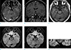
On second-admission, it was shown that the gadolinium-enhanced T1-weighted images showed a thickening and homogeneous enhancement of the right oculomotor nerve at the cisternal segment, as compared with the non-enhanced T1-weighted image (Fig. 1a-c). Upon review, the gadolinium-enhanced 3D-FLAIR image clearly demonstrates thickening and heterogeneous enhancement of the oculomotor nerve at the cisternal segment (Fig. 1d-f). At that time, the methylprednisolone was administered, and the symptoms were recovered six days later. Finally, the patient was diagnosed as RPON at that time.
DISCUSSION
In this respect, RPON is noted as common in children less than ten years of age and is seen as a very rare occurrence in adults (13). The diagnostic criteria as reviewed in the ICHD third edition are noted as recurrent unilateral headache and at least one paresis of the ocular motor nerve on the ipsilateral side at least two attacks without orbital, parasellar or found as a posterior fossa lesion on MRI (2). In this light, the neurological symptoms are characterized by a complete recovery within days to weeks (1).
In general, RPON was formerly referred to historically as an ophthalmoplegic migraine. Currently, it is reclassified as characteristically one of painful cranial neuropathies, other facial pains and other headaches, because this syndrome accompanies recurrent oculomotor cranial nerve paresis, mainly oculomotor nerve, rather than only the occurrence of a simple migraine. In addition, nerve thickening and enhancement of the oculomotor nerve can be demonstrable as noted on a gadolinium-enhanced MRI (2).
The pathophysiology of RPON is unclear. The proposed hypotheses are chronic inflammatory demyelination, simple nerve compression, reversible ischemic state of the cranial nerve and autoimmune disease (13).
Generally speaking, if there is contrast enhancement of the oculomotor nerve, the differential diagnose is known to also characteristically include tumorous conditions (schwannoma, hemangioma and lymphoma), inflammatory diseases (sarcoidosis and Tolosa-Hunt syndrome) and infectious conditions (syphilis and human immunodeficiency virus infection and meningitis) (456). To our knowledge, there is no RPON case report that shows a similar thickening and enhancement of the cranial nerve in a review of the Korean literature (7).
The gadolinium-enhanced 3D-FLAIR image is known to be more specific and accurate than the use of 3D-T1-weighted imaging in the diagnosis of a cranial neuritis (8). In our experience, the gadolinium-enhanced 3D-FLAIR images more clearly showed thickening and enhancement of the oculomotor nerve than is available to review on the gadolinium-enhanced T1-weighted image. In addition, the identified heterogeneous enhancement pattern of the oculomotor nerve on the gadolinium-enhanced 3D-FLAIR images could be explained, due to the fact that the 3D-FLAIR image is more sensitive to evaluate cranial nerves in the lower gadolinium concentrations, than as shown in the gadolinium-enhanced T1-weighted image (89). In this way, the gadolinium-enhanced 3D-FLAIR image could be useful to evaluate other cranial neuropathies.
This report is the first case of RPON with an enhancement of the oculomotor nerve on the gadolinium-enhanced 3D-FLAIR image.
 XML Download