

 PDF
PDF ePub
ePub Citation
 Print
Print


INTRODUCTION
Möbius syndrome is a rare congenital disorder, characterized by facial and abducens nerve palsy. Recently, it has been considered a type of congenital cranial dysinnervation disorder (1). In literature, most of the previous studies described morphological differences of the brainstem or cerebellum, without evaluating corresponding cranial nerves using conventional brain MRI (234). To the best of our knowledge, there are only 6 studies which have evaluated abnormalities of corresponding cranial nerves, on MRI in Möbius syndrome (156789). Herein, we present a case of Möbius syndrome in a 19-year-old male with limitation of lateral gaze, and weakness of facial expression, since the neonatal period. And, abnormalities of corresponding cranial nerves were clearly depicted, on high-resolution MR imaging. We also conducted relevant literature review on this disease.
CASE REPORT
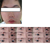
A 19-year-old male presented to our outpatient department with prolonged bilateral facial palsy, and eyeball deviation, since the neonatal period. He had no medical history of this condition. Physical examination and laboratory studies, revealed no gross abnormalities. On neurologic examination, he had difficulty blinking his eyes, smiling, or frowning when attempting to make facial expressions. Although his vertical gaze was intact, he showed deficits in horizontal gaze in both eyes, and the limitation was more prominent during abduction (Fig. 1). So, high-resolution MR imaging was performed using the 3T system (IngeniaCx; Philips Medical Systems, Best, The Netherlands). MR images included to evaluate cranial nerves were 3D T2-weighted sequence (volume isotropic turbo spin echo acquisition, VISTA), pre- and postcontrast 3D T1-fast field echo (FFE)-fat suppression sequences, and precontrast 3D fluid attenuated inversion recovery (FLAIR) VISTA sequence using a 32-channel head coil and Gd-DOTA (Dotarem; Guerbet, Paris, France) at 0.1 mmol/kg of body weight. Parameters for 3D T2 VISTA were as follow: repetition time (TR), 2000 ms; echo time (TE), 256 ms; flip angle, 90°; section thickness, 1.2 mm; spacing between sections, 0.6 mm; field of view (FOV), 18 × 18 cm; matrix size, 512 × 512; number of excitation (NEX), 1; sensitivity encoding (SENSE) factor, 2; and acquisition time, 4 minutes 2 seconds. Parameters for 3D T1-FFE-fat suppression were as follow: TR, 25 ms; TE, 4.6 ms; flip angle, 30°; FOV, 18 × 18 cm; matrix size, 512 × 512; section thickness, 0.6 mm; NEX, 1; SENSE factor, 2; and acquisition time, 3 minutes 43 seconds. Parameters for 3D FLAIR VISTA were as follow: TR, 8000 ms; TE, 268 ms; TI, 2400 ms; modulation of flip angle for refocusing pulses; section thickness, 0.6 mm; overcontiguous sections; 60 sections; FOV, 18 × 18 cm; matrix size, 512 × 512; NEX, 1; SENSE factor, 2; and acquisition time, 6 minutes 2 seconds.
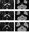
High-resolution MR images revealed absence of cisternal and canalicular segments of the left facial nerve, with severe hypoplasia of the right facial nerve (Fig. 2a–d). There was also severe hypoplasia of cisternal segments of bilateral abducens nerves, with overall poor delineation (Fig. 2). Posterior fossa showed morphological alterations in the brainstem and fourth ventricles, representing flattening of the posterior surface of the pons, due to absence of the medial colliculi, and straightening of the fourth ventricular floor (Fig. 3). There was no definite abnormality in other visualized intradural cranial nerves, and vascular structures. Extraocular muscles showed normal shape and size.
DISCUSSION
Möbius syndrome is a rare congenital and non-progressive neuromuscular disorder characterized by facial paralysis and limitation of ocular abduction. It is recently regarded as a type of congenital cranial dysinnervation (1).
Although Möbius syndrome is most frequently described as a sporadic pattern, exact etiology or pathogenesis of this syndrome remain unknown, and are likely to be diverse in nature. Among several proposed theories, two major pathogenetic explanations have been suggested (1011): (a) A primary genetic cause, implied as maldevelopment of the brainstem, and (b) A primary hypoxic-ischemic cause, possibly due to embryological or environmental toxic factors induced as interruption in the blood supply of the brainstem, during early embryologic development. Additionally, teratogenicity plays a key role in both pathogenetic mechanisms (10).
For diagnosis of this syndrome, clinical manifestations are not obligatory because clinical features are extremely variable, and the definition and criteria for diagnosis of this syndrome are debatable (10). In literature, Kumar suggested diagnostic criteria of this syndrome as follow (11): (a) complete or partial paralysis of the facial nerve as an essential criterion, (b) limb malformations (syndactyly, brachydactyly, absent digits, talipes) are commonly associated features, (c) other cranial nerve palsies such as abducens nerve may be observed, (d) orofacial malformations, ear deformities, and musculoskeletal deformities may also be observed. However, Verzijl et al. (10) recently proposed that facial palsy with impairment of ocular abduction, is primary criterion for this syndrome, regardless of association of other cranial nerve paralysis, musculoskeletal defects, or orofacial malformations.
Approximately 300 cases of this syndrome have been reported in literature. However, few studies have described imaging findings of the Möbius syndrome, and these studies usually focused on morphological alterations and differences of the posterior fossa structures, such as brainstem hypoplasia or flattening of the fourth ventricle, without evaluating abnormality of corresponding cranial nerves on the conventional brain MRI (234). As shown in these previous studies, conventional MRI was limited to delineate cranial nerves, because they are anatomically complex and small.
However, with technical advances, a new isotropic 3D turbo spin-echo sequence with variable flip angles (VISTA, sampling perfection with application optimized contrast using different flip angle evolution [SPACE], or Cube), was introduced on the 3T MR system. This sequence provides much higher spatial resolution, as well as excellent contrast resolution with submillimetric section thicknesses, because it can generate a strong signal in tissues with high T2/T1 ratio, such as cerebrospinal fluid and fat (12). So, this sequence is particularly valuable in depicting cisternal segments of cranial nerves clearly, in acceptable scan duration. To the best of our knowledge, there are only six studies to evaluate abnormality of cranial nerves in detail, using this high-resolution MRI in patients with Möbius syndrome (156789).
In this case, we diagnosed Möbius syndrome, based on clinical and radiological findings. Imaging findings of our case are consistent with the previous studies, by showing underdevelopment of bilateral facial and abducens nerves such as aplasia or hypoplasia, and flatting of the posterior pons, with straightening of the fourth ventricles, due to absence of facial folliculi (123456789). Although Möbius syndrome is usually diagnosed by characteristic clinical features in daily practice, this case provides that high-resolution MRI is valuable in ensuring clinical diagnosis, by demonstrating anatomical details and abnormalities of cranial nerves, as well as adjacent vascular structures, brainstem, and cerebellum. In contrast to the previous studies, we obtained the well-known 3D FLAIR VISTA sequence to allow recognition subtle tissue change of cranial nerves, or minimal subtle compositional changes of the inner ear, because it can minimize undesired inflow artifacts of CSF flow. In this case, the 3D FLAIR VISTA sequence is more valuable in tracing the abducens nerve in the prepontine cistern, than 3D T2 VISTA, due to suppression of background CSF flow despite severe hypoplasia (Fig. 1a, b, e, f). So, this sequence may be valuable in differentiating the pathologic condition of cranial nerves, between hypoplasia and aplasia.
In conclusion, we report a rare case of Möbius syndrome, caused by abducens and facial nerve abnormalities, revealed on high-resolution MRI. Through this case report, we hope to highlight the value of meticulous radiological review, for cranial nerves and structures of the posterior fossa on high-resolution MRI, as well as clinical features for diagnosing patients with Möbius syndrome.

 XML Download