

 PDF
PDF ePub
ePub Citation
 Print
Print


INTRODUCTION
Klebsiella pneumoniae (K. pneumoniae) has the ability to develop resistance to multiple classes of antibiotics (1). In K. pneumoniae, multidrug resistance is mostly due to the production of CTX-M-type extended-spectrum β-lactamases (ESBLs) (2). In particular, CTX-M-15 producing K. pneumoniae sequence type 11 (ST11) strains are disseminated worldwide, including in Korea, like the ST131 clone of Escherichia coli (E. coli) (34). It has been reported that plasmids harboring blaCTX-M-15 do not confer less competitiveness to their bacterial hosts (56). Thus, it has been suggested that toxin-antitoxin (TA) systems, such as VagCD, aid in plasmid maintenance (78), as TA systems are known to be associated with persister cell formation under stress conditions such as antibiotics, oxidative stress, and nutrient deficiency (9). Persisters are a small subpopulation of bacterial cells that are highly tolerant to antibiotics despite their susceptibility (10). It has been suggested that persisters might be one of the reasons for antibiotic treatment failure and might contribute to the evolution of antibiotic resistance (1112). For instance, there is a report that DNA damage of the persisters caused by fluoroquinolone induces RecA and SOS response that accelerates the development of antibiotic resistance (13). In this study, we investigated whether the TA systems in blaCTX-M-15 bearing plasmids could contribute to persister formation.
MATERIALS AND METHODS
Strains, plasmids, and antimicrobial susceptibility testing
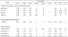
Plasmids were obtained from four CTX-M-15-producing K. pneumoniae ST11 clinical isolates that were collected from patients in Hong Kong, Malaysia, India, and Thailand (HK02-026, M16-13, IN03-01, and TH02-34, respectively). Their whole plasmid sequences were determined previously (GenBank accession numbers, KY751926, KY751925, KY499796, and KY499797) (14). While TA genes, such as pemIK, mok-hok, and vagCD, were identified on the blaCTX-M-15-bearing plasmids of HK02-026 and M16-13 (TA+), the blaCTX-M-15-bearing plasmids of IN03-01 and TH02-34 had no TA genes (TA−). Then, recovered plasmids were transferred to E. coli J53 cells, as described previously (6). In vitro antimicrobial susceptibility testing was performed by the broth microdilution method according to Clinical and Laboratory Standards Institute (CLSI) guidelines (15), and susceptibility was defined according to CLSI breakpoints. E. coli ATCC 25922 and Pseudomonas aeruginosa ATCC 27853 were used as control strains.
Persister cell assay
A persister cell formation assay was performed using aminoglycosides (amikacin and streptomycin) as previously described (16), with minor modifications. Briefly, cells were grown to exponential phase (optical density (OD) 600= 0.5) and then exposed to antibiotics (5×minimum inhibitory concentration (MIC) or 10×MIC) at 37℃, with shaking at 185 rpm for 3.5 hours (h). After incubation, samples were washed twice with phosphate-buffered saline (PBS), and then serial dilutions were spread on Luria-Bertani (LB) agar (MBcell, Seoul, Korea). After incubation, the colony-forming units (CFUs) of surviving persister cells were determined. To confirm their identity as persisters, two random colonies from each sample were re-inoculated into fresh LB broth for an overnight culture and in vitro antimicrobial susceptibility testing was performed to ensure the no change in MIC.
Quantitative reverse transcription PCR (qRT-PCR)
Expression levels of relA and spoT, which are known to be involved in (p)ppGpp synthesis, were determined by quantitative reverse transcription PCR (qRT-PCR) as described previously (17). Expression levels of three toxin genes (hok, pemK, and vagD) of TA systems were compared between wild-type (WT) and persister cells in two transconjugants with TA+ plasmids (J53/pHK02-026 and J53/pM16-13) by qRT-PCR. The expression levels of target genes were analyzed by the comparative threshold cycle (CT) method, and rpoB expression was assessed in parallel for normalization of target gene transcript levels. Experiments were repeated with three independent cultures, each tested in duplicate.
Statistical analysis
Data are presented as means ± standard deviations. Pairwise comparisons were performed with one-way analysis of variance (ANOVA), using Prism version 5.01 software for Windows (GraphPad Software, San Diego, CA, USA). P values less than 0.05 were considered to be statistically significant.
RESULTS
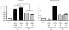
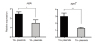
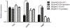
The transconjugants showed elevated MICs of β-lactam antibiotics because of the presence of CTX-M-15 on the plasmids. While three of the transconjugants were susceptible to both amikacin and streptomycin, J53/pHK02-026 showed an increased MIC of streptomycin (Table 1). Therefore, we did not perform a persister assay with J53/pHK02-026 in streptomycin. Persister formation in amikacin for all transconjugants showed significant increase when compared to that of the J53 strain (Fig. 1). In streptomycin, only J53/pM16-13 exhibited a significantly elevated rate of persister formation. In addition, the persister formation rates in transconjugants with TA+ plasmids (J53/pHK02-026 and J53/pM16-13) were higher than in transconjugants with TA− plasmids (J53/pIN03-01 and J53/pTH02-34) for both tested aminoglycosides (Fig. 1). Moreover, the expression levels of both relA and spoT were significantly higher in transconjugants with TA+ plasmids than in those with TA− plasmids (Fig. 2). While expression of hok did not increase in persister cells of both transconjugants with TA+ plasmids, pemK and vagD in persister cell of J53/pHK02-026 were overexpressed compared with WT (Fig. 3). However, WT and persister did not show different expression levels of all TA systems in another transconjugant, J53/pM16-13.
DISCUSSION
Previously, it was suggested that a plasmid harboring blaCTX-M-15 might not impose a fitness cost, even without antibiotic pressure, with respect to in vitro competition and serum resistance (56). Such a selective advantage may contribute to the prevalence of antibiotic-resistant clones. In this study, we further suggest that TA systems on plasmids are associated with a high persister formation rate and maintenance of CTX-M-15-producing K. pneumoniae strains even with high antibiotic pressure.
Although the two plasmids in K. pneumoniae isolates from Hong Kong and Malaysia had different structure (14), they harbored the same TA systems, pemIK, mok-hok, and vagCD. These TA systems have similar target mechanisms, i.e., translation inhibition, mostly by interrupting ribosome function (18). Once activated, TA systems can convert normal growing cells into persister cells without changing their drug susceptibility; thus helping the bacteria survive antibiotic treatment. Indeed, higher expression levels of relA and spoT, which are involved in (p)ppGpp synthesis (19), were observed in transconjugants with TA+ plasmids. In this study, transconjugants with TA+ plasmid showed elevated expression of pemK and vagD in persister cell, although another with TA+ plasmid showed no elevated expression of TA systems in persister cell. Thus, it was proposed that persister formation induced by TA systems on blaCTX-M-15-bearing plasmid may be one of the mechanisms contributing to both plasmid maintenance and bacterial survival, even in the presence of effective antibiotics.
 XML Download